Advancing single molecule microscopy with active stabilization
Speaker: Simao Pereira Coelho Moderator: Antoine de Morrée
Online live: Aug 16, 2022 12:00 PM ET | 9:00 AM PT Posted: Aug 31, 2022 Views: 6284
 WEBINAR SERIES
Abstract
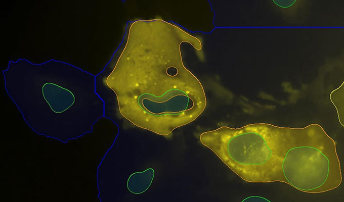
Single-molecule microscopy has greatly expanded our knowledge of structural organizations, functional conformations and dynamics of protein complexes within cellular environments. Remarkable progress has been made in single-molecule imaging methods to improve spatial resolution, penetration depth, and live-cell imaging capabilities. A key part of any super-resolution technique involves accurately correcting for mechanical motion of the sample and setup during acquisition. If left uncorrected, drift degrades the resolution of the final reconstructed image and can introduce unwanted artifacts. Here, I will describe the recent advances we have made including the application of Direct-laser writing to perform 3D single-molecule acquisitions over cellular volumes.
Speaker
Simao Pereira Coelho, Ph. D.
Research Associate, University of New South Wales / Instituto Gulbenkian de Ciência
Dr. Coelho has a PhD in Biophysics (King’s College London, 2015) funded by Cancer Research UK; an MRes in Biophysics (University of Liverpool, 2010...
View more ![]()
Moderator

Antoine de Morrée, Ph.D.
Tenure-track Assistant Professor, University of Aarhus
Antoine de Morrée, PhD is a tenure-track Assistant Professor at the department of Biomedicine, Aarhus University, where his View more ![]()
Keywords
Single molecule microscopy, Drift correction, Direct laser writing
References
Coelho, S., Baek, J., Gooding, J. J. and Gaus, K. (2021). Building a Total Internal Reflection Microscope (TIRF) with Active Stabilization (Feedback SMLM). Bio-protocol 11(13): e4074. DOI: 10.21769/BioProtoc.4074.
Do you have a question about this webinar?
Post your question, and we'll invite the webinar speaker to respond. You're welcome to join the discussion by answering or commenting on questions ( Note: Not all questions, especially those not directly relevant to the webinar topic, may be answered by the speaker. ).
![]()
+
35 Q&A

How can I track fluorescent particles over time? What do I have to consider for experimental setup, what software or algorithms to analyze the data?
Which method is used to attach fluorophores in single molecule fluorescence microscopy?
How can you differentiate between sample movement (drift) and movement of the particle of interest?

How can undrift algorithms be used for SMLM in live-cell imaging?


Can single-molecule microscopy be applied to the work of spatiotemporal-omics? Can it will be used to integrate genomics and imaging techniques?
Please, is it allowed to share the link to the webinar with colleagues and friends?
I work on microalgae can u ple How i can relate microscopy with microalgae? And we will also get the e - certificate for the workshop?
How to stain Cell with FITC?,how to visualize leukocyte transmigration by imaging?,how to detect microfilaments of DNA during Netosis process?
How to plan single molecule microscopy with "Bodipy" tagged vancomycin in bacterial cells?
 Jun 18, 2026
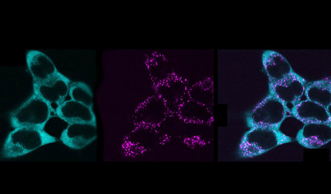 May 27, 2026
 Apr 28, 2026