
- Home
- Protocols

-

Surgical procedures and viral vector injections
Last updated date: Jan 19, 2022 Views: 794 Forks: 0
Thank you for your interest in our work. The followings are the responses to your requests.
a) The rate of each viral vector injection was approximately 1 µL/sec.
b) The wait time between vector injections in a single track was approximately 5 sec.
c) The incubation wait time was approximately 1 sec.
Our detailed protocol is as follows.
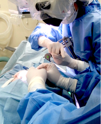
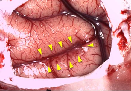
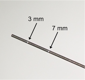
After retracting skin and galea, the frontal cortex was exposed by removing a bone flap and reflecting the dura mater. Handheld injections were made under visual guidance through an operating microscope (Leica M220, Leica Microsystems GmbH), with care taken to place the beveled tip of the Hamilton syringe containing the viral vector at an oblique angle to the brain surface (Fig. 1). Nine tracks were injected in each hemisphere; one was located 1 mm posterior to the caudal tip of the principal sulcus, and the others were located along the dorsal (4 tracks) and ventral (4 tracks) bank of the principal sulcus posterior to the rostral tip of the ascending limb of the arcuate sulcus (Fig. 2). The injection needle was inserted into the brain for 5-8 mm, and vector was injected with approximately 1.0-1.5 mm intervals, resulting that viral vectors were injected 3 to 5 µL per track depending on the depth. We used a customized needle with markers (Muromachi Kikai Co., Ltd.) so that the depth can be visually estimated (Fig. 3).
The angle and depth of each track was pre-determined before the surgery based on MR images. The injections for one hemisphere were completed within 30 min, minimizing damage to the exposed tissue. We consider that the handheld injection is an efficient method to fill viral vectors to cover a large volume of cortical tissue, like this case.
Additional requests or comments are cordially welcome.
- Oyama, K, Nagai, Y and Minamimoto, T(2022). Surgical procedures and viral vector injections. Bio-protocol Preprint. bio-protocol.org/prep1510.
- Oyama, K., Hori, Y., Nagai, Y., Miyakawa, N., Mimura, K., Hirabayashi, T., Inoue, K., Suhara, T., Takada, M., Higuchi, M. and Minamimoto, T.(2021). Chemogenetic dissection of the primate prefronto-subcortical pathways for working memory and decision-making. Science Advances 7(26). DOI: 10.1126/sciadv.abg4246
Category
Do you have any questions about this protocol?
Post your question to gather feedback from the community. We will also invite the authors of this article to respond.
