- Protocols

- Articles and Issues
- For Authors
- About
- Become a Reviewer

Separation and Detection of Phosphorylated and Nonphosphorylated BvgA, a Bordetella pertussis Response Regulator, in vivo and in vitro
Published: Vol 3, Iss 22, Nov 20, 2013 DOI: 10.21769/BioProtoc.970 Views: 11570
Reviewed by: Fanglian He
Original research article
The authors used this protocol in:

Apr 2013
Advertisement


Protocol Collections
Comprehensive collections of detailed, peer-reviewed protocols focusing on specific topics


Abstract
Protein phosphorylation plays a central role in signal transduction in bacteria. However, separation and detection of the phosphorylated protein from its nonphosphorylated form remain challenging. Here we describe a method to detect phosphorylation of the Bordetella pertussis response regulator BvgA, which is phosphorylated at an aspartate residue (Boulanger et al., 2013). This method is based on the proprietary adduct, Phos-tagTM, a dinuclear metal complex, which together with Zn2+ or Mn2+, forms a complex with a phosphomonoesterdianion, such as the phosphorylated aspartate of a response regulator (Barbieri and Stock, 2008; Kinoshita and Kinoshita-Kikuta, 2011). For in vivo detection, B. pertussis cells are lysed in mild formic acid at 4 °C to minimize the disruption of the phospho-aspartate bond, and the phosphorylated BvgA is separated from its nonphosphorylated form by electrophoresis (SDS-PAGE) containing Phos-tagTM. Both forms of BvgA are subsequently detected by Western Blot analysis. Quantification of the level of phosphorylated BvgA formed after treatment with acetyl phosphate in vitro is also easily accomplished. Thus, this technique allows one to readily assess the levels of BvgA phosphorylation in B. pertussis and in E. coli under different laboratory conditions in vivo or after phosphorylation under varying reaction conditions in vitro (this research was supported in part by the Intramural Research Program of the NIH, NIDDK).
Materials and Reagents
- Bordetella pertussis strain BP536 is the tet-resistant derivative of the clinical isolate Tohama I strain (laboratory inventory)
- Formic acid (> 95%) (Sigma-Aldrich, catalog number: F0507 )
- NaOH (Thermo Fisher Scientific, catalog number: SS255-1 )
- Tris base (MP Biomedicals, catalog number: 819620 )
- Bromophenol blue (Sigma-Aldrich, catalog number: B8026 )
- Glycerol (Invitrogen, catalog number: 15514-011 )
- Butanol (Sigma-Aldrich, catalog number: 15467-9 )
- 30% 29:1 Acrylamide/bis-acrylamide (Sigma-Aldrich, catalog number: A3574 )
- 10% SDS (Hoefer, catalog number: GR155-1 )
- Phos-tagTM acrylamide (Wako Pure Chemical Industries, catalog number: AAL-107 )
- Zn(NO3)2 (Sigma-Aldrich, catalog number: 228737 )
- Ammonium persulfate (Sigma-Aldrich, catalog number: 3678 )
- N,N,N’,N’-Tetramethylethylenediamine (TEMED) (Sigma-Aldrich, catalog number: T9281 )
- 0.5 M EDTA, pH 8.0 (Research Genetics, catalog number: 750009 )
- MOPS (Fluka, catalog number: 69947 )
- Sodium metabisulfite (Na2S2O5) (Sigma-Aldrich, catalog number: S9000 )
- Glycine (Sigma-Aldrich, catalog number: G7126-1KG )
- Methanol (EMD Millipore, catalog number: MX0485-7 )
- PBS buffer (Gibco, catalog number: 2014-10 )
- BactoTM proteose peptone (Becton, Dickinson and Company, catalog number: 211684 )
- DifcoTM Bordet Gengou agar base (Becton, Dickinson and Company, catalog number: 248200 )
- Defibrinated sheep blood (LAMPIRE® Biological Labs, catalog number: 7239001 )
- Lithium potassium acetyl phosphate (Sigma-Aldrich, catalog number: A0262-500MG )
- Colloidal Blue staining kit (Life Technologies, catalog number: LC6025 )
- Tween-20 (Bio-Rad Laboratories, catalog number: M3524 )
- Non-fat dry milk (Giant Food Store, catalog number: 688267078330 )
- Monoclonal anti-BvgA antibody (laboratory inventory)
- HRP-conjugated Secondary Antibody (Santa Cruz, catalog number: SC2005 )
- Amersham ECL Primer Western Blotting Detection System (General Electric Company, catalog number: RPN2232 )
- PVDF filter (Invitrogen, catalog number: LC6025 )
- BSA (Albumin from bovine serum) (Sigma-Aldrich, catalog number: A2153 )
- 1 M formic acid (see Recipes)
- 5 N NaOH (see Recipes)
- 1 M Tris-Cl solutions (see Recipes)
- 1% Bromophenol blue (see Recipes)
- 5x Loading Solution (see Recipes)
- Water-saturated butanol (see Recipes)
- 5 mM Phos-tagTM acrylamide (see Recipes)
- 10 mM Zn(NO3)2 (see Recipes)
- 10% APS (see Recipes)
- 1x MOPS Running Buffer, pH 7.8 (see Recipes)
- Transfer Buffer (see Recipes)
- BG agar plates (see Recipes)
- 200 mM acetyl phosphate (see Recipes)
- 1% or 5% BSA in PBS (see Recipes)
- 0.05% Tween-20 in PBS (see Recipes)
- 1% Non-fat milk in PBS (see Recipes)
Equipment
- Polyester-tipped applicator (Puritan Medical, catalog number: 25-806 1PD )
- Spectrophotometer
- Mini gel cassettes (1.0 mm) (Invitrogen, catalog number: NC2010 )
- 10, 12 or 15-well combs (1.0 mm) (Invitrogen, catalog number: NC3015 )
- XCell SureLock® Mini-Cell (Invitrogen, catalog number: EI0001 )
- Platform shaker
- Eppendorf microfuge
- Bio-Rad Mini-Protean II (Bio-Rad Laboratories)
Procedure
- Whole cell lysate preparation for in vivo detection of BvgA phosphorylation
Note: This protocol has been successfully used with another gram-negative bacteria, E. coli.
- To collect bacteria sample, Bordetella pertussis strain BP536, grown at 37 °C for 2 days on BG agar plate, is swabbed from the plate with a polyester-tipped applicator and resuspended in 1.5 ml of PBS.
- An aliquot from step A1 is used to determine the OD600 reading.
- A 0.3 ml aliquot from step A1 is centrifuged at 15,600 x g in Eppendorf microfuge for 1 min at room temperature. The supernatant is removed, and the resulting pellet is frozen in dry ice. It can be used directly or stored at -80 °C.
- The frozen pellet is treated as follows.
Note: Volumes are based on an OD600 reading of 0.5 and a pellet obtained from 0.3 ml, one should adjust the volumes proportionally based on the determined OD600.
First, 33 μl of ice-cold 1 M formic acid is added, and the pellet is disrupted by pipetting repeatedly. Immediately, a freshly made ice-cold solution (27 μl) containing 2 μl of 5 N NaOH (to neutralize the acid), 10 μl H2O, and 15 μl of 5x Loading Solution is added.
Note: The color of the solution turns yellow due to the acid, but the bromophenol blue changes back to blue once it enters the gel.
- 4 μl of the resulting cell lysate are loaded onto the Phos-tagTM gel for electrophoresis.
Note: Do not boil the treated cell lysate; the lysate should be kept on ice before loading.
- To collect bacteria sample, Bordetella pertussis strain BP536, grown at 37 °C for 2 days on BG agar plate, is swabbed from the plate with a polyester-tipped applicator and resuspended in 1.5 ml of PBS.
- Phos-tagTM SDS-PAGE gel electrophoresis (Recipe makes one mini gel.)
4% stacking gel
30% Acrylamide/bis-acrylamide
417.5 μl
1 M Tris pH 6.8
1,093 μl
10% SDS
31.3 μl
10% APS (freshly prepared)
25 μl
TEMED
5 μl
H2O
1,570 μl
10% resolving gel
30% Acrylamide/bis-acrylamide
2,085 μl
1 M Tris pH 6.8
2,185 μl
10% SDS
62.5 μl
5 mM Phos-tagTM
93.8 μl
10 mM Zn(NO3)2
93.8 μl
10% APS (freshly prepared)
25 μl
TEMED
5 μl
H2O
1,715 μl
- For resolving gel: Acrylamide, Tris, SDS, Phos-tagTM, Zn(NO3)2, and H2O are mixed. 10% APS and TEMED are added, and the solution is immediately poured into the gel cassette. The solution is then overlaid with 1 ml of water-saturated butanol. The resolving gel polymerizes in ~40 min at room temperature.
- The butanol is poured off of the polymerized resolving gel, and the gel surface is then washed with water. Excess water is removed using absorbent paper.
- For stacking gel: Acrylamide, Tris, SDS, and H2O are mixed. 10% APS and TEMED are added, and the solution is immediately poured into the gel cassette. The comb is quickly inserted (avoiding the introduction of air bubbles) and the stacking gel is allowed to polymerize for 20 min at room temperature.
- After insertion of the gel cassette into the electrophoresis apparatus, the ice-cold 1x MOPS Running Buffer is added. The entire apparatus is then placed into an ice-filled bucket and the bucket is placed on a platform shaker so that it can be gently rotated until the temperature of the Running Buffer in the chamber has cooled to 4 °C. This typically takes at least 30 min.
Note: Place only one gel cassette in one XCell SureLock® Mini-Cell to ensure that the temperature remains cold during electrophoresis.
- The shaker is turned off and the samples are loaded using a pipetman.
- After resumption of gentle shaking, electrophoresis is performed with constant 100 V for 30 min, 120 V for 30 min, and then 150 V until the bromophenol blue dye is ~ 1 cm from the bottom of the gel. (Temperature within the inner gel chamber will rise to 10 °C to 15 °C.)
- For Western blot analysis, the gel is washed with 100 ml Transfer Buffer containing 1 mM EDTA for 10 min to remove Zn2+ from the gel, followed by washing again with 100 ml Transfer Buffer for 20 min.
- The gel is transferred to a PVDF filter, which was previously rinsed with methanol and then with Transfer Buffer, in a Bio-Rad Mini-Protean II apparatus filled with ice-cold Transfer Buffer at 100 constant voltage for 1 h at 4 °C.
- The antibody detection of BvgA is carried out at room temperature as follows: After the transfer, the PVDF filter is blocked by washing with 5% BSA in PBS for 1 h, and then incubated with monoclonal anti-BvgA antibody (1:5,000) in 1% BSA in PBS for 1 h. Three 10-min washes with 0.05% Tween-20 in PBS are conducted before incubation with HRP-conjugated Secondary Antibody (1:2,000) in 1% non-fat milk in PBS for 1 h. The PVDF filter is washed three times for 10 min, each, with 0.05% Tween-20 in PBS, and then developed with Amersham ECL Primer Western Blotting Detection System, according to the manufacture’s instruction.
- For resolving gel: Acrylamide, Tris, SDS, Phos-tagTM, Zn(NO3)2, and H2O are mixed. 10% APS and TEMED are added, and the solution is immediately poured into the gel cassette. The solution is then overlaid with 1 ml of water-saturated butanol. The resolving gel polymerizes in ~40 min at room temperature.
- Separation of in vitro phosphorylated purified protein
- To phosphorylate in vitro, 9 μl purified BvgA (from ~1 to 25 pmol) is mixed with 1 μl 200 mM acetyl phosphate on ice and then incubated for the desired amount of time at 37 °C.
- Reaction is stopped by placing the sample on dry ice.
- Sample is mixed with 2.5 μl 5x Loading Solution (final concentration of 1x), separated on PhostagTM acrylamide gel, washed, and detected by Western analysis as described above.
- Alternately, gel can be stained. In this case, the gel is first washed 3 times in 100 ml of water for 10 min. The gel is then treated with Colloidal Blue following manufacturer’s instructions.
Note: For best results, use either Colloidal Blue or classic Coomassie blue staining rather than other types of protein stains.
Note: The optimal level of purified protein for loading is ~ 1 pmol for Western analysis and ~25 pmol for gel staining
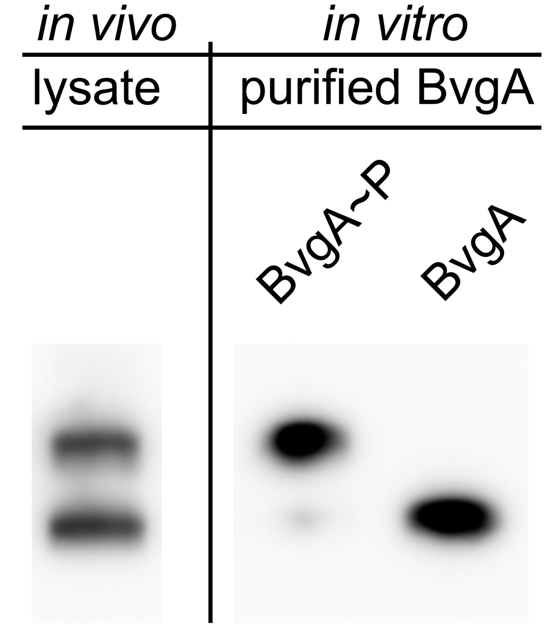
Figure 1. Separation of phosphorylated BvgA from unphosphorylated BvgA by Phos-tagTM SDS-PAGE, analyzed by Western blot. First lane shows the in vivo lysate of B. pertussis, which contains both phosphorylated BvgA (BvgA~P) and nonphosphorylated BvgA (BvgA). Lanes 2 and 3 show purified BvgA, which has been phosphorylated in vitro by the addition of acetyl phosphate or incubated in the absence of acetyl phosphate, respectively.
- To phosphorylate in vitro, 9 μl purified BvgA (from ~1 to 25 pmol) is mixed with 1 μl 200 mM acetyl phosphate on ice and then incubated for the desired amount of time at 37 °C.
Recipes
- 1 M formic acid
5 ml of formic acid (> 95%) in 95 ml of H2O
Stored at 4 °C
- 5 N NaOH
5 ml 10 N NaOH
5 ml H2O
Stored at 4 °C
- 1 M Tris-Cl solutions (for 1 L)
121.14 g Tris base adjusted to pH 6.8 or to pH 8.0 with HCl at 4 °C
Filter sterilized
- 1% Bromophenol blue
1 g bromophenol blue in 100 ml H2O
- 5x Loading Solution
1% SDS
65 mM Tris-Cl (pH 6.8)
25% glycerol
0.02% bromophenol blue
Stored at 4 °C
- Water-saturated butanol
4 ml water added to 40 ml butanol
Mix by shaking
Butanol will be the top layer
- 5 mM Phos-tagTM acrylamide
10 mg Phos-tagTM acrylamide dissolved in 100 μl methanol
Solution is pipetted repeatedly to dissolve the acrylamide
Add 3.2 ml H2O
Solution is centrifuged at 2,000 x g for 10 min to remove white precipitate
Stored at 4 °C in dark
- 10 mM Zn(NO3)2
2.97 g Zn(NO3)2 dissolved in 1 L H2O
Filter sterilized
- 10% APS
1 g ammonium persulfate dissolved in 10 ml H2O
Solution is freshly prepared
- 1x MOPS Running Buffer, pH 7.8
100 mM Tris-CL, pH 7.8
100 mM MOPS, pH 7.8
0.1% SDS
5 mM sodium metabisulfite
For 1 L
12.1 g Tris base
20.9 g MOPS
10 ml 10% SDS
0.95 g sodium metabisulfite (Na2S2O5)
Solution is adjusted to pH 7.8 with 10 N HCl at 4 °C
Stored at 4 °C
- Transfer Buffer
3 g Tris base
14.4 g glycine
200 ml of methanol
800 ml of H2O
- BG agar plates
10 g DifcoTM Bordet Gengou agar base
3.3 g BactoTM proteose peptone
3.3 ml glycerol added to 350 ml H2O
The mixture is autoclaved
Cooled to 46 °C before adding 50 ml defibrinated sheep blood
Pour warm solution into petri dishes and allow to cool to room temperature.
Stored at 4 °C
- 200 mM acetyl phosphate
0.0039 g lithium potassium acetyl phosphate dissolved in 105.7 μl 20 mM Tris-Cl, pH 8 Solution is freshly made
Stored on ice
- 1% or 5% BSA in PBS
1 g or 5 g BSA
100 ml PBS
- 0.05% Tween-20 in PBS
0.05 ml of Tween-20
100 ml of PBS
- 1% non-fat milk in PBS
1 g non-fat dry milk
100 ml of PBS
Acknowledgments
This protocol is based on the previously published paper Boulanger et al. (2013). The research was supported in part by the Intramural Research Program of the NIH, NIDDK.
References
- Barbieri, C. M. and Stock, A. M. (2008). Universally applicable methods for monitoring response regulator aspartate phosphorylation both in vitro and in vivo using Phos-tag-based reagents. Anal Biochem 376(1): 73-82.
- Boulanger, A., Chen, Q., Hinton, D. M. and Stibitz, S. (2013). In vivo phosphorylation dynamics of the Bordetella pertussis virulence-controlling response regulator BvgA. Mol Microbiol 88(1): 156-172.
- Kinoshita, E. and Kinoshita-Kikuta, E. (2011). Improved Phos-tag SDS-PAGE under neutral pH conditions for advanced protein phosphorylation profiling. Proteomics 11(2): 319-323.
Article Information
Copyright
© 2013 The Authors; exclusive licensee Bio-protocol LLC.
How to cite
Chen, Q., Boulanger, A., Hinton, D. M. and Stibitz, S. (2013). Separation and Detection of Phosphorylated and Nonphosphorylated BvgA, a Bordetella pertussis Response Regulator, in vivo and in vitro. Bio-protocol 3(22): e970. DOI: 10.21769/BioProtoc.970.
Category
Microbiology > Microbial signaling > Phosphorylation
Cell Biology > Cell signaling > Phosphorylation
Biochemistry > Protein > Modification
Do you have any questions about this protocol?
Post your question to gather feedback from the community. We will also invite the authors of this article to respond.