


- Protocols

- Articles and Issues
- For Authors
- About
- Become a Reviewer


Elimination of cis-cleavage in CRISPR Diagnostics for One-Pot Rapid Nucleic Acid Detection

(*contributed equally to this work) Published: Vol 16, Iss 23, Dec 5, 2026 DOI: 10.21769/BioProtoc.5870 Views: 30
Reviewed by: Navnita DuttaGyan Prakash SrivastavaSAPTARSHI MAJI
 PDF
 Ask a question

Original research article
The authors used this protocol in:

Apr 2026

Protocol Collections
Comprehensive collections of detailed, peer-reviewed protocols focusing on specific topics



Abstract
CRISPR/Cas12a-based nucleic acid detection enables rapid molecular diagnostics, yet conventional two-step assays are slow and contamination-prone, while one-pot methods suffer from Cas12a cis-cleavage of amplicons. This protocol presents a one-pot, cis-cleavage-free SCas12a system using a bipartite split crRNA—comprising a scaffold and 20-nt spacer—that directs Cas12a to PAM-free double-stranded DNA with no detectable cis-cleavage under the tested conditions while maintaining trans-cleavage of ssDNA reporters. We detail RNA/primer design, ortholog expression and purification, RNP assembly, and integration with RPA, RT-RPA, or LAMP for unified detection and single-nucleotide variant (SNV) genotyping. The original study demonstrated improved sensitivity and single-base discrimination in selected model assays, with clinical application examples involving HPV16, SARS-CoV-2, and TP53. These results support potential point-of-care adaptation; broader clinical performance and portable readout remain to be validated.
Key features
• PAM-independent split-crRNA design frees target-site selection from canonical and suboptimal PAM sequence constraints.
• Absence of detectable cis-cleavage under the tested conditions prevents amplicon degradation, enabling simultaneous isothermal amplification and CRISPR detection in a single tube.
• Compatible with RPA/RT-RPA using AsCas12a at 37 °C and LAMP using thermostable CtCas12a at 55 °C; LbCas12a was also examined in the original biochemical characterization.
• Attomolar sensitivity within 30 min and single-base resolution (up to 91-fold distinguishing factor) for SNV genotyping; these values apply to the reported model assays.
Keywords: CRISPR/Cas12a Split crRNA
SCas12a
One-pot detection
Isothermal amplification
RPA
LAMP
Point-of-care diagnostics
Graphical overview
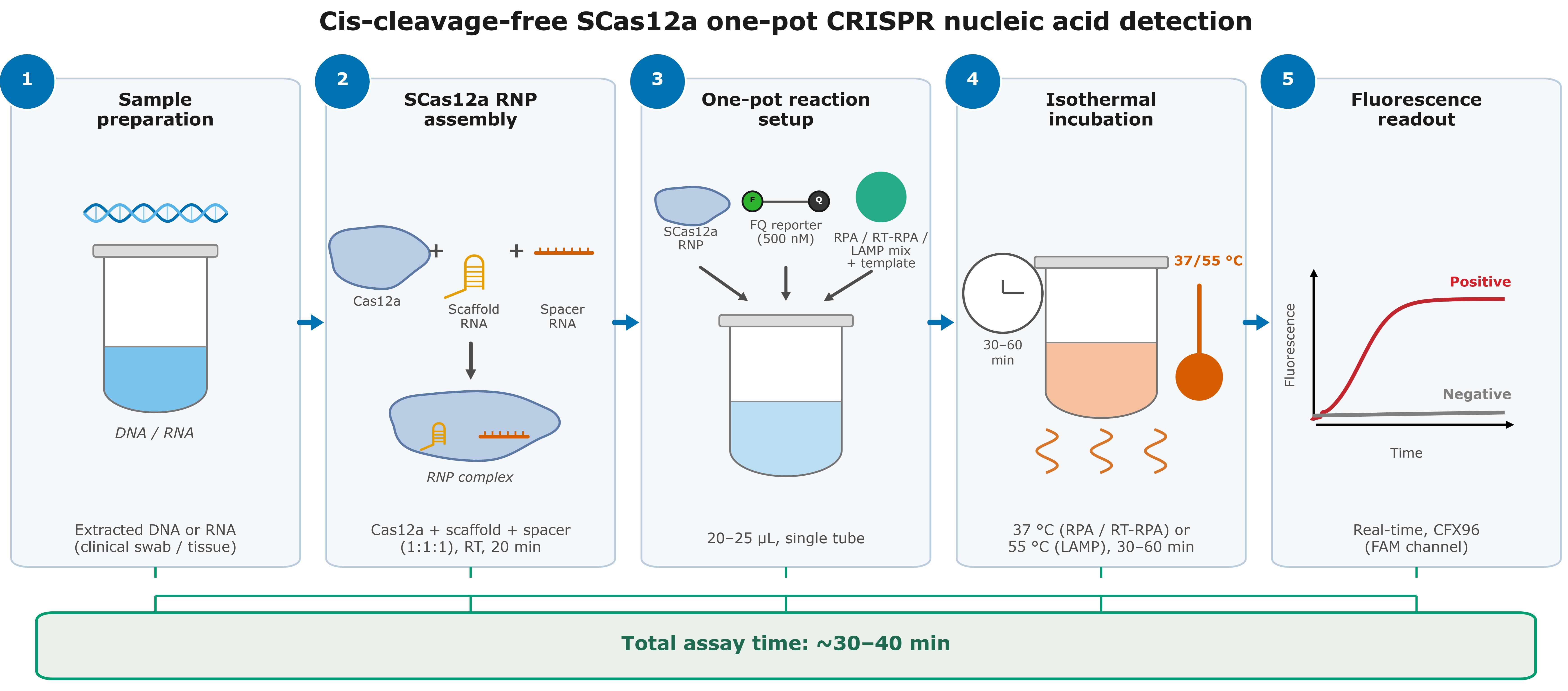
Workflow of the cis-cleavage-free SCas12a one-pot nucleic acid detection assay. The diagram is schematic: The experimental Cas12a:scaffold RNA:spacer RNA input ratio is 1:2:2 (section B). DNA and RNA templates follow sections C and D, respectively; RPA/RT-RPA uses AsCas12a at 37 °C, whereas LAMP uses CtCas12a at 55 °C.
Background
Nucleic acid diagnostics are essential for controlling infectious diseases, genetic screening, and environmental monitoring, yet gold-standard methods such as PCR and sequencing remain confined to centralized laboratories. Isothermal nucleic acid amplification technologies, including recombinase polymerase amplification (RPA) [1] and loop-mediated isothermal amplification (LAMP) [2], eliminate the need for thermocycling and enable portable, cost-effective testing. The discovery of the collateral cleavage activity of CRISPR effectors, such as Cas12a, has enabled highly specific detection of amplified products, giving rise to diagnostic platforms such as DETECTR [3], HOLMES [4], and SHERLOCK [5]. However, these early CRISPR diagnostics employ a two-step procedure—nucleic acid amplification followed by the addition of CRISPR reagents—which increases procedural complexity, prolongs testing time, and heightens the risk of contamination from nucleic acid aerosols.
Combining isothermal amplification and Cas12a-mediated detection in a single tube (one-pot assay) is highly desirable, but a major challenge remains: the cis-cleavage activity of Cas12a, triggered upon PAM-dependent target recognition, degrades amplicons as they are generated and thereby severely interferes with amplification. Several mitigation strategies have been developed, each with limitations. Targeting suboptimal PAM sequences (e.g., NTTV, TTTN) attenuates LbCas12a cis-cleavage kinetics and improves one-pot performance [6], but PAM compatibility constraints and residual cis-cleavage remain [6,7]. Sodium heparin can tune Cas12a cis-cleavage by disrupting Cas12a–crRNA binding, allowing amplicon accumulation [8]; photocaged crRNAs permit light-controlled activation of Cas12a after amplification [9]; and spatial segregation of amplification and detection reagents using glycerol or sucrose phases [10], or physical separation of components in all-in-one formats [11,12], can reduce premature cleavage. Despite these advances, existing one-pot systems still face prolonged detection times, operational complexity, and residual collateral damage, because the nuclease remains inherently capable of cis-cleavage.
Our group previously developed the SCas12a assay, which integrates Cas12a with a split crRNA—comprising a scaffold RNA and a separate spacer RNA—to enable direct RNA detection without reverse transcription [13,14]. In the underlying study of this protocol [15], we reevaluated the system for DNA detection and unexpectedly found that SCas12a maintains robust trans-cleavage activity on double-stranded DNA (dsDNA) even in the complete absence of canonical or reported suboptimal PAM sequences, while exhibiting negligible cis-cleavage activity toward dsDNA targets. The split-crRNA architecture therefore renders Cas12a simultaneously PAM-independent and cis-cleavage-free, effectively decoupling signal generation from target degradation in one-pot reactions (Figure 1). No target cis-cleavage was detectable under the tested PAM-free conditions (Figure 1 and Supplementary Figure S1 from [15]). Strand invasion is a proposed mechanism: terminal sites and lower duplex stability may facilitate spacer access and preserve amplification; the pathway has not been directly resolved.
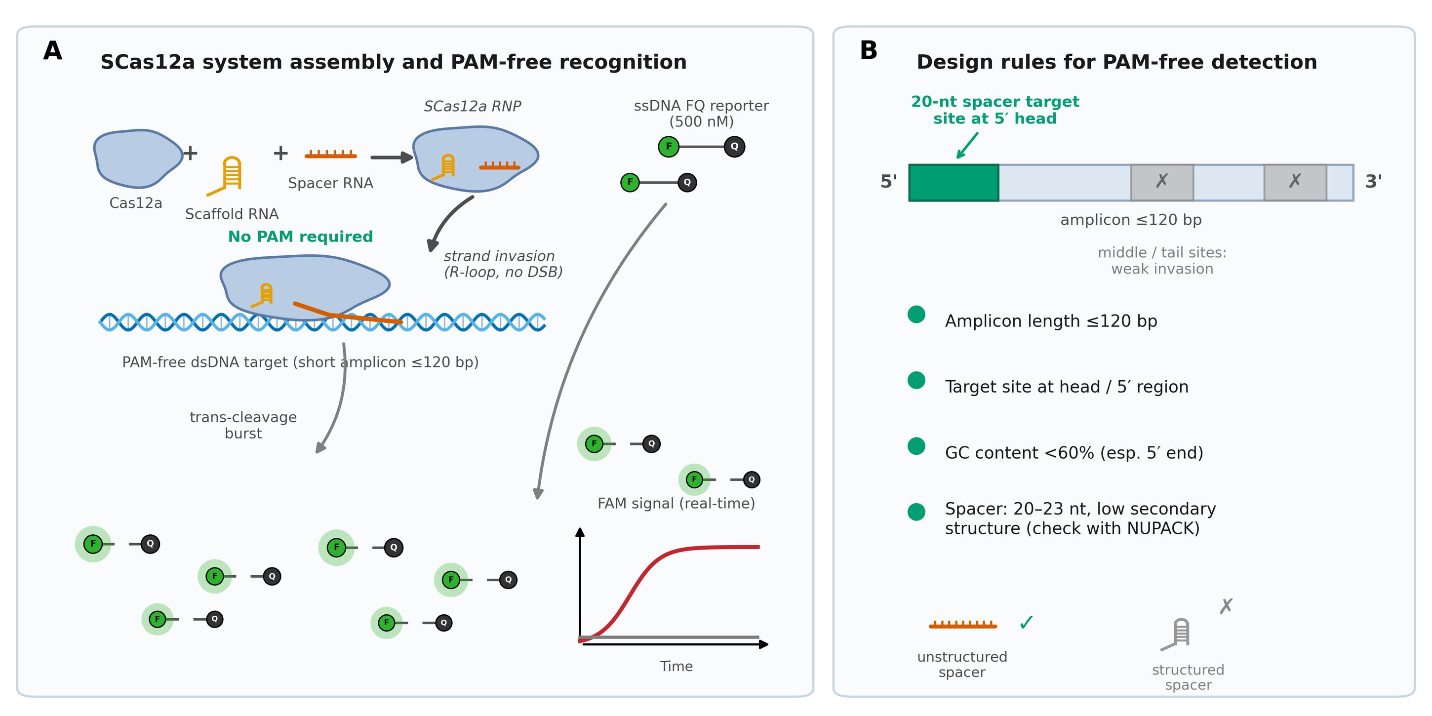
Figure 1. Assembly and design rules of the SCas12a system. (A) The SCas12a RNP is assembled from the Cas12a protein, a scaffold RNA, and a 20-nt spacer RNA. (B) For PAM-free dsDNA detection, the spacer preferably targets a PAM-free site located at the head (5′) region of an amplicon of ≤120 bp with GC content below 60%; spacer secondary structure should be minimized as predicted by NUPACK. The length and GC values are empirical design preferences, not universal cutoffs [15, Figure 2].
Materials and reagents
Reagents
1. RPA kit and RT-RPA (Weifang Amp-Future Biotech, Changzhou, China, catalog number: WLB8201KIT); contains lyophilized RPA pellets and buffer A; store lyophilized pellets at -20 °C
2. WarmStart® LAMP Kit (DNA & RNA) (New England Biolabs, catalog number: E1700S); store at -20 °C
3. 10× NEBuffer r2.1 (New England Biolabs, catalog number: B6002S); store at -20 °C
4. SuperScript IV reverse transcriptase (Thermo Fisher Scientific, catalog number: 18090010); store at -20 °C
5. RNase H (New England Biolabs, catalog number: M0297S); the original study used a 50 U/μL working stock; verify the supplied activity concentration; store at -20 °C
6. Proteinase K (Takara, catalog number: 9034); store at -20 °C
7. qPCR Detection kit (GenePharma, Suzhou, China, catalog number: QPG-040); used for orthogonal qPCR/RT-qPCR validation; store at -20 °C
8. FAM–BHQ1 ssDNA FQ reporter, 5′-FAM-TTATT-BHQ1-3′ or 5′-FAM-TTATTATT-3′-BHQ1 [synthesized by Sangon Biotech, Shanghai, China; HOLMES-Fluo ssDNA reporter 1 (FAM) is available from TOLOBIO, catalog number: 31101]; store the 100 μM stock at -20 °C in the dark. Reconstitute lyophilized reporter in nuclease-free water to 100 μM; for a supplied solution, use the stated concentration. Use the same reporter sequence throughout a comparison
9. Scaffold RNA, spacer RNAs, full-length crRNAs, RPA and LAMP primers, and dsDNA targets/gene fragments (synthesized by Sangon Biotech, Shanghai, China; HPLC- or PAGE-purified); dissolve in DEPC-treated water or nuclease-free water and store at -20 °C (RNA stocks may be stored at -80 °C for extended periods); the sequences used in the original study are listed in Supplementary Table S1 of [15]
10. 6×His-pET-28a(+) expression plasmids encoding AsCas12a, LbCas12a, and CtCas12a; constructs are available from Yi Liu
11. Ni-NTA resin (Qiagen, catalog number: 36113)
12. 100 kDa molecular weight cutoff (MWCO) centrifugal filters (Merck Millipore, Amicon Ultra, catalog number: UFC910024)
13. GoldView nucleic acid stain (Yeasen, catalog number: 10201ES)
14. Ultrapure water (18.2 MΩ·cm, from a Milli-Q purification system; used throughout all buffer and solution preparation)
15. Tris base (Trizma® base) (Sigma-Aldrich, catalog number: T1503); used to prepare 1 M Tris-HCl stocks (pH 7.9 and pH 8.8) and 50× TAE buffer; store at room temperature
16. 1 M Tris-HCl, pH 7.5 (Invitrogen, catalog number: 15567027); alternatively prepared in-house from Tris base adjusted with HCl; store at room temperature
17. Hydrochloric acid (HCl) (Sigma-Aldrich, catalog number: H1758); used for pH adjustment of Tris-HCl stocks; store at room temperature
18. 5 M NaCl, RNase-free (Invitrogen, catalog number: AM9759); store at room temperature
19. Glycerol, molecular biology grade (Sigma-Aldrich, catalog number: G5516); store at room temperature
20. 1 M MgCl2, RNase-free (Invitrogen, catalog number: AM9530G); store at room temperature
21. Bovine serum albumin (BSA), 20 mg/mL, molecular biology grade (New England Biolabs, catalog number: B9000S); store at -20 °C
22. Ammonium sulfate [(NH4)2SO4] (Sigma-Aldrich, catalog number: A4418); store at room temperature
23. 1 M MgSO4 solution, for molecular biology (Sigma-Aldrich, catalog number: M3409); also used for the additional 8 mM MgSO4 supplementation in the one-pot reaction; store at room temperature
24. Tween® 20, molecular biology grade (Sigma-Aldrich, catalog number: P9416); store at room temperature
25. Nuclease-free water, not DEPC-treated (Invitrogen, catalog number: AM9937); store at room temperature
26. Glacial acetic acid (Fisher Scientific, catalog number: A38-212); store at room temperature
27. 0.5 M EDTA, pH 8.0 (Invitrogen, catalog number: 15575020); store at room temperature
Storage of critical items: Purified Cas12a proteins are flash-frozen in liquid nitrogen and stored at -80 °C; a quantitative shelf life has not been established. Oligonucleotides (DNA primers, dsDNA targets) are stored at -20 °C; RNA oligonucleotides (scaffold RNA, spacer RNAs, crRNAs) are stored at -20 °C for routine use and at -80 °C for long-term storage. Lyophilized RPA/RT-RPA pellets and kit enzymes are stored at -20 °C.
Solutions
1. Cas12a storage buffer (see Recipes)
2. 10× NEBuffer r2.1 equivalent (see Recipes)
3. 10× LAMP reaction buffer (see Recipes)
4. 10 μM FQ reporter working stock (see Recipes)
5. 50× TAE buffer (see Recipes)
Recipes
1. Cas12a storage buffer (100 mL)
| Reagent | Final concentration | Quantity or volume |
|---|---|---|
| 1 M Tris-HCl, pH 7.5 | 20 mM | 2 mL |
| 5 M NaCl | 250 mM | 5 mL |
| Glycerol | 5% (v/v) | 5 mL |
| Ultrapure water | n/a | to 100 mL |
| Total | n/a | 100 mL |
Sterilize by filtration (0.22 μm) and store at 4 °C for up to 3 months.
2. 10× NEBuffer r2.1 equivalent (50 mL)
| Reagent | Final concentration | Quantity or volume |
|---|---|---|
| 5 M NaCl | 500 mM | 5 mL |
| 1 M Tris-HCl, pH 7.9 | 100 mM | 5 mL |
| 1 M MgCl2 | 100 mM | 5 mL |
| Recombinant albumin (e.g., 20 mg/mL BSA stock) | 100 μg/mL | 250 μL |
| Ultrapure water | n/a | to 50 mL |
| Total | n/a | 50 mL |
Adjust to pH 7.9 at 25 °C. Sterilize by filtration (0.22 μm), aliquot, and store at -20 °C for up to 1 year. This is also commercially available (NEB, catalog number: B6002S).
3. 10× LAMP reaction buffer (50 mL)
| Reagent | Final concentration | Quantity or volume |
|---|---|---|
| 1 M Tris-HCl, pH 8.8 | 200 mM | 10 mL |
| (NH4)2SO4 | 100 mM | 0.66 g |
| 1 M MgSO4 | 20 mM | 1 mL |
| Tween 20 | 1% (v/v) | 500 μL |
| Ultrapure water | n/a | to 50 mL |
| Total | n/a | 50 mL |
Adjust to pH 8.8 at 25 °C. Sterilize by filtration (0.22 μm), aliquot, and store at -20 °C for up to 1 year. Note that the additional 8 mM MgSO4 supplemented in the LAMP/SCas12a one-pot reaction (section E) is provided separately and is not part of this buffer. The nominal total MgSO4 concentration is therefore 10 mM, including 2 mM from the 1× buffer.
4. 10 μM FQ reporter working stock (100 μL)
| Reagent | Final concentration | Quantity or volume |
|---|---|---|
| 100 μM FQ reporter stock | 10 μM | 10 μL |
| Nuclease-free water | n/a | 90 μL |
| Total | n/a | 100 μL |
Prepare in a low-binding microtube, protect from light, and store at -20 °C in aliquots. Avoid repeated freeze/thaw cycles.
5. 50× TAE buffer (1 L)
| Reagent | Final concentration | Quantity or volume |
|---|---|---|
| Tris base | 2 M | 242 g |
| Glacial acetic acid | 1 M | 57.1 mL |
| 0.5 M EDTA, pH 8.0 | 50 mM | 100 mL |
| Ultrapure water | n/a | to 1 L |
| Total | n/a | 1 L |
Store at room temperature. Dilute to 1× TAE in ultrapure water before use for agarose gel electrophoresis.
Laboratory supplies
1. Pipette tips (filtered) (Thermo, catalog numbers: TF112-1000-Q, T104RS-Q, TF140-200-Q)
2. 0.2 mL PCR tubes (Thermo, catalog number: 431-MIXED-Q)
3. 1.5 mL microtubes (AXYGEN, catalog number: MCT-150-L-C)
Equipment
1. Real-time PCR system (Bio-Rad, model: CFX96 touch, as used in [15]; alternative instruments require verification of thermal control and FAM detection)
2. Microcentrifuge (mySPINTM 6 Mini, Thermo, model: 75004061 or equivalent)
3. Thermal cycler (VeritiPro PCR, Thermo, model: A48141)
4. Vortex mixer (Eppendorf mixer, Eppendorf, model: 5382000074)
5. Variable volume pipettes (P10, P200, P1000)
6. Freezer (-80 °C) (Thermo, model: 905)
7. Standard laboratory equipment (analytical balance, pH meter, etc.)
Software and datasets
1. CFX Maestro software (Bio-Rad, version 2.3 or compatible; supplied with the CFX96 Touch system; requires a license bundled with the instrument) for real-time fluorescence acquisition and baseline analysis
2. NUPACK (http://www.nupack.org/; accessed January 12, 2026; free for academic use) for screening spacer RNA sequences for minimal secondary structure. Free alternatives: RNAfold (http://rna.tbi.univie.ac.at/cgi-bin/RNAWebSuite/RNAfold.cgi) and Mfold (http://unafold.rna.albany.edu/?q=mfold)
3. AlphaFold3 Server (https://alphafoldserver.com/; free for non-commercial use; optional) for predictive structural modeling of the Cas12a/split crRNA/DNA complex
4. GraphPad Prism (version 8 or later; commercial license required) for plotting fluorescence kinetics, bar charts, and heatmaps. Alternatives: Microsoft Excel (commercial), R/RStudio, or Python (matplotlib). Record the actual software versions and prediction settings used; the listed versions are reference implementations
5. Oligonucleotide sequences (scaffold RNA, spacer RNAs, primers, and targets) used in the original study are listed in Supplementary Table S1 of Yin et al. [15] (Nucleic Acids Research, DOI: 10.1093/nar/gkag267). The complete source list is accessible through the Supplementary data link at https://doi.org/10.1093/nar/gkag267
Procedure
Protein preparation: Express His-tagged Cas12a in E. coli BL21(DE3); induce at OD600 = 0.8 with 0.5 mM IPTG for 16 h at 18 °C. Purify by Ni-NTA affinity and Superdex 200 size-exclusion chromatography, concentrate with a 100 kDa MWCO filter, quantify by Bradford assay, and store in Cas12a storage buffer (see Recipes) at -80 °C [15]. SDS-PAGE and matched-target trans-cleavage controls are recommended for quality checks; numerical yield, purity, specific-activity, and lot-equivalence criteria are not established.
A. Design of spacer RNA, scaffold RNA, and amplification primers
1. Select the target region within the gene of interest (e.g., the L1 gene of HPV16, the Orf1ab or E genes of SARS-CoV-2, or the TP53 locus containing rs215029805).
2. Choose a 20-nt spacer sequence targeting a site that lacks any canonical (TTTV) or suboptimal (NTTV, TTTN) PAM in its vicinity; a canonical PAM is not required, but inspect flanking sequences to reproduce the tested PAM-free configuration.
Note: PAM-free spacer selection expands the pool of usable target sites relative to WT Cas12a designs.
3. Preferentially, position the spacer-targeted site at the head (5′) region of an amplicon of ≤120 bp. This empirical preference derives from target-length and positioning comparisons (Figure 2 from [15]). Longer targets may reduce detection efficiency, but no universal quantitative decline beyond 120 bp is established. Optimize new targets individually; this length preference does not define a LAMP product-size limit.
4. Select target regions with GC content below 60%, particularly avoiding GC-rich sequences at the 5′ end of the target, because high duplex stability disfavors strand invasion by the spacer RNA and reduces sensitivity. The <60% GC criterion is an empirical preference based on the comparisons in Figure 2 from [15], not a validated cutoff. GC content alone does not predict performance; no universal quantitative decline above 60% is established.
5. Screen candidate spacer RNA sequences for minimal secondary structure using NUPACK (or RNAfold/Mfold). Prefer spacers with limited predicted folding; prediction alone does not establish RNP activity.
6. Use the validated scaffold RNA (S0 scaffold) sequence from [15]. For SNV genotyping, design a wild-type (WT)-specific spacer and a mutant-specific spacer differing by the single discriminating nucleotide (see section F).
7. Design RPA primers (forward and reverse, typically 30–35 nt) flanking the spacer target site according to standard TwistAmp-style rules, preferentially producing an amplicon of ≤120 bp. For LAMP, design a six-primer set (F3, B3, FIP, BIP, LF, and LB) using primer design software (e.g., PrimerExplorer), again placing the spacer target site at the head region of the amplicon.
8. Order the spacer RNAs, scaffold RNA, and primers as HPLC- or PAGE-purified oligonucleotides. Reconstitute RNAs in DEPC-treated water and DNAs in nuclease-free water to 100 μM stocks; store at -80 °C (RNA) or -20 °C (DNA).
Note: All spacer RNA, scaffold RNA, primer, and target sequences validated in the original study are listed in Supplementary Table S1 of [15].
B. Assembly of the SCas12a RNP complex
1. Thaw the Cas12a protein, scaffold RNA, and spacer RNA aliquots on ice. Briefly vortex and centrifuge all oligonucleotide stocks.
2. In a 1.5 mL low-binding microtube, combine the Cas12a protein, scaffold RNA, and spacer RNA at a 1:2 enzyme:RNA molar ratio (i.e., Cas12a:scaffold RNA:spacer RNA = 1:2:2; for example, 100 nM Cas12a with 200 nM scaffold RNA and 200 nM spacer RNA in the final assay) in 1× NEBuffer r2.1.
Critical: Prepare the RNP at working concentrations such that the final one-pot reaction contains 100 nM Cas12a RNP. For sections C–E, prepare a pre-mix containing the RNP and the FQ reporter at concentrations that yield 100 nM RNP and 500 nM FQ reporter after all components are combined. The 1:2:2 input ratio provides RNA excess to favor assembly and does not describe bound-complex stoichiometry. The 100 nM Cas12a and 500 nM reporter inputs are adopted working conditions, not universal optima.
3. Incubate the mixture for 20 min at room temperature to allow RNP complex formation.
4. Place the assembled RNP on ice and use it the same day.
Critical: Assemble the SCas12a RNP fresh immediately before each experiment; the preassembled-RNP storage stability and freeze/thaw tolerance have not been established.
Note: The same assembly procedure applies to As, Lb, and Ct Cas12a and to WT Cas12a/full-length crRNA controls. For the standalone trans-cleavage assays, 250 nM Cas12a with 500 nM of each split-RNA component was used as described in the original study [15].
C. One-pot RPA/SCas12a assay for DNA targets (HPV16 L1 model; 20 μL)
1. Prepare the RPA reaction mix by resuspending one lyophilized RPA pellet in 29.4 μL of buffer A (supplied with the kit), 14.1 μL of nuclease-free water, 2 μL of 10 μM RPA forward primer, and 2 μL of 10 μM RPA reverse primer, as described in [15]. Mix gently until the pellet is fully dissolved and keep on ice.
Note: One resuspended pellet provides enough RPA mix for approximately four 20 μL one-pot reactions (10 μL per reaction). Scale up proportionally for larger batches.
2. Assemble the AsCas12a SCas12a RNP as described in section B and prepare an RNP/FQ reporter pre-mix such that 6 μL of the pre-mix delivers the amounts required for final concentrations of 100 nM AsCas12a RNP and 500 nM FQ ssDNA reporter in the 20 μL reaction.
3. Set up each 20 μL one-pot reaction in a well of a 96-well qPCR plate (or a 0.2 mL PCR tube) on ice according to Table 1. Combine 10 μL of RPA reaction mix, 6 μL of the RNP/FQ reporter pre-mix, and 2 μL of 10× reaction buffer. The added buffer contributes a nominal 1×; account separately for buffer carried over with RPA and RNP stocks.
Table 1. One-pot RPA/SCas12a reaction setup (20 μL per reaction)
| Component | Stock | Volume | Final concentration |
|---|---|---|---|
| RPA reaction mix (resuspended pellet with 2 × 2 μL of 10 μM primers per pellet; recipe per the manufacturer) | n/a | 10 μL | 1× |
| RNP/FQ reporter pre-mix | Prepared to deliver working amounts | 6 μL | 100 nM AsCas12a RNP; 500 nM FQ ssDNA reporter |
| Reaction buffer | 10× | 2 μL | 1× |
| dsDNA activator/template | variable | 2 μL | Variable (e.g., 1 pM positive control) |
| Total | n/a | 20 μL | n/a |
4. Add 2 μL of dsDNA activator/template (e.g., HPV16 L1 dsDNA or extracted sample DNA) last.
Critical: Always add the template last to prevent premature amplification. Prepare all reactions on ice.
Caution: RPA amplicons are a potent contamination source. Prepare reaction mixes in a dedicated pre-amplification area physically separated from template handling and post-amplification analysis and use filtered tips throughout.
5. Include a positive control (e.g., 1 pM HPV16 target plasmid) and a no-template control (NTC; 2 μL of nuclease-free water instead of template) in every run.
6. Seal the plate with an optical adhesive seal, mix gently, and centrifuge briefly to collect the contents. Keep tubes closed during fluorescence acquisition and disposal.
7. Immediately place the plate in the CFX96 Touch Real-Time PCR System. Incubate at 37 °C and record fluorescence in the FAM channel every 1 min for 30–60 min.
Note: Define the readout time before testing and apply it consistently to controls and samples. Representative results are shown in Figure 2.
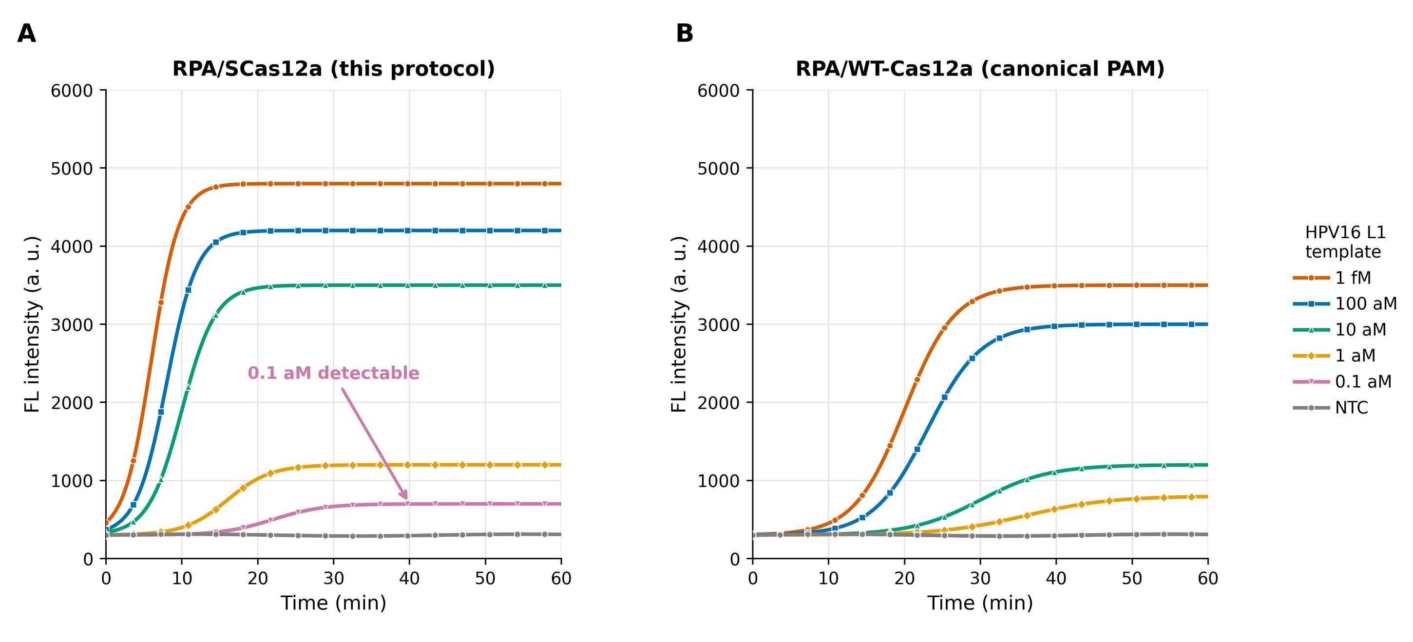
Figure 2. Representative one-pot detection results. (A) One-pot RPA/SCas12a detection of the HPV16 L1 gene using a PAM-free target: immediate fluorescence increase across a broad range of target concentrations, with a reported nominal analytical sensitivity of 0.1 aM for the purified model target. (B) One-pot RPA/WT-Cas12a detection of the same target with a canonical PAM (TTTA): minimal fluorescence within the first 10 min and a higher reported model-target detection limit, due to cis-cleavage-mediated amplicon degradation (representative curves adapted from Yin et al. [15], Nucleic Acids Res, Figure 3d, e). This value does not establish a clinical or 95% detection-probability limit.
D. One-pot RT-RPA/SCas12a assay for RNA targets (SARS-CoV-2 E/Orf1ab model)
1. Prepare the RT-RPA reaction mix as in step C1 and additionally add 0.9 μL of RNase H (50 U/μL stock) and 0.45 μL of SuperScript IV reverse transcriptase per resuspended RPA pellet. This corresponds to 45 U RNase H per pellet; adjust enzyme and water volumes if the supplied stock activity differs.
2. For the reported nasal-swab workflow, heat the specimens at 95 °C for 5–10 min before testing; use the lysate directly as the template.
Caution: Handle clinical specimens in an appropriate biosafety cabinet following institutional biosafety guidelines. This is a specimen-specific preparation, not a universal viral-inactivation procedure. Lysate inhibition and equivalence to purified RNA were not established; process and inhibition controls are recommended for new matrices.
3. Assemble the reaction as described in steps C2–6, using 2 μL of template (RNA extract or lysate) per 20 μL reaction.
4. Incubate at 37 °C on the CFX96 Touch Real-Time PCR System and record FAM fluorescence every 1 min for 30 min.
Note: With reagents prepared in advance, the reported specimen workflow requires approximately 40 min (5–10 min for heating/lysis and 30 min for the one-pot RT-RPA/SCas12a reaction). This excludes the 20-min RNP assembly and protein preparation.
E. One-pot LAMP/SCas12a assay with thermostable CtCas12a (SARS-CoV-2 E gene model; 25 μL)
1. Assemble the CtCas12a SCas12a RNP as described in section B.
Critical: Use the thermostable CtCas12a ortholog for LAMP-coupled one-pot assays at 55 °C; AsCas12a and LbCas12a are not thermostable and must not be substituted at this temperature.
2. Set up each 25 μL one-pot reaction on ice according to Table 2, combining 1× LAMP reaction buffer (Recipe 3), dNTPs (1.4 mM each), MgSO4 (8 mM, additional), taurine (50 mM), Bst 2.0 WarmStart DNA polymerase (0.32 U/μL), the LAMP primer mix (0.2 μM F3/B3, 0.4 μM LF/LB, and 1.6 μM FIP/BIP final), CtCas12a RNP (100 nM final), and FQ ssDNA reporter (500 nM final).
Table 2. One-pot LAMP/SCas12a reaction setup (25 μL per reaction)
| Component | Stock | Volume | Final concentration |
|---|---|---|---|
| 10× LAMP reaction buffer (Recipe 3) | 10× | 2.5 μL | 1× [20 mM Tris-HCl, 10 mM (NH4)2SO4, 2 mM MgSO4, 0.1% Tween 20, pH 8.8] |
| dNTP mix | 10 mM each | 3.5 μL | 1.4 mM each |
| MgSO4 | 100 mM | 2 μL | 8 mM (additional) |
| Taurine | 500 mM | 2.5 μL | 50 mM |
| LAMP primer mix (F3/B3/LF/LB/FIP/BIP) | 10× (2 μM F3/B3, 4 μM LF/LB, 16 μM FIP/BIP) | 2.5 μL | 0.2 μM F3/B3; 0.4 μM LF/LB; 1.6 μM FIP/BIP |
| CtCas12a RNP + FQ reporter pre-mix | Prepared to deliver working amounts | Variable | 100 nM CtCas12a RNP; 500 nM FQ ssDNA reporter |
| Bst 2.0 WarmStart DNA polymerase | 8 U/μL | 1 μL | 0.32 U/μL |
| Template | Variable | 2 μL | Variable |
| Nuclease-free water | n/a | to 25 μL | n/a |
| Total | n/a | 25 μL | n/a |
3. Add 2 μL of template last, seal, mix gently, and centrifuge briefly.
Critical: Add template last and keep reactions on ice during setup, as in section C. The reported LAMP model used DNA corresponding to the E gene; include a matched positive DNA control and an NTC.
4. Incubate at 55 °C on the CFX96 Touch Real-Time PCR System and record FAM fluorescence every 1 min for 60 min.
F. SNV genotyping application (TP53 rs215029805 model)
1. Design two spacer RNAs against the TP53 locus containing rs215029805 (A>T): a WT-specific spacer fully complementary to the WT allele and a mutant-specific spacer fully complementary to the A>T mutant allele, following the design rules in section A.
2. Run the one-pot RPA/SCas12a assay exactly as described in section C, using genomic DNA extracted from the sample (e.g., OSCC tissue) as the template, with a separate reaction for each spacer.
3. Include WT and mutant dsDNA/plasmid controls and an NTC in every run.
4. Record FAM fluorescence at 37 °C every 1 min for 30 min.
5. Compute the distinguishing factor (DF) for each sample as DF = (FWT - FWT0)/(FSNV - FSNV0), where FWT and FSNV are the endpoint fluorescence signals generated with the WT-specific and mutant-specific spacers, and FWT0 and FSNV0 are the corresponding initial (baseline) fluorescence signals.
6. Use the empirical thresholds DF > 4 (WT-associated response) and DF < 0.25 (mutant-associated response). Treat intermediate values as indeterminate; weak signals or a nonpositive denominator preclude interpretation.
Note: The entire genotyping process is completed within 30 min, substantially faster than two-step methods such as SHERLOCK (>2 h), and achieved 100% concordance with Sanger sequencing on nine OSCC tissue samples in the original study [15]. The assay provides up to 91-fold single-base selectivity for PAM-distal mismatches. The nine-specimen concordance does not establish performance in larger cohorts, heterozygotes, or low-variant-fraction samples.
Data analysis
1. Fluorescence data processing: Export the raw FAM fluorescence trajectories from CFX Maestro. Subtract the baseline (initial) fluorescence value of each well from its kinetic trace to obtain background-corrected fluorescence curves. Perform baseline subtraction and kinetic or endpoint analysis in CFX Maestro, Microsoft Excel, GraphPad Prism, or an equivalent tool.
2. Replicates and reporting: Perform a minimum of n = 3 technical replicates per condition and report data as mean ± SD (standard deviation). Include a positive control and an NTC in every run; exclude runs in which the positive control fails or the NTC produces a positive call. A valid run requires the matched positive control to respond within the specified readout period and the NTC to remain below the decision threshold. Technical replication does not establish between-day, operator, lot, or laboratory reproducibility.
3. Limit of detection (LOD): Estimate the analytical LOD from a dilution series of the DNA or RNA target using the formula LOD = 3σ/slope, where σ is the standard deviation of three blank (NTC) measurements, and the slope is the slope of the calibration line relating fluorescence signal to target concentration. This is an exploratory estimate with limited blank replication; it does not establish a 95% detection-probability LOD. Report template type, input volume, dilution basis, endpoint, and replicate detection counts.
4. Positive/negative calling: For exploratory analysis, provisionally call a sample positive when its baseline-corrected endpoint signal exceeds the mean + 3 SD of the NTC; results from failed controls or discordant low-signal replicates remain unresolved. For clinical samples, compute sensitivity and concordance against the orthogonal reference method (qPCR/RT-qPCR for HPV16 and SARS-CoV-2, Sanger sequencing for TP53 SNVs). Fix the endpoint and threshold before testing; this rule has not been independently validated as a clinical cutoff.
5. SNV genotyping: Calculate the distinguishing factor DF = (FWT - FWT0)/(FSNV - FSNV0) from baseline-corrected endpoint signals obtained with the WT-specific and mutant-specific spacers and assign genotypes using the thresholds DF > 4 (WT) and DF < 0.25 (mutant), as adopted from the original model study [15]. These thresholds require verification for other instruments, matrices, reagent lots, and laboratories; apply the interpretation safeguards in section F.
6. Statistical analysis: Plot kinetic curves, bar charts, and heatmaps in GraphPad Prism (or an alternative such as R or Excel). In the original study, no statistical method was used to predetermine sample size, all data were included without exclusion, and experiments were not randomized [15]. No specialized computational skills beyond standard spreadsheet/plotting software are required; NUPACK is used only at the design stage. The absence of randomization and predetermined sample-size calculations limits assessment of robustness and generalizability.
Validation of protocol
This protocol describes methods used in the following published study; no new independent validation dataset is presented:
• Yin et al. [15]. Elimination of cis-cleavage in CRISPR diagnostics for one-pot rapid nucleic acid detection. Nucleic Acids Research (2026), DOI: 10.1093/nar/gkag267.
Supporting data include target recognition/cleavage (Figure 1 and Supplementary Figure S1), design comparisons (Figure 2), one-pot detection (Figure 3), and clinical examples (Figures 4–6) in [15]. Supplementary Figure S4 reports 10 experiments with 2 replicates per run under identical conditions; broader inter-assay and inter-laboratory reproducibility remain unestablished.
General notes and troubleshooting
General notes
1. RNase-free practice: Spacer RNAs and scaffold RNA are sensitive to RNase contamination. Use RNase-free tubes and filtered tips, DEPC-treated or nuclease-free water, and clean gloves and workspaces; keep RNA stocks on ice during use.
2. Amplicon contamination control: RPA and LAMP products are extremely potent contaminants. Maintain physically separated areas for reagent preparation, template addition, and post-amplification analysis, use dedicated pipettes and filtered tips, and never open reaction vessels after amplification. The reported assay uses closed-tube fluorescence acquisition; enzymatic carry-over prevention was not evaluated.
3. Fresh RNP assembly: Assemble the SCas12a RNP immediately before each experiment and keep it on ice; storage stability of preassembled RNP has not been established. Purified Cas12a proteins are stored at -80 °C; verify activity with matched controls before use.
4. Spacer design rules: Target a PAM-free site at the head (5′) region of an amplicon of ≤120 bp, keep target GC content below 60% (especially at the 5′ end), and screen spacers with NUPACK to exclude stable secondary structures. These empirical preferences are target-dependent and should not be used as strict acceptance criteria (section A).
5. Ortholog/temperature matching: Use AsCas12a at 37 °C (the demonstrated RPA/RT-RPA workflow) and the thermostable CtCas12a at 55 °C (LAMP). Do not use As/Lb Cas12a at elevated temperatures.
6. Reagent storage: Store lyophilized RPA/RT-RPA pellets, enzymes, and oligonucleotide stocks at -20 °C; protect the FQ reporter from light; store purified Cas12a at -80 °C.
7. Scope and limitations: Design preferences, RNP/reporter inputs, DF thresholds, and sensitivity values are adopted from [15] and are target-dependent. Clinical examples do not establish general diagnostic performance across matrices or populations. The demonstrated readout uses the CFX96 Touch; transferring to portable readers requires independent verification. For adaptation, assess primer/spacer design, then vary RNP input at 1:2:2, and finally reporter input; compare time to signal, separation from NTCs, and replicate consistency. No universal optimal concentration range is established.
Troubleshooting
The following are possible causes and recommended checks, not independently validated remedies. Use the run-acceptance criteria in Data analysis; invalidate runs with positive NTCs or failed positive controls.
Problem 1: Weak or no fluorescence signal in positive samples.
Possible causes: Suboptimal spacer design (PAM-free site in the middle/tail of the amplicon, amplicon >120 bp, or GC-rich target), degraded RNA components, or inactive Cas12a protein.
Solutions: Redesign the spacer to target the head region of a short, low-GC amplicon, using the preferences in section A and re-screen with NUPACK; use fresh RNA aliquots and freshly thawed or freshly prepared Cas12a protein; verify RNP activity with a positive-control dsDNA target.
Problem 2: High background fluorescence in the no-template control (NTC).
Possible causes: Amplicon carry-over contamination, reporter degradation, or nonspecific amplification (primer-dimers).
Solutions: Decontaminate workspaces and pipettes, open a fresh aliquot of every reagent, and strictly separate pre- and post-amplification areas; prepare fresh FQ reporter working stock; redesign primers to eliminate primer-dimers.
Problem 3: No amplification/signal with the matched-target positive-control reaction.
Possible causes: Loss of enzyme activity, incorrect buffer or missing Mg2+, or expired lyophilized RPA pellets.
Solutions: Check the activity of each component individually (Cas12a RNP with a defined dsDNA activator and reporter; RPA mix with a positive-control template); use freshly resuspended pellets and verify that 1× buffer is present in the final reaction. The matched positive control must respond within the predefined readout period while the NTC remains negative; no universal specific-activity or amplification-efficiency cutoff is established.
Problem 4: Inconsistent signals between technical replicates.
Possible causes: Inadequate mixing, pipetting inaccuracy at small volumes, or partially assembled RNP.
Solutions: Prepare a master mix for all shared components, mix gently but thoroughly by pipetting, centrifuge plates/tubes briefly before loading, and allow the full 20 min for RNP assembly; use calibrated pipettes. Inspect individual traces and investigate discordance above the sensitivity limit; no universal fluorescence-CV cutoff is established, particularly near background.
Problem 5: Nonspecific amplification in the LAMP/SCas12a assay.
Possible causes: Suboptimal LAMP primer set, excessive MgSO4, or incubation above 55 °C with a non-thermostable ortholog.
Solutions: Redesign or screen alternative LAMP primer sets (F3/B3/FIP/BIP/LF/LB), titrate MgSO4 and taurine, confirm the incubation temperature is 55 °C, and ensure that thermostable CtCas12a (not As/Lb Cas12a) is used.
Problem 6: Failure of RT-RPA/SCas12a with RNA samples that are positive by RT-qPCR.
Possible causes: RNA degradation during sample handling, insufficient reverse transcription (missing SuperScript IV or RNase H), or inhibitors carried over from crude lysates.
Solutions: Process samples promptly with RNase-free technique, verify that SuperScript IV (0.45 μL) and RNase H (0.9 μL, 50 U/μL) were added per pellet resuspension, and check process/inhibition controls. Lysate dilution or longer heating requires separate evaluation and is not a validated standard remedy.
Acknowledgments
This work was supported by the Hubei Provincial Department of Education Scientific Research Program (B2025436), the Science and Technology Innovation Talent Plan of Hubei Province (2023DJC136 to Y.L.), and the Natural Science Foundation of Hubei Province (2025AFB825 to J.Q.).
This protocol was adapted from the original research article Yin et al. [15], Nucleic Acids Research (2026) 54: gkag267, DOI: 10.1093/nar/gkag267, and was developed from our prior SCas12a work (Chen et al. [13]; Zhang et al. [14]). Cas12a purification is described in [15]; a related CRISPR ribonucleoprotein-production method is cited in [18].
Author contributions
Protocol Development, L.H., Y.Z., R.Y., J.Z., and Y.L.; Protocol Optimization or Validation, Z.H., Y.P., and T.T.; Original Draft, Y.L.; Review & Editing, Y.L.; Funding acquisition, L.H.; Supervision, Y.L.
Competing interests
The authors declare no competing interests.
Ethical considerations
Human nasal swab specimens for SARS-CoV-2 detection were collected and provided under a protocol approved by the ethics committee of Wuhan Polytechnic University. Human vaginal swab samples for HPV16 detection were obtained following an approved ethics protocol of Zhengzhou University (samples provided via Henan Cancer Hospital). Human oral cancer tissue samples were collected at the First Affiliated Hospital of Zhengzhou University under a protocol approved by its ethics committee. Written informed consent was obtained from all participants. The clinical specimens are used in this protocol only as validation examples; researchers must obtain approval from their own institutional ethics committee and informed consent from all participants before working with human specimens.
References
- Piepenburg, O., Williams, C. H., Stemple, D. L. and Armes, N. A. (2006). DNA Detection Using Recombination Proteins. PLoS Biol. 4(7): e204. https://doi.org/10.1371/journal.pbio.0040204
- Notomi, T. (2000). Loop-mediated isothermal amplification of DNA. Nucleic Acids Res. 28(12): 63e–63. https://doi.org/10.1093/nar/28.12.e63
- Chen, J. S., Ma, E., Harrington, L. B., Da Costa, M., Tian, X., Palefsky, J. M. and Doudna, J. A. (2018). CRISPR-Cas12a target binding unleashes indiscriminate single-stranded DNase activity. Science. 360(6387): 436–439. https://doi.org/10.1126/science.aar6245
- Li, S. Y., Cheng, Q. X., Wang, J. M., Li, X. Y., Zhang, Z. L., Gao, S., Cao, R. B., Zhao, G. P. and Wang, J. (2018). CRISPR-Cas12a-assisted nucleic acid detection. Cell Discov. 4(1): e1038/s41421–018–0028–z. https://doi.org/10.1038/s41421-018-0028-z
- Gootenberg, J. S., Abudayyeh, O. O., Lee, J. W., Essletzbichler, P., Dy, A. J., Joung, J., Verdine, V., Donghia, N., Daringer, N. M., Freije, C. A., et al. (2017). Nucleic acid detection with CRISPR-Cas13a/C2c2. Science. 356(6336): 438–442. https://doi.org/10.1126/science.aam9321
- Lu, S., Tong, X., Han, Y., Zhang, K., Zhang, Y., Chen, Q., Duan, J., Lei, X., Huang, M., Qiu, Y., et al. (2022). Fast and sensitive detection of SARS-CoV-2 RNA using suboptimal protospacer adjacent motifs for Cas12a. Nat Biomed Eng. 6(3): 286–297. https://doi.org/10.1038/s41551-022-00861-x
- Yamano, T., Zetsche, B., Ishitani, R., Zhang, F., Nishimasu, H. and Nureki, O. (2017). Structural Basis for the Canonical and Non-canonical PAM Recognition by CRISPR-Cpf1. Mol Cell. 67(4): 633–645.e3. https://doi.org/10.1016/j.molcel.2017.06.035
- Cheng, Z. H., Luo, X. Y., Yu, S. S., Min, D., Zhang, S. X., Li, X. F., Chen, J. J., Liu, D. F. and Yu, H. Q. (2025). Tunable control of Cas12 activity promotes universal and fast one-pot nucleic acid detection. Nat Commun. 16(1): e1038/s41467–025–56516–3. https://doi.org/10.1038/s41467-025-56516-3
- Hu, M., Qiu, Z., Bi, Z., Tian, T., Jiang, Y. and Zhou, X. (2022). Photocontrolled crRNA activation enables robust CRISPR-Cas12a diagnostics. Proc Natl Acad Sci USA. 119(26): e2202034119. https://doi.org/10.1073/pnas.2202034119
- Lin, M., Yue, H., Tian, T., Xiong, E., Zhu, D., Jiang, Y. and Zhou, X. (2022). Glycerol Additive Boosts 100-fold Sensitivity Enhancement for One-Pot RPA-CRISPR/Cas12a Assay. Anal Chem. 94(23): 8277–8284. https://doi.org/10.1021/acs.analchem.2c00616
- Ding, X., Yin, K., Li, Z., Lalla, R. V., Ballesteros, E., Sfeir, M. M. and Liu, C. (2020). Ultrasensitive and visual detection of SARS-CoV-2 using all-in-one dual CRISPR-Cas12a assay. Nat Commun. 11(1): e1038/s41467–020–18575–6. https://doi.org/10.1038/s41467-020-18575-6
- Joung, J., Ladha, A., Saito, M., Kim, N. G., Woolley, A. E., Segel, M., Barretto, R. P., Ranu, A., Macrae, R. K., Faure, G., et al. (2020). Detection of SARS-CoV-2 with SHERLOCK One-Pot Testing. N Engl J Med. 383(15): 1492–1494. https://doi.org/10.1056/nejmc2026172
- Chen, Y., Wang, X., Zhang, J., Jiang, Q., Qiao, B., He, B., Yin, W., Qiao, J. and Liu, Y. (2024). Split crRNA with CRISPR-Cas12a enabling highly sensitive and multiplexed detection of RNA and DNA. Nat Commun. 15(1): 8342. https://doi.org/10.1038/s41467-024-52691-x
- Zhang, J., Yin, W., Jiang, Q., Mao, W., Deng, W., Jin, S., Wang, X., He, R., Qiao, J., Liu, Y., et al. (2025). Precise amplification-free detection of highly structured RNA with an enhanced SCas12a assay. Commun Biol. 8(1): 366. https://doi.org/10.1038/s42003-025-07806-5
- Yin, W., Jin, Z., Jiang, Q., Jin, S., Wang, X., He, R., Qiao, B., Qiao, J., Zhang, X., Liu, Y., et al. (2026). Elimination of cis -cleavage in CRISPR diagnostics for one-pot rapid nucleic acid detection. Nucleic Acids Res. 54(6): e1093/nar/gkag267. https://doi.org/10.1093/nar/gkag267
- Rananaware, S. R., Vesco, E. K., Shoemaker, G. M., Anekar, S. S., Sandoval, L. S. W., Meister, K. S., Macaluso, N. C., Nguyen, L. T. and Jain, P. K. (2023). Programmable RNA detection with CRISPR-Cas12a. Nat Commun. 14(1): e1038/s41467–023–41006–1. https://doi.org/10.1038/s41467-023-41006-1
- Liu, Q., Jiang, Z., Li, S., Li, Y., Wan, Y., Hu, Z., Ma, S., Zou, Z. and Yang, R. (2025). Nonequilibrium hybridization-driven CRISPR/Cas adapter with extended energetic penalty for discrimination of single-nucleotide variants. Nucleic Acids Res. 53(7): e1093/nar/gkaf287. https://doi.org/10.1093/nar/gkaf287
- Qiao, J., Li, W., Lin, S., Sun, W., Ma, L. and Liu, Y. (2019). Co-expression of Cas9 and single-guided RNAs in Escherichia coli streamlines production of Cas9 ribonucleoproteins. Commun Biol. 2(1): e1038/s42003–019–0402–x. https://doi.org/10.1038/s42003-019-0402-x
Article Information
Publication history
Received: Aug 21, 2026
Accepted: Oct 14, 2026
Available online: Oct 28, 2026
Published: Dec 5, 2026
Copyright
© 2026 The Author(s); This is an open access article under the CC BY license (https://creativecommons.org/licenses/by/4.0/).
How to cite
Hu, L., Hu, Z., Pei, Y., Hu, T., Zhang, Y., Yu, R., Zhao, J. and Liu, Y. (2026). Elimination of cis-cleavage in CRISPR Diagnostics for One-Pot Rapid Nucleic Acid Detection. Bio-protocol 16(23): e5870. DOI: 10.21769/BioProtoc.5870.

 Download Citation in RIS Format
Do you have any questions about this protocol?
Post your question to gather feedback from the community. We will also invite the authors of this article to respond.
 Post a Question


