


- Protocols

- Articles and Issues
- For Authors
- About
- Become a Reviewer


Escherichia coli-Based Production and Purification From Inclusion Bodies of Hetero-Oligomeric Mitochondrial Protease ClpXP in Active Form


Published: Vol 16, Iss 22, Nov 20, 2026 DOI: 10.21769/BioProtoc.5865 Views: 14
Reviewed by: Alejandro Avilés-ReyesAnonymous reviewer(s)
 PDF
 Ask a question


Protocol Collections
Comprehensive collections of detailed, peer-reviewed protocols focusing on specific topics



Abstract
Obtaining a functionally active mitochondrial protease complex ClpXP using the Escherichia coli–based expression system is complicated by two factors: (i) complex hetero-oligomeric structure of this multi-subunit complex requires correct assembly of the subunits in heterological environment, while (ii) restoration of the catalytic activity during the expression is toxic for the host cells. This paper presents a simple and low-cost approach that combines separate expression of two ClpXP subunits in insoluble form, their combined purification from inclusion bodies by Ni-NTA metal affinity chromatography, and refolding of the active hetero-oligomeric protein directly on the column by stepwise decreasing the urea concentration. Separate expression allows individual optimization of expressional conditions. The insoluble state of expressed subunits prevents host toxicity and increases the yield. The combined denaturation and immobilization on the column promote the formation of a native hetero-oligomeric structure during refolding, enabling the isolation of the enzyme in its active form. Elution with imidazole ensures the release of correctly folded protein immediately after the removal of denaturing stress, minimizing the time the protease remains in a partially denatured state. The proposed protocol is reproducible and can be applied to the production of other hetero-oligomeric recombinant proteases in active forms.
Key features
• Isolation of active ClpXP complexes by separately expressing insoluble ClpX and ClpP subunits in E. coli, then purifying them together via gravity chromatography.
• Individually expressed subunits are purified and refolded on a Ni-NTA column using urea and imidazole gradients, sequentially removing the denaturant via competitive elution.
• Controlled removal of denaturing stress on the solid phase prevents subunit aggregation and promotes native hetero-oligomeric formation, yielding a catalytically active enzyme.
• Ni-NTA sorbent with urea-imidazole gradient ensures highly selective isolation of the multimeric complex with confirmed activity, indicating the method’s suitability for purification of hetero-oligomeric proteases.
Keywords: Recombinant hetero-oligomeric protein Inclusion body
Refolding
Immobilized metal affinity chromatography (IMAC)
Purification of multimeric protein in active form
Background
Heterologous expression of recombinant proteins is a fundamental technique in modern biotechnology. However, its efficiency is significantly limited by the mismatch between the molecular apparatus of the host cell and the structural and functional requirements of the target product [1,2]. This problem is most acute in the production of complex proteins characterized by a multidomain organization, the presence of multiple disulfide bonds, cofactor-dependent regions, or an oligomeric quaternary structure [3,4]. Such molecules are evolutionarily adapted to the specific microenvironment of donor cells, and their reproduction in heterologous systems is associated with fundamental biophysical barriers.
A key limitation of prokaryotic systems, particularly Escherichia coli, remains the inability to ensure the correct folding of complex polypeptides. The intracellular environment of bacteria is characterized by a reducing potential that prevents the formation of stable disulfide bonds, as well as the absence of specialized chaperone systems necessary for the assembly of multisubunit complexes [5,6], resulting in the accumulation of the target protein within inclusion bodies—insoluble aggregates consisting of a mixture of misfolded conformers [7]. Proteins containing prosthetic groups (heme, iron-sulfur clusters, flavin cofactors) pose an additional challenge, as their functional activity directly depends not only on the correct folding of the polypeptide chain but also on the correct incorporation of the cofactor, which is not always achieved in heterologous systems [8,9].
At the same time, the formation of inclusion bodies rescues host cells from the toxicity of heterologous proteins [10]. For example, most proteases become inherently dangerous when expressed in E. coli cells due to their ability to hydrolyze host cell proteins, while their autoproteolytic capacity reduces the production level [11]. In this case, the production of recombinant proteases in the form of inactive inclusion bodies followed by isolation under denaturing conditions is an effective isolation strategy provided that there are refolding conditions that restore the native protein structure [12].
Removal of target proteins from inclusion bodies requires the development of individual refolding protocols for each molecular target. Methods based on dilution or dialysis are characterized by low reproducibility when working with multisubunit protein complexes due to their high tendency to aggregate during intermediate stages of denaturant removal [13]. Chromatographic strategies that allow immobilization of the target protein on a solid phase and controlled removal of denaturing stress are considered more promising. This is particularly critical for oligomeric complexes, where spatial separation of molecules minimizes nonspecific association [14]. However, even under optimal refolding conditions, restoration of the native conformation for proteins with complex architecture remains a process difficult to predict, requiring empirical selection of parameters for each specific target.
Human mitochondrial protease complex ClpXP consists of two subunits: ClpX, an ATP-dependent chaperone that recognizes protein substrates and unfolds them using energy derived from ATP hydrolysis (UniProtKB O76031), and proteolytic subunit ClpP (UniProtKB Q16740) [15–17]. ClpXP is a key player in the mitochondrial proteostasis system, being involved in the maintenance of cellular energy supply; dysfunction of this complex is associated with a wide range of pathologies [18]. The study of human ClpXP is of particular importance, not only because of its biomedical significance but also because of the unique structural and regulatory organization of the complex, as revealed in recent cryo-electron studies [19]. ClpX forms a homohexameric ring with axial processing, and ClpP forms a tetradecameric structure of two heptameric rings within which 14 catalytic centers are located [20,21]. Expression of the upper components in E. coli leads to the accumulation of the target protein in inclusion bodies, and traditional refolding methods (dialysis, dilution) do not lead to the correct assembly of the multimeric structure [22]. A fundamental breakthrough in ClpXP purification was achieved when the use of a catalytically inactive mutant of ClpP (ClpP S97A in E. coli or S98A in S. aureus), called ClpPtrap, was proposed [22–25]. ClpPtrap retained the ability to bind substrates but was unable to cleave them, allowing co-purification of the ClpXP complex together with the captured protein substrates. At the same time, the E. coli extract-based cell-free expression system was used for successfully obtaining the active ClpXP protease complex [26].
Here, we report a novel, efficient method for obtaining functionally active ClpXP protease. The method is based on the separate expression of ClpX and ClpP in Escherichia coli in an insoluble state and their united purification in denaturing conditions with consequent refolding. The purification protocol includes immobilized metal affinity chromatography (IMAC) on the Ni-NTA gravity column, during which the stepwise increasing imidazole concentration (for removing unspecific bound proteins) was combined with stepwise decreasing urea concentration (refolding). Controlled removal of denaturing stress on the solid phase prevents nonspecific subunit aggregation and facilitates the formation of a native hetero-oligomeric structure, enabling the isolation of the enzyme in a catalytically active conformation. Key advantages of this approach include the integration of solubilization, affinity purification, and renaturation in a single cycle, which provides high selective isolation of the multimeric complex and reproducibility of the results. The presented strategy can be adapted to produce other hetero-oligomeric recombinant proteases.
Materials and reagents
Biological materials
1. E. coli Rosetta 2 (DE3) pLysS (Novagen, catalog number: 71403)
2. Encyclo Polymerase kit (Evrogen, catalog number: PK002S)
3. HS Taq DNA-polymerase (Evrogen, catalog number: PK017L)
4. Human cDNA generated by reverse transcription of RNA from HeLa cell line (BioinnLabs, catalog number: bn-hcdna)
5. Oligonucleotides (Table 1)
Table 1. Sequences of the used oligonucleotides
| Name | Sequence1, 5′→3′ |
| CLPP AgeI fr | caccaCCGGTATGggaTGGCCCGGAATATTGGTAGG |
| CLPP XhoI rev | ccacctcgagCTAGGTGCTAGCTGGGACAGGTTCTGC |
| CLPX AgeI fr | caccACCGGTATGggaCCCAGCTGCGGTGCTTGTACTTG |
| CLPX NheI rev | ccacGCTAGCCTAGCTGTTTGCAGCATCTGCTTGGC |
1All oligonucleotides were synthesized in Evrogen, Moscow
5. QIAquick Gel Purification kit (Qiagen, catalog number: 28704)
6. QIAquick PCR Purification kit (Qiagen, catalog number: 28104)
7. QIAprep Spin Miniprep kit (Qiagen, catalog number: 28XXX)
8. Restriction endonuclease AgeI (NEB, catalog number: R0552S)
9. Restriction endonuclease NheI (NEB, catalog number: R0111S)
10. Restriction endonuclease XhoI (NEB, catalog number: R0146S)
11. T4 DNA ligase (Evrogen, catalog number: LK101S)
12. T7 Promoter (T7uni) primer (10 μM) (Evrogen, catalog number: PP006)
13. T7 Terminal primer (10 μM) (Evrogen, catalog number: PP007)
14. Vector plasmid: modified pET32b (Novagen, catalog number: 69016-3)
Reagents
1. β-casein from bovine milk (Bioultra, catalog number: C6905-250mg)
2. β-Mercaptoethanol (Sigma-Aldrich, catalog number: M6250)
3. Acetic acid, glacial (Panreac, catalog number: 131008.1214)
4. Acrylamide (PanEco, catalog number: Х123)
5. Adenosine 5′-triphosphate (Sigma-Aldrich, catalog number: A-7699)
6. Agarose (Millipore, catalog number: 1.01236)
7. Ammonium acetate (Supelco, catalog number: 1.01116)
8. Ammonium persulfate (APS) (Bio–Rad, catalog number: 161-0700)
9. Ampicillin sodium salt (Sigma-Aldrich, catalog number: A9518)
10. BactoTM yeast extract (Thermo Scientific, catalog number: 212750)
11. Bradford reagent (Sigma-Aldrich, catalog number: B6916)
12. Bromophenol blue (Sigma-Aldrich, catalog number: B0126)
13. Calcium chloride, dihydrate (CaCl2·2H2O) (Sigma-Aldrich, catalog number: 21102)
14. Chloramphenicol (Sigma-Aldrich, catalog number: C0378)
15. Coomassie Brilliant Blue R-250 (Thermo Scientific, catalog number: 20278)
16. DifcoTM tryptone (Thermo Scientific, catalog number: 211921)
17. DNA 1 kb ladder (Evrogen, catalog number: NL001)
18. Ethylenediaminetetraacetic acid (EDTA) (Sigma-Aldrich, catalog number: E9884)
19. Ethanol, 96% (SAFC, catalog number: 1.00971)
20. Ethidium bromide solution, 10 mg/mL (VWR International LCC, catalog number: X328-10ML)
21. Glycerol (Sigma-Aldrich, catalog number: G5516)
22. Glycine (Sigma-Aldrich, catalog number: G7126)
23. Hydrochloric acid (HCl) (Supelco, catalog number: 1.00317)
24. Imidazole (Acros Organics, catalog number: 12202-5000)
25. Isopropyl β-D-1-thiogalactopyranoside (IPTG) (Sigma-Aldrich, catalog number: I6758)
26. Magnesium chloride solution, 1 M (Sigma-Aldrich, catalog number: M1028)
27. Manganese chloride, dihydrate (MnCl2·2H2O) (Sigma-Aldrich, catalog number: 1.05934)
28. N,N’-methylenebisacrylamide (Bio-Rad, catalog number: 161-0201)
29. Ni-NTA agarose (Quigen, catalog number: 30210)
30. Prestained protein molecular weight marker, 20–120 kDa (Thermo Scientific, catalog number: 26612)
31. Sodium chloride (NaCl) (PanEco, catalog number: Х085/130315)
32. Sodium dodecyl sulfate (SDS) (Panreac, catalog number: 142363.1211)
33. Sodium hydroxide (Sigma-Aldrich, catalog number: 221465)
34. TEMED (Fluka, catalog number: 87690)
35. Tris acetate-EDTA buffer (Sigma-Aldrich, catalog number: T9650)
36. Tris(hydroxymethyl)aminomethane (Tris base) (Merck, catalog number: T1503)
37. TritonTM X-100 (Bio-Rad, catalog number: 161-0407)
38. Urea (Sigma-Aldrich, catalog number: U5128)
39. Xylene cyanol (Sigma-Aldrich, catalog number: X4126)
40. Zinc sulfate heptahydrate (Sigma-Aldrich, catalog number: Z0251)
Solutions
1. 0.5 M EDTA pH 8.0 (see Recipes)
2. 0.5 M Tris-HCl, pH 6.8 (see Recipes)
3. 1 M IPTG (see Recipes)
4. 1.2% agarose gel (see Recipes)
5. 10% APS (see Recipes)
6. 10% SDS (see Recipes)
7. 10% (15%) SDS-PAGE (see Recipes)
8. 10% Triton X-100 solution (see Recipes)
9. 1 M Tris-HCl, pH 7.5 (see Recipes)
10. 1.5 M Tris-HCl, pH 8.8 (see Recipes)
11. 2 M imidazole, pH 7,5 (see Recipes)
12. 3 M NaCl (see Recipes)
13. 30% Acrylamide:Bis solution (see Recipes)
14. 4× SB buffer with β-Mercaptoethanol (see Recipes)
15. 5× SB buffer (see Recipes)
16. 5× SDS-PAGE running buffer (see Recipes)
17. 6× Loading buffer (Lb) (see Recipes)
18. 8 M urea (see Recipes)
19. 50× TAE Buffer (see Recipes)
20. Ampicillin (100 μg/mL) (see Recipes)
21. Assay buffer (AB) (see Recipes)
22. Bovine β-casein solution (10 μg/L) (see Recipes)
23. Buffer 1 (see Recipes)
24. Buffer 2 (see Recipes)
25. Buffer 3 (see Recipes)
26. Buffer 4 (see Recipes)
27. Buffer 5 (see Recipes)
28. Buffer 6 (see Recipes)
29. Buffer 7 (see Recipes)
30. Buffer 8 (see Recipes)
31. Buffer RF1 (see Recipes)
32. Buffer RF2 (see Recipes)
33. Chloramphenicol (25 μg/mL) (see Recipes)
34. Coomassie Brilliant Blue staining solution (see Recipes)
35. Fixing solution (see Recipes)
36. LB medium (see Recipes)
Recipes
Notes:
1. All chemicals should be handled according to the best laboratory practices and safety data sheets.
2. Unless otherwise specified, solutions should be filtered through a 0.22 μm membrane filter and stored at room temperature.
1. 0.5 M EDTA, pH 8.0
| Reagent | Final concentration | Quantity or volume |
|---|---|---|
| EDTA disodium salt dihydrate | 0.5 M | 93.06 g |
| Milli-Q water | n/a | to 500 mL |
Dissolve 93.06 g of EDTA disodium salt dihydrate in approximately 400 mL of Milli-Q water, adjust the pH to 8.0 using NaOH (EDTA will not fully dissolve until the pH approaches 8.0), and adjust the final volume to 500 mL.
2. 0.5 M Tris-HCl, pH 6.8
| Reagent | Final concentration | Quantity or volume |
|---|---|---|
| Tris base | 0.5 M | 30.3 g |
| Milli-Q water | n/a | to 500 mL |
Dissolve 30.3 g of Tris base in approximately 400 mL of Milli-Q water. While stirring, carefully adjust the pH to 6.8 using concentrated HCl. Once the pH is stable at 6.8 and the Tris base is completely dissolved, adjust the final volume to 500 mL.
3. 1 M IPTG
| Reagent | Final concentration | Quantity or volume |
|---|---|---|
| IPTG | 1 M | 2.38 g |
| Milli-Q water | n/a | to 10 mL |
Dissolve 2.38 g of IPTG in approximately 8 mL of Milli-Q water. After complete dissolution, adjust the final volume to 10 mL. Sterilize the solution by filtration through a sterile 0.22 μm membrane filter. Aliquot into sterile tubes and store at –20 °C.
4. 1.2% agarose gel
| Reagent | Final concentration | Quantity or volume |
|---|---|---|
| 1× TAE | 1× | 50 mL |
| Ethidium bromide | 0.5 μg/mL | 2.5 μL |
| Agarose | 1.2% | 0.6 g |
Mix 0.6 g of agarose and 50 mL of 1× TAE buffer and melt until clear. Cool the mixture to 50–55 °C and add 2.5 µL of ethidium bromide solution. Mix thoroughly and pour into the mold.
Note: Ethidium bromide solution is toxic; wear gloves when handling.
5. 10% APS
| Reagent | Final concentration | Quantity or volume |
|---|---|---|
| Ammonium persulfate | 10% (w/v) | 1 g |
| Milli-Q water | n/a | 10 mL |
Dissolve 1 g of ammonium persulfate in 10 mL of Milli-Q water. Mix thoroughly until completely dissolved. Aliquot into small volumes (e.g., 0.5–1 mL) and store at -20 °C. The solution is stable for at least 6 months when stored frozen. For best results in polymerization reactions, use freshly prepared solution or aliquots that have undergone less than five freeze-thaw cycles.
6. 10% SDS
| Reagent | Final concentration | Quantity or volume |
|---|---|---|
| Sodium dodecyl sulfate | 10% (w/v) | 50 g |
| Milli-Q water | n/a | to 500 mL |
Dissolve 50 g of SDS in approximately 400 mL of Milli-Q water with gentle stirring and heating to 50 °C to facilitate dissolution. Once fully dissolved, adjust the final volume to 500 mL.
Caution: Wear appropriate personal protective equipment, including a mask, when handling SDS powder to avoid inhalation.
7. 10% (15%) SDS-PAGE (for 2 mini-gels)
A. Resolving gel (10 mL)
| Reagent | Final concentration | Quantity or volume |
|---|---|---|
| 30% Acrylamide/Bis solution | 10% (15%) | 3.3 (5) mL |
| 1.5 M Tris-HCl, pH 8.8 | 0.375 M | 2.5 mL |
| 10% SDS | 0.1% | 100 μL |
| 10% APS | 0.1% | 100 μL |
| TEMED | 0.01% | 10 μL |
| Milli-Q water | n/a | 4 (2.3) mL |
Combine 3.3 (5) mL of 30% acrylamide/bis solution, 2.5 mL of 1.5 M Tris-Base (pH 8.8), 100 μL of 10% SDS, 4 (2.3) mL of Milli-Q water, and 100 μL of 10% APS in a 50 mL Falcon tube. Mix gently by swirling the tube. Immediately before pouring, add 10 μL of TEMED, mix gently but thoroughly, and pour the solution into the gel cassette using the 5 mL pipette. Leave appropriate space for the stacking gel and carefully overlay with isopropanol to ensure a flat interface. Allow gel to polymerize completely (approximately 15–30 min).
Caution: Acrylamide is a neurotoxin. Wear appropriate personal protective equipment when handling unpolymerized solutions.
B. 5% Stacking gel (2 mL)
| Reagent | Final concentration | Quantity or volume |
|---|---|---|
| 30% Acrylamide/Bis solution | 5% | 330 μL |
| 0.5 M Tris-HCl, pH 6.8 | 0.125 M | 500 μL |
| 10% SDS | 0.1% | 20 μL |
| 10% APS | 0.1% | 20 μL |
| TEMED | 0.01% | 2 μL |
| Milli-Q water | n/a | 1150 μL |
After polymerization of the resolving gel, remove the overlay and rinse the gel surface with Milli-Q water. Combine 330 μL of 30% acrylamide/bis solution, 500 μL of 0.5 M Tris-Base (pH 6.8), 20 μL of 10% SDS, 1150 μL of Milli-Q water, and 20 μL of 10% APS in a Falcon tube. Mix gently by swirling the tube. Add 2 μL of TEMED, mix gently but thoroughly, and pour immediately on top of the polymerized resolving gel using the 1 mL pipette. Immediately insert a clean comb, avoiding air bubbles. Allow to polymerize completely (approximately 10–15 min).
8. 10% Triton X-100 solution
| Reagent | Final concentration | Quantity or volume |
|---|---|---|
| Triton X-100 | 10% (v/v) | 10 mL |
| Milli-Q water | n/a | to 100 mL |
Add 10 mL of Triton X-100 to approximately 80 mL of Milli-Q water. Mix thoroughly using a magnetic stirrer until the solution becomes clear, and adjust the final volume to 100 mL. Store at 4 °C.
9. 1 M Tris-HCl, pH 7.5
| Reagent | Final concentration | Quantity or volume |
|---|---|---|
| Tris base | 1 M | 121.14 g |
| Milli-Q water | n/a | to 1 L |
Dissolve 121.14 g of Tris base in 900 mL of Milli-Q water with vigorous stirring. Once completely dissolved, adjust pH to 7.5 using concentrated HCl and adjust the final volume to 1 L.
10. 1.5 M Tris-HCl, pH 8.8
| Reagent | Final concentration | Quantity or volume |
|---|---|---|
| Tris base | 1.5 M | 90.86 g |
| Milli-Q water | n/a | to 500 mL |
Dissolve 90.86 g of Tris base in approximately 400 mL of Milli-Q water. While stirring, adjust the pH to 8.8 using concentrated HCl. Once the pH is stable at 8.8 and the Tris base is completely dissolved, adjust the final volume to 500 mL.
11. 2 M imidazole, pH 7.5
| Reagent | Final concentration | Quantity or volume |
|---|---|---|
| Imidazole | 2 M | 136.2 g |
| Milli-Q water | n/a | to 1 L |
Dissolve 136.2 g of imidazole in approximately 800 mL of Milli-Q water. While stirring, adjust the pH to 7.5 using concentrated HCl. Once the pH is stable and the imidazole is completely dissolved, adjust the final volume to 1 L.
12. 3 M NaCl
| Reagent | Final concentration | Quantity or volume |
|---|---|---|
| NaCl | 3 M | 175.3 g |
| Milli-Q water | n/a | to 1 L |
Dissolve 175.3 g of NaCl in approximately 800 mL of Milli-Q water with stirring. Once completely dissolved, adjust the final volume to 1 L.
13. 30% Acrylamide/Bis solution 29:1
| Reagent | Final concentration | Quantity or volume |
|---|---|---|
| Acrylamide | 29% (w/v) | 145 g |
| N,N’-methylenebisacrylamide | 1% (w/v) | 5 g |
| Milli-Q water | n/a | to 500 mL |
Dissolve 145 g of acrylamide and 5 g of N,N’-methylenebisacrylamide in approximately 400 mL of Milli-Q water with gentle stirring. Once completely dissolved, adjust the final volume to 500 mL. Store the solution protected from light at 4 °C.
Caution: Acrylamide is a potent neurotoxin and a suspected carcinogen. Always wear appropriate personal protective equipment (gloves, lab coat, safety glasses) when handling unpolymerized acrylamide. Avoid inhalation of powder and skin contact. For safety reasons, the self-prepared solution can be replaced with commercially available reagents (e.g., Acrylamide/Bis Solution (29:1) 30%, Sigma-Aldrich, catalog number: A3574).
14. 4× SB buffer with β-mercaptoethanol
| Reagent | Final concentration | Quantity or volume |
|---|---|---|
| 5× SB buffer | 4× | 0.8 mL |
| β-Mercaptoethanol | 20% (2.86 M) | 0.2 mL |
Combine 0.8 mL of 5× SB buffer and 0.2 mL of β-Mercaptoethanol. Mix thoroughly by pipetting. β-mercaptoethanol has a strong, unpleasant odor and is toxic; add it only in a fume hood.
15. 5× SB buffer
| Reagent | Final concentration | Quantity or volume |
|---|---|---|
| 0.5 М Tris-HCl, pH 6.8 | 312.5 mМ | 3.125 mL |
| SDS | 10% | 1.0 g |
| Glycerol | 50% | 5 mL |
| Bromophenol blue | 0.05% | 5 mg |
| Milli-Q water | n/a | To 10 mL |
Transfer 3.125 mL of 0.5 M Tris-HCl (pH 6.8) to a clean 15 mL beaker. Add 5 mL of glycerol, 1.0 g of SDS, 5 mg of bromophenol blue, and distilled water to bring the total volume to 10 mL. Mix thoroughly until the SDS and dye are completely dissolved. Store at room temperature.
Note: SDS is an irritant; wear gloves when handling it and avoid inhaling dust.
16. 5× SDS-PAGE running buffer
| Reagent | Final concentration | Quantity or volume |
|---|---|---|
| Tris base | 0.125 M | 15 g |
| Sodium dodecyl sulfate | 1.0% | 5 g |
| Glycine | 0.96 M | 72 g |
| Milli-Q water | n/a | to 1 L |
Add approximately 800 mL of Milli-Q water to a 1 L graduated cylinder or beaker. Add 15 g of Tris base, 5 g of sodium dodecyl sulfate, and 72 g of glycine. Mix thoroughly using a magnetic stirrer until the solution becomes clear and homogeneous. Adjust the final volume to 1 L.
17. 6× Lb
| Reagent | Final concentration | Quantity or volume |
|---|---|---|
| Glycerol | 30% | 3 mL |
| Bromophenol blue | 0.25% | 25 mg |
| Xylene cyanol | 0.25% | 25 mg |
| Milli-Q water | n/a | To 10 mL |
Place 3 mL of glycerol in a 15 mL graduated tube. Add 25 mg of bromophenol blue, 25 mg of xylene cyanol, and distilled water to make up to 10 mL. Mix thoroughly until the dyes are completely dissolved. Store at 4 °C.
18. 8 M urea
| Reagent | Final concentration | Quantity or volume |
|---|---|---|
| Urea | 8 M | 480.45 g |
| Milli-Q water | n/a | to 1 L |
Dissolve 480.45 g of urea in approximately 800 mL of Milli-Q water. Once the urea is completely dissolved, adjust the final volume to 1 L.
19. 50× TAE buffer
| Reagent | Final concentration | Quantity or volume |
|---|---|---|
| Tris base | 2 M | 242 g |
| Glacial acetic acid | 5.7% (v/v) | 57.1 mL |
| 0.5 M EDTA, pH 8.0 | 0.05 M | 100 mL |
| Milli-Q water | n/a | to 1 L |
Dissolve 242 g of Tris base in approximately 700 mL of deionized water. Carefully add 57.1 mL of glacial acetic acid and 100 mL of 0.5 M EDTA, pH 8.0. Adjust the final volume to 1 L.
Caution: Prepare this solution in a well-ventilated area or fume hood due to the volatile and corrosive nature of glacial acetic acid. Wear appropriate personal protective equipment.
20. Ampicillin (100 mg/mL)
| Reagent | Final concentration | Quantity or volume |
|---|---|---|
| Ampicillin sodium salt | 100 mg/mL | 50 mg |
| Sterile Milli-Q water | n/a | to 50 mL |
Dissolve 50 mg of ampicillin sodium salt in approximately 40 mL of sterile Milli-Q water. After complete dissolution, adjust the final volume to 50 mL. Sterilize the solution by filtration through a 0.22 μm membrane filter. Aliquot into sterile tubes and store at -20 °C.
21. Assay buffer (AB)
| Reagent | Final concentration | Quantity or volume |
|---|---|---|
| 3M NaCl | 150 mM | 500 μL |
| 1 M Tris-HCl, pH 7.5 | 50 mM | 500 μL |
| 10% Triton X-100 solution | 0.1% | 100 μL |
| 1 M MgCl2 | 5 mM | 50 μL |
| 10 mM ZnSO4 | 25 μM | 25 μL |
| Milli-Q water | n/a | to 10 mL |
Combine 500 μL of 3 M NaCl, 500 μL of 1 M Tris-HCl, pH 7.5, 100 μL of 10% Triton X-100 solution, 50 μL of 1 M MgCl2 solution, 25 μL of 10 mM ZnSO4 solution, and 100 μL of 1 M ATP. Adjust the final volume to 10 mL with deionized water and mix briefly by vortexing. Due to the instability of ATP solutions, prepare the AB right before use, keep it refrigerated until use, and discard the remnants.
22. Bovine β-casein solution (10 μg/μL)
| Reagent | Final concentration | Quantity or volume |
|---|---|---|
| Casein powder | 10 mg/mL | 10 mg |
| 1 M Tris-HCl, pH 7.5 | 10 mM | 10 μL |
| Milli-Q water | n/a | to 1 L |
Weigh 10 mg of casein powder in a 1.5 mL Eppendorf tube. Add 900 μL of deionized water and 10 μL of 1 M Tris-HCl solution, pH 7.5. Mix gently and gradually increase the temperature to 80–85 °C for 10 min until you have a homogenous dispersion. Adjust the volume to 1 mL.
Critical: Do not boil the solution, as this may denature the protein. Cool to room temperature, aliquot, and store at -20 °C.
23. Buffer 1
| Reagent | Final concentration | Quantity or volume |
|---|---|---|
| 1 M Tris-HCl, pH 7.5 | 50 mM | 50 mL |
| Urea | 8 M | 480.45 g |
| Milli-Q water | n/a | to 1 L |
Dissolve 480.45 g of urea in 950 mL of Milli-Q water, add 50 mL of 1 M Tris-HCl solution, pH 7.5, and mix thoroughly. Store at 4 °C.
24. Buffer 2
| Reagent | Final concentration | Quantity or volume |
|---|---|---|
| 1 M Tris-HCl, pH 7.5 | 47.1 mM | 50 mL |
| 2 M imidazole | 20 mM | 10 mL |
| 8 M urea | 7.5 M | 1 L |
Dissolve 480.45 g of urea in 940 mL of Milli-Q water, add 50 mL of 1 M Tris-HCl solution, pH 7.5, and 10 mL of 2 M imidazole solution (pH 7.5), mix thoroughly, and store at 4 °C.
25. Buffer 3
| Reagent | Final concentration | Quantity or volume |
|---|---|---|
| 1 M Tris-HCl, pH 7.5 | 50 mM | 50 mL |
| 3 M NaCl | 150 mM | 50 mL |
| 8 M urea | 6 M | 750 mL |
| 2 M imidazole | 25 mM | 12.5 mL |
| Milli-Q water | n/a | to 1 L |
Add 50 mL of 1 M Tris solution, pH 7.5, 50 mL of 3 M NaCl stock solution, and 12.5 mL of 2 M imidazole solution (pH 7.5) to 750 mL of 8 M urea solution. Adjust the final volume to 1 L with Milli-Q water. Mix thoroughly and store at 4 °C.
26. Buffer 4
| Reagent | Final concentration | Quantity or volume |
|---|---|---|
| 1 M Tris-HCl, pH 7.5 | 50 mM | 50 mL |
| 3 M NaCl | 150 mM | 50 mL |
| 8 M urea | 4 M | 500 mL |
| 2 M imidazole | 30 mM | 15 mL |
| Milli-Q water | n/a | to 1 L |
Add 50 mL of 1 M Tris solution, pH 7.5, 50 mL of 3 M NaCl stock solution, and 15 mL of 2 M imidazole solution (pH 7.5) to 500 mL of 8 M urea solution. Adjust the final volume to 1 L with Milli-Q water. Mix thoroughly and store at 4 °C.
27. Buffer 5
| Reagent | Final concentration | Quantity or volume |
|---|---|---|
| 1 M Tris-HCl, pH 7.5 | 50 mM | 50 mL |
| 3 M NaCl | 150 mM | 50 mL |
| 8 M urea | 2 M | 250 mL |
| 2 M imidazole | 35 mM | 17.5 mL |
| Milli-Q water | n/a | to 1 L |
Add 50 mL of 1 M Tris solution, pH 7.5, 50 mL of 3 M NaCl stock solution, and 17.5 mL of 2 M imidazole solution (pH 7.5) to 250 mL of 8 M urea solution. Adjust the final volume to 1 L with Milli-Q water. Mix thoroughly and store at 4 °C.
28. Buffer 6
| Reagent | Final concentration | Quantity or volume |
|---|---|---|
| 1 M Tris-HCl, pH 7.5 | 50 mM | 50 mL |
| 3 M NaCl | 150 mM | 50 mL |
| 8 M urea | 1 M | 125 mL |
| 2 M imidazole | 40 mM | 20 mL |
| Milli-Q water | n/a | to 1 L |
Add 50 mL of 1 M Tris solution, pH 7.5, 50 mL of 3 M NaCl stock solution, and 20 mL of 2 M imidazole solution (pH 7.5) to 125 mL of 8 M urea solution. Adjust the final volume to 1 L with Milli-Q water. Mix thoroughly and store at 4 °C.
29. Buffer 7
| Reagent | Final concentration | Quantity or volume |
|---|---|---|
| 1 M Tris-HCl, pH 7.5 | 50 mM | 50 mL |
| 3 M NaCl | 150 mM | 50 mL |
| 2 M imidazole | 50 mM | 25 mL |
| 10% Triton X-100 | 0.1% | 10 mL |
| Milli-Q water | n/a | to 1 L |
Combine 50 mL of 1 M Tris solution, pH 7.5, 50 mL of 3 M NaCl stock solution, 25 mL of 2 M imidazole solution (pH 7.5), and 10 mL of 10% Triton X-100 solution. Adjust the final volume to 1 L with Milli-Q water. Mix thoroughly and store at 4 °C.
30. Buffer 8
| Reagent | Final concentration | Quantity or volume |
|---|---|---|
| 1 M Tris-HCl, pH 7.5 | 50 mM | 50 mL |
| 3 M NaCl | 150 mM | 50 mL |
| 2 M imidazole | 400 mM | 200 mL |
| 10% Triton X-100 | 0.1% Triton | 10 mL |
| Milli-Q water | n/a | to 1 L |
Combine 50 mL of 1 M Tris solution, pH 7.5, 50 mL of 3 M NaCl stock solution, 200 mL of 2 M imidazole solution (pH 7.5), and 10 mL of 10% Triton X-100 solution. Adjust the final volume to 1 L with Milli-Q water. Mix thoroughly and store at 4 °C.
31. Buffer RF1
| Reagent | Final concentration | Quantity or volume |
|---|---|---|
| KCl | 4.9 mM | 0.6 g |
| CH3COOH 1 M | 1.5 mM | 1.5 mL |
| CaCl2·2H2O | 0.51 mM | 0.075 g |
| MnCl2·2H2O | 3.1 mM | 0.5 g |
| Glycerol | 6.52 mM | 0.6 g |
| Milli-Q water | n/a | to 1 L |
Dissolve 0.6 g of KCl, 0.075 g of CaCl2·2H2O, 0.5 g of MnCl2·2H2O, and 0.6 g of glycerol in approximately 800 mL of Milli-Q water. Mix thoroughly until all components are completely dissolved. Add 1.5 mL of CH3COOH and mix thoroughly again. Adjust the final volume to 1 L. Sterilize the solution by filtration through a sterile 0.22 μm membrane filter. Aliquot into sterile tubes and store at 4 °C.
32. Buffer RF2
| Reagent | Final concentration | Quantity or volume |
|---|---|---|
| KCl | 0.1 mM | 0.012 g |
| CaCl2·2H2O | 0.748 mM | 0.11 g |
| Glycerol | 16.3 mM | 1.5 g |
| Milli-Q water | n/a | to 1 L |
Dissolve 0.012 g of KCl, 0.11 g of CaCl2·2H2O, and 1.5 g of glycerol in approximately 800 mL of Milli-Q water. Mix thoroughly until all components are completely dissolved. Adjust the final volume to 1 L. Sterilize the solution by filtration through a sterile 0.22 μm membrane filter. Aliquot into sterile tubes and store at 4 °C.
33. Chloramphenicol (25 mg/mL)
| Reagent | Final concentration | Quantity or volume |
|---|---|---|
| Chloramphenicol | 25 mg/mL | 12.5 mg |
| Ethanol | 96% (v/v) | 50 mL |
Dissolve 12.5 mg of chloramphenicol in 50 mL of 96% ethanol. Mix thoroughly until completely dissolved. The solution is sterile and does not require filtration. Aliquot and store at -20 °C protected from light.
34. Coomassie Brilliant Blue staining solution
| Reagent | Final concentration | Quantity or volume |
|---|---|---|
| Coomassie Brilliant Blue R-250 | 0.1% (w/v) | 1.0 g |
| Ethanol, 96% | 43.2% (v/v) | 450 mL |
| Glacial acetic acid | 10% (v/v) | 100 mL |
| Milli-Q water | n/a | 450 mL |
Dissolve 1.0 g of Coomassie Brilliant Blue R-250 in a mixture of 450 mL of 96% ethanol and 100 mL of glacial acetic acid until the dye is completely dissolved. Add 450 mL of Milli-Q water and mix thoroughly. Filter the solution through filter paper before use to remove any undissolved particles. Store in a tightly sealed container.
Caution: Glacial acetic acid is corrosive and volatile. Prepare this solution in a well-ventilated area or fume hood and wear appropriate personal protective equipment.
35. Fixing solution
| Reagent | Final concentration | Quantity or volume |
|---|---|---|
| Glacial acetic acid | 7% (v/v) | 70 mL |
| Milli-Q water | n/a | to 1 L |
Add 70 mL of glacial acetic acid to 800 mL of Milli-Q water in a 1 L graduated cylinder placed in a well-ventilated area or fume hood, mix thoroughly, and adjust the final volume to 1 L. Store in a tightly sealed container to prevent evaporation.
36. LB medium
| Reagent | Final concentration | Quantity or volume |
|---|---|---|
| DifcoTM tryptone peptone | 10 g/L | 10 g |
| BactoTM yeast extract | 5 g/L | 5 g |
| NaCl | 5 g/L | 5 g |
| Milli-Q water | n/a | to 1 L |
Dissolve 10 g of DifcoTM tryptone, 5 g of BactoTM yeast extract, and 5 g of NaCl in 800 mL of Milli-Q water, mix thoroughly, and adjust the final volume to 1 L. Sterilize by autoclaving at 121 °C for 20 min.
Critical: The bacterial culture is inoculated into a medium cooled to room temperature. If necessary, the required antibiotics should be added immediately before inoculation.
Laboratory supplies
1. Falcon conical centrifuge tubes, 15 mL (Accumax, catalog number: Ac-ACT-15-R-S)
2. Falcon conical centrifuge tubes, 50 mL (Accumax, catalog number: Ac-ACT-50-S-W)
3. Centrifugal filters JetSpin, 2 mL 100 KDa MWCO (JetBioFil, catalog number: FTT100105)
4. Petri dishes (NEST, catalog number: N-752001)
5. Pipette dispensers (various volumes)
6. Pipette tips (various volumes)
7. Volumetric cylinders
8. Beakers
9. Erlenmeyer flasks
10. Round-bottomed flasks
11. Laboratory stands
12. Safe-lock tubes, 2.0 mL (SSI, catalog number: SSI-1310-00)
13. Safe-lock tubes, 0.5 mL (NEST, catalog number: N-605001)
14. Safe-lock tubes, 1.5 mL (SSI, catalog number: SSI-1260-00)
15. Safe-lock tubes, 0.2 mL (SSI, catalog number: SSI-3225-00)
Equipment
1. Autoclave VK-7501 (Tyumen medical, catalog number: 00003592)
2. Biological Safety Cabinet Labconco Purifier Logic+ Class II, Type A2 (Labconco, catalog number: 302481101)
3. ChemiDocTM MP Imaging System (Bio-Rad, catalog number: IBRD12003154)
4. CLARIOstar® Plus multi-mode plate reader (BMG Labtech, catalog number: 430-501S-F)
5. Electrophoresis Power Supply Bio-Rad PowerPac Basic (Bio-Rad, catalog number: 1645050)
6. Glass vacuum filtration device (Whatman, catalog number: WHA10441001)
7. High-Speed benchtop centrifuge Eppendorf Centrifuge 5804R (Eppendorf, catalog number: 5805000010)
8. Horizontal Electrophoresis Unit (Labotronics, catalog number: LB-10HEU)
9. Lab Balance Mettler Toledo XPE204 (Mettler Toledo, catalog number: 30087643)
10. Lab freezer (-20 °C)
11. Lab refrigerator (2–8°C)
12. Magnetic Stirrer Heidolph MR Hei-Standard (Heidolph, catalog number: 505-20000-00)
13. Milli-Q Direct Water Purification System (Millipore, catalog number: ZR0Q008US)
14. Multiskan FC with thermostat (Thermo Fisher Scientific, catalog number: 51119100)
15. New Brunswick Innova® 44/44R Stackable Incubator Shaker (Eppendorf, catalog number: M1282-0002)
16. pH-meter Mettler Toledo S20 SevenEasyTM (Mettler Toledo, catalog number: S20)
17. Solid-state thermostat TT-1 Gnome (DNA Technology, catalog number: 00010014049)
18. Standard Incubator Binder BD 56 (Binder, catalog number: 9010-0323)
19. Heated Lid Thermal Cycler (MJ Research, catalog number: PTC-100)
20. Ultra-low (-70 °C) temperature freezer Sanyo MDF-U53V (Sanyo, catalog number: SM9910073)
21. Ultrasonic disintegrator (Ningbo Scientz Biotechnology Co., China, model: JY92-IIN)
22. Vertical Polyacrylamide Gel Electrophoresis System Bio-Rad Mini-PROTEAN Tetra Cell (Bio-Rad, catalog number: 1658005EDU)
Software and datasets
1. Unipro UGENE 53.1 [27–29], to analyze sequences and design oligonucleotides
2. Image J [30], for densitometry analysis
Procedure
A. Production of plasmid constructs and producing clones for prokaryotic expression of ClpXP protease
Note: All bacterial cultivation operations are carried out under sterile conditions. Unless otherwise specified, all overnight cultures are incubated for 16 h (overnight) at 37 °C and 200 rpm on an orbital thermoshaker.
1. Amplify the coding regions of the ClpX and ClpP proteases using polymerase chain reaction (PCR) on human cDNA using the oligonucleotides listed in Table 1 and Encyclo polymerase. Introduce restriction sites for NheI, AgeI, and XhoI at the 5′ ends of the oligonucleotides. Select PCR conditions according to the Encyclo polymerase manufacturer’s recommendations. The thermal cycling includes initial denaturation at 94 °C for 3 min, 25 cycles of [denaturation at 95 °C for 25 s, annealing at 60 °C for 30 s, and extension at 72 °C for 1 min (ClpP) and 2 min (ClpX)], and final extension at -72 °C for 5 min.
2. Validate PCR products using agarose gel electrophoresis (see section A1 below) and purify using the QIAquick PCR Purification Kit according to the manufacturer’s recommendations.
3. Digest purified PCR products and vector plasmid pET32b with the appropriate restriction endonucleases, with consequent purification of the restriction products from agarose gel using the QIAquick Gel Purification kit, according to the manufacturer’s recommendations.
4. Incubate the ligation mixtures (prepared according to the T4 DNA-ligase manufacturer’s recommendations) overnight (16 h) at 10 °C. After incubation, use the ligation mixtures to transform competent E. coli cells, strain MH-1 (see section A2). Plate transformed cells on LB Petri dishes containing 100 μg/mL ampicillin.
5. Select individual colonies for plasmid DNA isolation by the sizes of the inserts using PCR with T7-universal primers and HS Taq DNA-polymerase.
6. Isolate plasmid DNAs from 3 mL of overnight cultures of positive (insert-containing) clones using the QIAprep Spin Miniprep kit, according to the manufacturer’s recommendations.
7. Test purified plasmid DNAs for the absence of cloning-related mutations using automated sequencing. In our work, Sanger sequencing was performed on an Applied Biosystems 3500xL genetic analyzer (Evrogen, Moscow).
8. After successful validation, transform sequenced plasmids into E. coli cells, strain Rosetta 2 (DE3) pLysS (see section A2). Plate transformed cells on LB Petri dishes containing 100 μg/mL ampicillin and 25 μg/mL chloramphenicol. Use single colonies for expression of ClpX and ClpP proteases.
A1. Agarose gel electrophoresis
1. Attach a comb to an electrophoresis tray, then pour a layer of 1.2% agarose in 1× TAE buffer (containing 0.5 μg/mL ethidium bromide) into the tray and allow the gel to solidify.
2. Load 10 μL of loading DNA samples (prepared by combining 8 μL of DNA sample and 2 μL of 6× Lb) into the wells of the gel. Perform the electrophoresis in 1× TAE at 5 V/cm.
A2. Preparation of chemically competent E. coli cells and plasmid DNA transformation
1. Dilute the overnight culture grown in LB medium 100-fold and grow the cells to OD600 = 0.4–0.5 at 37 °C on an orbital thermoshaker (180 rpm).
2. Transfer the cells to ice and pellet in a centrifuge at 4,000× g for 5 min.
3. Discard the supernatant, resuspend the pellet in cold RF1 buffer (3.5 mL per 10 mL of culture), and incubate the resuspended pellet on ice for 15 min.
4. Pellet the cells as described in step A2.2.
5. Discard the supernatant, resuspend the pellet in cold RF2 buffer (0.8 mL per 10 mL of culture), and incubate the resuspended pellet on ice for 15 min.
6. Prepare100 µL aliquots of cells and store them at -70 °C.
7. For transformation, add 100 ng of expression plasmid to ice-thawed competent cells and incubate the mixture for 60 min on ice.
8. Then, heat shock the cells at 42 °C for 2 min and incubate them on ice for 2 min.
9. Next, add 500 μL of LB medium, incubate at 37 °C for 60 min without shaking, and plate the transformants on Petri dishes with solid LB medium containing the indicated antibiotic(s) with consequent overnight (16 h) incubation at 37 °C.
B. Inducible expression of recombinant proteins
Note: For any particular protein, the optimal expression strain and conditions (e.g., induction temperature and inducer concentration) may vary. Therefore, before proceeding with this protocol, users should perform their own small-scale expression tests to identify the conditions that maximize the level of the target protein expression. In our case, the E. coli strain Rosetta 2 (DE3) pLysS was used for the expression of both ClpX and ClpP proteases, while the optimal induction temperature was 24 and 37 °C for ClpX and ClpP, respectively.
1. Inoculate a single well-formed colony into a 50-mL Falcon tube containing 5–10 mL of LB medium with 25 μg/mL chloramphenicol and 100 μg/mL ampicillin and incubate for 16 h (overnight) at 37 °C with 200 rpm rotation speed on the orbital thermoshaker.
2. Next morning, dilute the overnight cultures of ClpP and ClpX 50-fold in LB medium containing 100 μg/mL ampicillin and incubate for 2 h at 37 °C to an OD600 of 0.4–0.6.
3. Then, induce expression by adding IPTG to a concentration of 1 mM. Culture the ClpP-expressing cells at 37 °C for 5 h, and culture the ClpX-expressing cells at 25 °C for 16 h.
4. Collect the cells by centrifugation at 4,000× g for 10 min. Freeze and store the collected cells at -20 °C for subsequent protein isolation.
C. Cell lysis and inclusion body isolation
Note: Unless otherwise specified, all procedures should be performed at 4 °C (on ice or in a cold room). Use pre-cooled buffers and centrifuge rotors. The sonication should be performed in a beaker placed on an ice bath until the supernatant becomes clear.
1. Harvest the cells from the 400 mL culture (200 mL of ClpP and 200 mL of ClpX) by centrifugation at 4,000× g for 15 min. Carefully decant and discard the supernatant. Either immediately use the cell pellet for purification or freeze it at -20 or -80 °C for long-term storage.
2. Completely resuspend the cell pellet in 35 mL of Buffer 1 by short-time vortexing to keep the lysate cool.
3. Disintegrate the cellular membranes and DNA using an ultrasonic disintegrator with the following settings to minimize heating: Tip 6 mm, amplitude 100%; 10 s pulse-on, 10 s pulse-off; for a total time of 600 s.
4. Transfer the lysate into an appropriate centrifuge tube and centrifuge it at 10,000× g for 20 min at 4 °C to pellet the urea-insoluble fraction.
5. For protein isolation, use the supernatant.
D. Resin preparation, affinity chromatography, and on-column refolding
Notes:
1. Prepare the resin at room temperature just before the protein purification. Either the batch method or the gravity column method can be used for equilibration. The prepacked column can be stored at 4 °C with the end caps closed (with a preservative, e.g., 20% ethanol, for long-term storage).
2. In this particular case of the ClpXP purification from 400 mL of cell culture, we used 1 mL of Ni-NTA resin, the batch method for protein binding, and a self-made gravity column for washing and elution. Washing conditions (e.g., proposed step gradient of urea and imidazole) were optimal to eliminate unspecific binding without losing the target protein. Buffers 7 and 8 contained Triton X-100 to prevent protein aggregation. However, the optimal chromatography conditions may differ for other proteins and other types of resins used. Therefore, users should conduct their own small-scale protein purification experiments to find optimal conditions.
3. All protein fractions collected during purification were analyzed by SDS-PAGE.
4. We recommend performing the purification in a refrigerated cabinet, cold room, or, at least, using pre-cooled buffers.
5. The resin can be regenerated as recommended by the manufacturer.
6. If protein isolation needs to be scaled up or if procedure automation is desired, any FPLC chromatograph can be used. The protein can be loaded onto the resin in a single step, and the step gradient can be replaced with a linear gradient. It is recommended to perform all manipulations in a cold room or using prechilled buffers.
1. Gently mix the bottle with Ni-NTA resin, place 2 mL of Ni-NTA resin suspension (50% slurry in 20% EtOH) in a 2 mL tube, and centrifuge at 8,000× g for 30 s. Discard the supernatant. Wash the resin with 1 volume of water and then with 2 volumes of Buffer 1.
2. Adjust the volume of the clarified lysate to 49 mL with Buffer 1 and add imidazole to a concentration of 20 mM (per 50 mL).
Note: Line 2 in Figure 1A illustrates the 8 M urea-soluble protein fraction loaded onto Ni-NTA resin.
3. Add 1 mL of the equilibrated Ni-NTA resin to the protein lysate and incubate the mixture overnight at 8 °C with permanent rotation, providing optimal conditions for protein binding.
4. After incubation, transfer the suspension to the empty column and leave the column with its end caps open for removing the flowthrough fraction by gravity.
Note: Line 4 in Figure 1A illustrates the flowthrough fraction from the Ni-NTA resin.
5. To eliminate the unspecific binding and refold the denatured ClpXP subunits, perform stepwise decreasing the urea concentration from 8 to 0 M and increasing the imidazole from 20 to 50 mM. To do this, consequently wash the resin with 5 volumes of Buffers 2–7 by gravity.
Note: Line 5 in Figure 1A illustrates the combined washing fraction.
6. After the washing/refolding step, perform the elution by gravity with Buffer 8. Collect 1–2 mL fractions manually. Monitor the elution efficiency using the Bradford reagent according to the manufacturer’s recommendations.
7. Pool together the ClpXP-containing fractions and concentrate the protein solution to 2–5 mg/mL using a centrifuge concentrator with a 100 kDa cutoff.
Note: Lines 6 and 8 in Figure 1A illustrate the purified ClpXP after the concentration step, while lines 7 and 9 illustrate the flowthrough from the 100 kDa concentrator.
8. Since ClpXP was a protease, in order to avoid autoproteolysis, add glycerol to a final concentration of 50% and keep the resulting protein solution at -20 °C.
E. Purity analysis by SDS-PAGE
1. Prepare the protein samples (e.g., 10–20 μg of protein or 10–15 µL of fractions) by mixing them 3:1 by volume with 4× SB and denaturing at 90 °C for 3 min.
2. Load the samples into wells of a 10% or 15% polyacrylamide gel.
3. Perform the electrophoresis in a chamber filled with electrophoresis buffer at a constant voltage of 120 V until the dye front reaches the bottom of the gel.
4. Place the gel in fixing solution for 5–10 min.
5. Transfer the gel to hot (50–60 °C) Coomassie Brilliant Blue staining solution for 10–20 min.
6. Remove the excess dye by rinsing the gel with hot fixing solution (50–60 °C) until clear protein bands appear on a transparent background.
Data analysis
A. Protein purity assessment
1. All protein fractions (protein lysate, flowthrough, column wash, and elution fraction) were analyzed by SDS-PAGE using 10% polyacrylamide gels (Figure 1).
2. Protein bands were visualized by Coomassie Brilliant Blue R-250 staining (Figure 1A).
3. Protein purity was confirmed by densitometric analysis of gel images using ImageJ [28]. As shown in Figure 1B and C, approximately 63.8% and 74.1% of the total protein was attributed to the ClpX subunits in elution fractions 1 and 2, respectively, while 27.3% and 20.9% of the total protein was attributed to the ClpP subunits in elution fractions 1 and 2, respectively. That indicates that the purity of the purified ClpXP complexes was approximately 91.1% and 95% in elution fractions 1 and 2, respectively. In both cases, satisfactory protein purity (greater than 90%) was achieved.
B. Assessment of protein yield and formation of high-molecular-weight complex
1. Protein concentration was determined using Bradford reagent according to the manufacturer’s recommendations, with bovine serum albumin as the standard.
2. Total protein yield was calculated on the basis of protein concentration in elution fractions. 6.75 mg of ClpXP protease complex was obtained from 400 mL of E. coli culture (200 mL of ClpP-expressed cells and 200 mL of ClpX-expressed cells).
3. SDS-PAGE analysis confirmed the absence of the individual subunits of the ClpXP complex in the flowthrough fraction from the centrifuge concentrator device with a 100 kDa cutoff (Figure 1A: line 7 vs. 6, and line 9 vs. 8). That indicates successful formation of the high-molecular-weight complex.
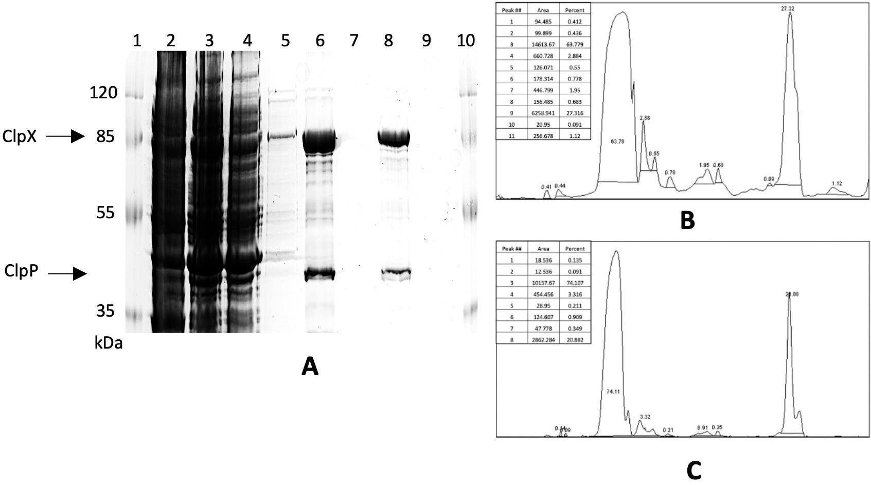
Figure 1. Purification of ClpXP and analysis of the collected protein fractions. (A) 10% SDS-PAGE analysis of the ClpXP purification: 1, prestained protein ladder; 2, 8 M urea-soluble fraction; 3, 8 M urea-unsoluble fraction; 4, flowthrough fraction; 5, wash fraction; 6 and 8, elution fractions 1 and 2, respectively, concentrated with a centrifuge concentrator with a 100 kDa cutoff; 7 and 9, flowthrough fractions from a centrifuge concentrator with a 100 kDa cutoff obtained after concentrating elution fractions 1 and 2, respectively; 10, prestained protein ladder. ClpX and ClpP positions are indicated by arrows at the left. (B, C) Purity assessment of elution fractions 1 and 2 by densitometric analysis using ImageJ software.
C. Assessment of the desirable stoichiometry of the ClpXP complex
Using the above protocol, high-molecular-weight mitochondrial protease complex ClpXP was obtained after separate expression in E. coli of two His-tagged subunits and their combined purification from inclusion bodies using Ni-NTA IMAC in denatured conditions with consequent on-column refolding. Separate expression of the two subunits allowed individual optimization of the temperature conditions that provided the maximum yield of the final complex.
Figure 2A shows that combined protein purification after expression of both ClpP and ClpX at the same temperature (either 24 or 37 °C) yielded undesirable stoichiometry of the subunits of the ClpXP complex and a higher level of contaminants, whereas Figure 2B shows that combined protein isolation after their individual expression at different induction times and temperatures (5 h of induction at 37 °C for ClpP and 16 h of induction at 24 °C for ClpX) yielded the optimal combination of complex stoichiometry and purity.
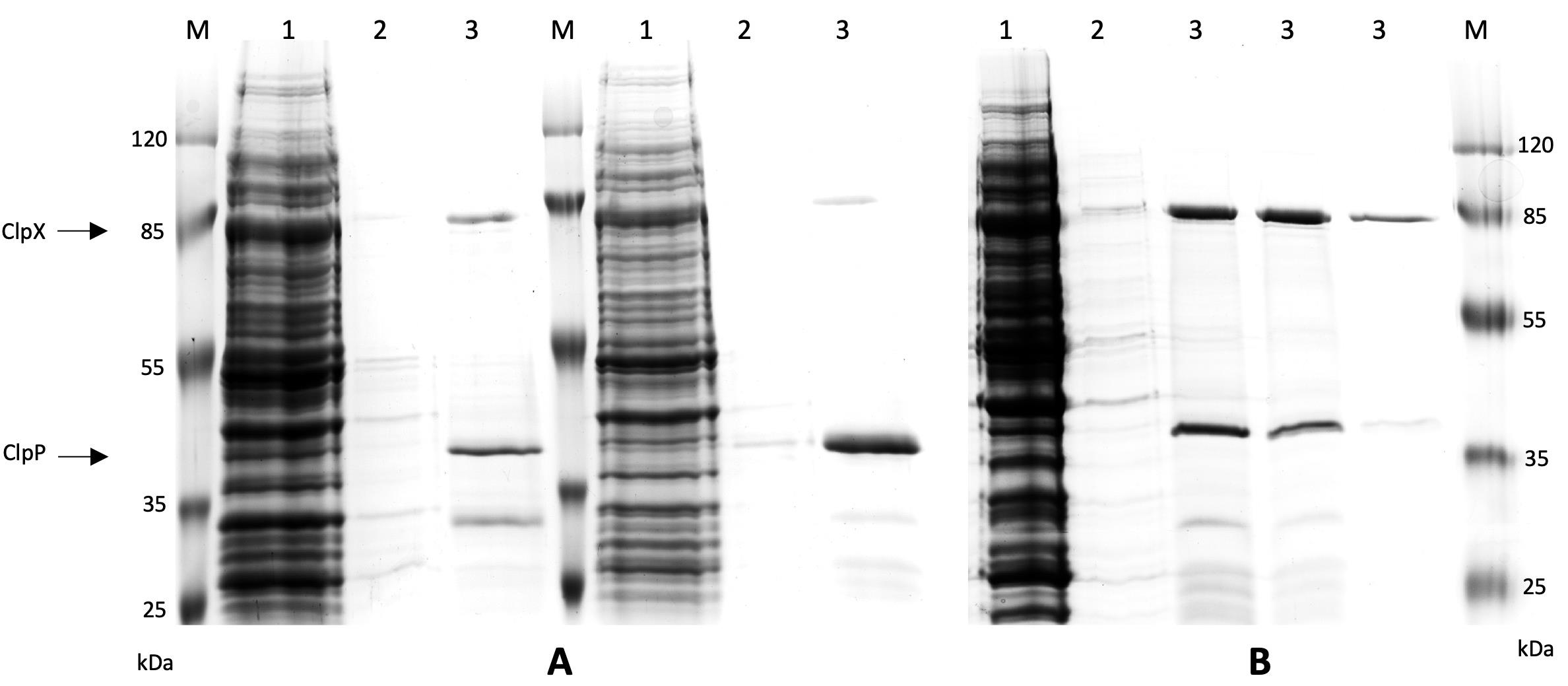
Figure 2. Analysis of the ClpXP complex purification upon expression of the two subunits using similar or different conditions. (A) Expression of both subunits either at 24 °C (left) or 37 °C (right). (B) Expression of the ClpX and ClpP subunits at their optimal expression conditions: for ClpP, it was 5 h of induction at 37 °C; for ClpX, it was 16 h of induction at 24 °C. M, prestained protein ladder; 1, flowthrough fraction; 2, combined wash fraction; 3, elution fraction. ClpX and ClpP positions are indicated by arrows at the left.
D. Quality control criteria
1. The purity of the obtained ClpXP fraction, determined by densitometry, should be over 90% of total protein.
2. Protein fraction with significant proteolytic degradation or smearing on SDS-PAGE should be excluded.
3. The obtained protein must have functional activity.
Validation of protocol
Final validation of the presented protocol required assessment of the functional activity of the human ClpXP protease complex obtained in the heterologous expression system. The proteolytic activity of the purified ClpXP complex was analyzed by its ability to digest casein. Casein is widely used as the standard model substrate for evaluating mitochondrial proteases (such as Lon and ClpP). Since mitochondrial matrix proteases function primarily as quality-control enzymes, casein, as an intrinsically disordered protein, structurally mirrors a denatured or unfolded mitochondrial protein [31]. Protocol validation was performed using three independent protein preparations (biological replicates). Figure 3A illustrates the typical pattern of beta-casein hydrolysis by the purified ClpXP complex. Figure 3B shows the mean percentage of casein hydrolysis by ClpXP across three independent experiments.
A. Assessment of the proteolytic activity of the purified ClpXP complex
1. All points of the assay were done in triplicate.
2. The assay was carried out in 0.5 mL tubes; the assay volume was 30 μL.
3. The assay buffer (AB) was prepared and well mixed as described in Recipes.
4. The required volume of 10 μg/μL bovine β-casein solution (2 μg per reaction) was added to the appropriate AB volume. AB volume was calculated as follows: (30 μL × N of assays) × 1.1. The β-casein volume was calculated as follows: (0.2 μL × N of assays) × 1.1. The mixture was mixed by pipetting and aliquoted at 30 μL into 0.5 mL tubes in accordance with the number of reactions.
5. Enzyme serial dilutions in AB were added to the reaction replicates (the maximum added volume should not exceed 3 μL), mixed, spun briefly, and incubated at 37 °C for 3 h.
6. 15 μL of 4× SB were added to the hydrolysis samples. The samples were incubated at 90 °C for 5 min (to halt hydrolysis) and spun briefly. The samples were loaded by 20 μL into the wells of 15% SDS-PAAGE and resolved by electrophoresis. The gel was stained with Coomassie Brilliant Blue (see Figure 3A).
7. The stained gels were digitized using a Bio-Rad gel documentation system, and the casein bands were quantified via densitometric analysis using ImageJ software.
Figure 3A shows SDS-PAGE analysis of ClpXP-dependent hydrolysis of 2 µg of bovine β-casein. In the left line, where the maximum amount of enzyme (50 ng) was used, all casein was digested. In subsequent lines, the hydrolysis efficiency decreases in accordance with the decreasing amounts of ClpXP. The right line shows unhydrolyzed casein in the absence of the protease complex. Figure 3B shows statistical analysis of the three independent purification/hydrolysis experiments.
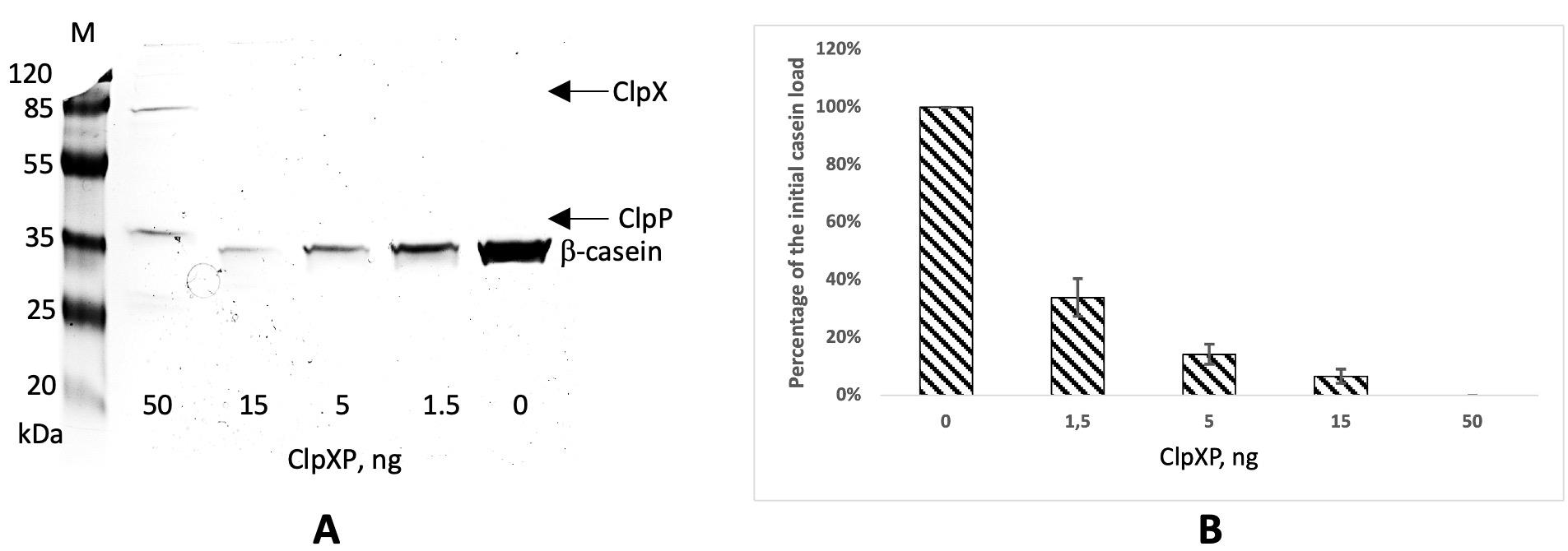
Figure 3. Analysis of ClpXP-dependent hydrolysis of β-casein. (A) 15% SDS-PAGE analysis of the bovine β-casein (2 μg) hydrolysis by the purified ClpXP complex: the ClpXP amount is indicated on the bottom; ClpX, ClpP, and casein positions are indicated by arrows at the right. M, prestained protein ladder. (B) Densitometric analysis of β-casein bands using the ImageJ software. Data are presented as the mean percentage of the remaining (unhydrolyzed) β-casein, where the initial load (2 μg) is taken as 100%, ± SD (n = 3, i.e. ClpXP was produced in three independent expression experiments).
General notes and troubleshooting
General notes
1. Protein storage:
a. Refolded protein should be stored at –20 °C into 50% glycerol.
b. Optimal protein concentration is 1–2.5 mg/mL.
2. Buffer and gradient optimization:
a. The chaotropic agent for the dilution of the inclusion bodies can be optimized (8 M urea or 6 M guanidine hydrochloride).
b. The stepwise gradient for decreasing urea concentration from 8 to 0 M and increasing imidazole from 20 to 50 mM can be optimized.
c. Ionic strength and pH conditions in the buffer solutions should be adapted to the nature of the protein purified.
d. For proteins requiring specific redox agents, such as β-mercaptoethanol or DTT, these agents should be included in the buffer solutions.
e. Different mild detergents or their combination could be tested.
3. System applicability: In case of different affinity tags (GST, MBP), inclusion bodies should be washed, solubilized, and refolded before the affinity purification step.
4. Scale considerations:
a. When the production volume is increased, the volumes of the resin and buffer solutions should be scaled up proportionally.
b. For larger scales, automation is recommended: any FPLC chromatograph with appropriate columns can be used.
5. Quality control:
a. All protein fractions (protein lysate, flowthrough, column wash, and elution fraction) should be analyzed by SDS-PAGE using appropriate percentage polyacrylamide gels.
b. Freshly prepared buffer solution, regular cleaning of the working area, and temperature control during the purification process are strongly recommended.
Troubleshooting
| Problem | Possible cause | Solution |
| Low protein yield and quality | Low expression of the recombinant protein | Optimize the expression conditions |
| Insufficient solubilization of inclusion bodies | Optimize the chaotropic agent or extend the solubilization time | |
| Poor binding to Ni-NTA resin | Verify His-tag integrity by western blotting | |
| Optimize binding buffer composition to prevent protein aggregation | ||
| Extend binding time | ||
| Protein aggregation during refolding and elution | Optimize the stepwise gradient | |
| Optimize elution buffer composition | ||
| Add stabilizing additives (arginine 0.5 M, sucrose 0.4 M) to refolding and elution buffers | ||
| Inconsistent protein yield and quality between different experiments | Variations in experimental protocols and conditions | Standardize experimental protocol and buffer compositions |
| Use freshly prepared buffers for each experiment | ||
| Control and document protein yield and quality in all steps of each experiment |
Acknowledgments
The work was carried out with the financial support of the Russian Science Foundation project 24–74-10107.
Author contributions
Conceptualization, E.V.S.; Investigation, O.S.E., E.V.S., T.V.R.; Writing—Original Draft, O.S.E., E.V.S., T.V.R.; Writing—Review & Editing, A.A.K., A.A.B.; Funding acquisition, A.A.K., A.A.B.
Competing interests
The authors declare that they have no competing interests.
References
- Rosano, G. L. and Ceccarelli, E. A. (2014). Recombinant protein expression in Escherichia coli: advances and challenges. Front Microbiol. 5: e00172. https://doi.org/10.3389/fmicb.2014.00172
- Gupta, S. K. and Shukla, P. (2015). Advanced technologies for improved expression of recombinant proteins in bacteria: perspectives and applications. Crit Rev Biotechnol. 36(6): 1089–1098. https://doi.org/10.3109/07388551.2015.1084264
- Gasser, B., Saloheimo, M., Rinas, U., Dragosits, M., Rodríguez-Carmona, E., Baumann, K., Giuliani, M., Parrilli, E., Branduardi, P., Lang, C., et al. (2008). Protein folding and conformational stress in microbial cells producing recombinant proteins: a host comparative overview. Microb Cell Fact. 7(1): 11. https://doi.org/10.1186/1475-2859-7-11
- Ortega, C., Prieto, D., Abreu, C., Oppezzo, P. and Correa, A. (2018). Multi-Compartment and Multi-Host Vector Suite for Recombinant Protein Expression and Purification. Front Microbiol. 9: e01384. https://doi.org/10.3389/fmicb.2018.01384
- de Marco, A. (2009). Strategies for successful recombinant expression of disulfide bond-dependent proteins in Escherichia coli. Microb Cell Fact. 8(1): 26. https://doi.org/10.1186/1475-2859-8-26
- Wang, L., Maji, S. K., Sawaya, M. R., Eisenberg, D. and Riek, R. (2008). Bacterial Inclusion Bodies Contain Amyloid-Like Structure. PLoS Biol. 6(8): e195. https://doi.org/10.1371/journal.pbio.0060195
- Singh, A., Upadhyay, V., Upadhyay, A. K., Singh, S. M. and Panda, A. K. (2015). Protein recovery from inclusion bodies of Escherichia coli using mild solubilization process. Microb Cell Fact. 14(1): 41. https://doi.org/10.1186/s12934-015-0222-8
- Lill, R. and Mühlenhoff, U. (2008). Maturation of Iron-Sulfur Proteins in Eukaryotes: Mechanisms, Connected Processes, and Diseases. Annu Rev Biochem. 77(1): 669–700. https://doi.org/10.1146/annurev.biochem.76.052705.162653
- Khoury, G. A., Baliban, R. C. and Floudas, C. A. (2011). Proteome-wide post-translational modification statistics: frequency analysis and curation of the swiss-prot database. Sci Rep. 1(1): 90. https://doi.org/10.1038/srep00090
- Saida, F., Uzan, M., Odaert, B. and Bontems, F. (2006). Expression of Highly Toxic Genes in E. coli: Special Strategies and Genetic Tools. Curr Protein Pept Sci. 7(1): 47–56. https://doi.org/10.2174/138920306775474095
- Kramer, R. A., Zandwijken, D., Egmond, M. R. and Dekker, N. (2000). In vitro folding, purification and characterization of Escherichia coli outer membrane protease OmpT. Eur J Biochem. 267(3): 885–893. https://doi.org/10.1046/j.1432-1327.2000.01073.x
- Hung, C. C. and Chiou, S. H. (2000). Expression of a Kallikrein-like Protease from the Snake Venom: Engineering of Autocatalytic Site in the Fusion Protein to Facilitate Protein Refolding. Biochem Biophys Res Commun. 275(3): 924–930. https://doi.org/10.1006/bbrc.2000.3411
- Jungbauer, A. and Kaar, W. (2007). Current status of technical protein refolding. J Biotechnol. 128(3): 587–596. https://doi.org/10.1016/j.jbiotec.2006.12.004
- Li, M., Su, Z. G. and Janson, J. C. (2004). In vitro protein refolding by chromatographic procedures. Protein Expression Purif. 33(1): 1–10. https://doi.org/10.1016/j.pep.2003.08.023
- Kang, S. G., Ortega, J., Singh, S. K., Wang, N., Huang, N. n., Steven, A. C. and Maurizi, M. R. (2002). Functional Proteolytic Complexes of the Human Mitochondrial ATP-dependent Protease, hClpXP. J Biol Chem. 277(23): 21095–21102. https://doi.org/10.1074/jbc.m201642200
- Lowth, B. R., Kirstein-Miles, J., Saiyed, T., Brötz-Oesterhelt, H., Morimoto, R. I., Truscott, K. N. and Dougan, D. A. (2012). Substrate recognition and processing by a Walker B mutant of the human mitochondrial AAA+ protein CLPX. J Struct Biol. 179(2): 193–201. https://doi.org/10.1016/j.jsb.2012.06.001
- Yien, Y. Y., Ducamp, S., van der Vorm, L. N., Kardon, J. R., Manceau, H., Kannengiesser, C., Bergonia, H. A., Kafina, M. D., Karim, Z., Gouya, L., et al. (2017). Mutation in human CLPX elevates levels of δ- aminolevulinate synthase and protoporphyrin IX to promote erythropoietic protoporphyria. Proc Natl Acad Sci USA. 114(38): e1700632114. https://doi.org/10.1073/pnas.1700632114
- Szczepanowska, K. and Trifunovic, A. (2021). Mitochondrial matrix proteases: quality control and beyond. FEBS J. 289(22): 7128–7146. https://doi.org/10.1111/febs.15964
- Chen, W., Lander, G. C. and Yang, J. (2026). Cryo-EM structures of human ClpXP reveal mechanisms of assembly and proteolytic activation. Nat Commun. 17(1): 1064. https://doi.org/10.1038/s41467-025-67010-1
- Corydon, T. J., Wilsbech, M., Jespersgaard, C., Andresen, B. S., Børglum, A. D., Pedersen, S., Bolund, L., Gregersen, N. and Bross, P. (2000). Human and mouse mitochondrial orthologs of bacterial ClpX. Mamm Genome. 11(10): 899–905. https://doi.org/10.1007/s003350010173
- Feng, Y., Nouri, K. and Schimmer, A. D. (2021). Mitochondrial ATP-Dependent Proteases—Biological Function and Potential Anti-Cancer Targets. Cancers (Basel). 13(9): 2020. https://doi.org/10.3390/cancers13092020
- Wojtkowiak, D., Georgopoulos, C. and Zylicz, M. (1993). Isolation and characterization of ClpX, a new ATP-dependent specificity component of the Clp protease of Escherichia coli.. J Biol Chem. 268(30): 22609–22617. https://doi.org/10.1016/s0021-9258(18)41572-4
- Joshi, S. A., Hersch, G. L., Baker, T. A. and Sauer, R. T. (2004). Communication between ClpX and ClpP during substrate processing and degradation. Nat Struct Mol Biol. 11(5): 404–411. https://doi.org/10.1038/nsmb752
- Neher, S. B., Villén, J., Oakes, E. C., Bakalarski, C. E., Sauer, R. T., Gygi, S. P. and Baker, T. A. (2006). Proteomic Profiling of ClpXP Substrates after DNA Damage Reveals Extensive Instability within SOS Regulon. Mol Cell. 22(2): 193–204. https://doi.org/10.1016/j.molcel.2006.03.007
- Feng, J., Michalik, S., Varming, A. N., Andersen, J. H., Albrecht, D., Jelsbak, L., Krieger, S., Ohlsen, K., Hecker, M., Gerth, U., et al. (2013). Trapping and Proteomic Identification of Cellular Substrates of the ClpP Protease in Staphylococcus aureus. J Proteome Res. 12(2): 547–558. https://doi.org/10.1021/pr300394r
- Shi, X., Wu, T., M. Cole, C., K. Devaraj, N. and Joseph, S. (2018). Optimization of ClpXP activity and protein synthesis in an E. coli extract-based cell-free expression system. Sci Rep. 8(1): e1038/s41598–018–21739–6. https://doi.org/10.1038/s41598-018-21739-6
- Okonechnikov, K., Golosova, O., Fursov, M. and UGENE Team. (2012). Unipro UGENE: a unified bioinformatics toolkit. Bioinformatics. 28(8): 1166–1167. https://doi.org/10.1093/bioinformatics/bts091
- Golosova, O., Henderson, R., Vaskin, Y., Gabrielian, A., Grekhov, G., Nagarajan, V., Oler, A. J., Quiñones, M., Hurt, D., Fursov, M., et al. (2014). Unipro UGENE NGS pipelines and components for variant calling, RNA-seq and ChIP-seq data analyses. PeerJ. 2: e644. https://doi.org/10.7717/peerj.644
- Rose, R., Golosova, O., Sukhomlinov, D., Tiunov, A. and Prosperi, M. (2018). Flexible design of multiple metagenomics classification pipelines with UGENE. Bioinformatics. 35(11): 1963–1965. https://doi.org/10.1093/bioinformatics/bty901
- Schneider, C. A., Rasband, W. S. and Eliceiri, K. W. (2012). NIH Image to ImageJ: 25 years of image analysis. Nat Methods. 9(7): 671–675. https://doi.org/10.1038/nmeth.2089
- Lau, E., Wang, D., Zhang, J., Yu, H., Lam, M. P., Liang, X., Zong, N., Kim, T. Y. and Ping, P. (2012). Substrate- and Isoform-Specific Proteome Stability in Normal and Stressed Cardiac Mitochondria. Circ Res. 110(9): 1174–1178. https://doi.org/10.1161/circresaha.112.268359
Article Information
Publication history
Received: Jul 21, 2026
Accepted: Oct 8, 2026
Available online: Oct 30, 2026
Published: Nov 20, 2026
Copyright
© 2026 The Author(s); This is an open access article under the CC BY-NC license (https://creativecommons.org/licenses/by-nc/4.0/).
How to cite
Elmeeva, O. S., Smirnova, E. V., Rakitina, T. V., Kudriaeva, A. A. and Belogurov, A. A. (2026). Escherichia coli-Based Production and Purification From Inclusion Bodies of Hetero-Oligomeric Mitochondrial Protease ClpXP in Active Form. Bio-protocol 16(22): e5865. DOI: 10.21769/BioProtoc.5865.

 Download Citation in RIS Format
Do you have any questions about this protocol?
Post your question to gather feedback from the community. We will also invite the authors of this article to respond.
 Post a Question


