


- Protocols

- Articles and Issues
- For Authors
- About
- Become a Reviewer


MORECOVERY: A Swab-Based Surface Sampling Protocol Incorporating a Nutrient-Free Resuscitation Step for the Detection of Clinically Relevant Gram-Negative Pathogens in the Viable but Non-Culturable State

(*contributed equally to this work) Published: Vol 16, Iss 16, Aug 20, 2026 DOI: 10.21769/BioProtoc.5782 Views: 267
Reviewed by: Alba BlesaRodrigo de Siqueira MeloAnonymous reviewer(s)
 PDF
PDF  Ask a question
Original research article
The authors used this protocol in:
Mar 2026

Protocol Collections
Comprehensive collections of detailed, peer-reviewed protocols focusing on specific topics




Abstract
Difficult-to-treat Gram-negative bacteria are a major cause of healthcare-associated infections due to multidrug resistance and limited therapeutic options. The hospital environment plays a central role in the persistence and transmission of infection, making environmental monitoring an essential component of infection prevention and control plans. Standard surface sampling techniques, including swabs, contact plates, and sponges, are widely used for environmental surveillance and are all based on culture-dependent methods. However, these techniques may underestimate the actual level of bacterial contamination since they fail to detect bacteria in the viable but non-culturable (VBNC) state, a reversible physiological condition in which bacterial cells remain viable but do not grow on conventional culture media. An innovative environmental sampling protocol, herein named MORECOVERY, has been developed to improve the detection of VBNC bacteria. The protocol integrates an essential resuscitation step, which improves the recovery of VBNC Gram-negative bacteria by a few orders of magnitude, into the standard swab-based sampling workflow. Following sample collection, swabs are incubated for 24 h at 37 °C in a carbon-free resuscitation buffer before plating, enabling the recovery of VBNC bacterial pathogens that would otherwise remain undetectable. By improving the recovery of VBNC cells, the MORECOVERY protocol allows a more accurate assessment of bacterial contamination of critical surfaces in healthcare settings. Its simplicity and minimal variation from standard workflows facilitate easy implementation in routine environmental monitoring.
Key features
• Enables the detection of Gram-negative bacteria in the viable but non-culturable (VBNC) state, allowing the recovery of otherwise undetectable bacterial contaminants.
• Is compatible with standard swab-based environmental sampling workflows and routine microbiological procedures.
• Relies on low-cost materials and standard laboratory equipment and can easily be implemented in healthcare facilities and other settings requiring careful monitoring of bacterial contamination.
• Enhances the sensitivity of culture-based environmental surveillance methods through a simple resuscitation step prior to plating.
• Combines a carbon-free resuscitation buffer and 24-h incubation at 37 °C, preventing bacterial growth while ensuring VBNC resuscitation without bacterial replication.
Keywords: Biocontamination Culturability
Environmental sampling
Gram-negative bacteria
Resuscitation
Viable but non-culturable
Difficult-to-treat healthcare-associated infections
Swab-based sampling
Culturability
Environmental sampling
Gram-negative bacteria
Resuscitation
Viable but non-culturable
Difficult-to-treat healthcare-associated infections
Swab-based sampling
Graphical overview
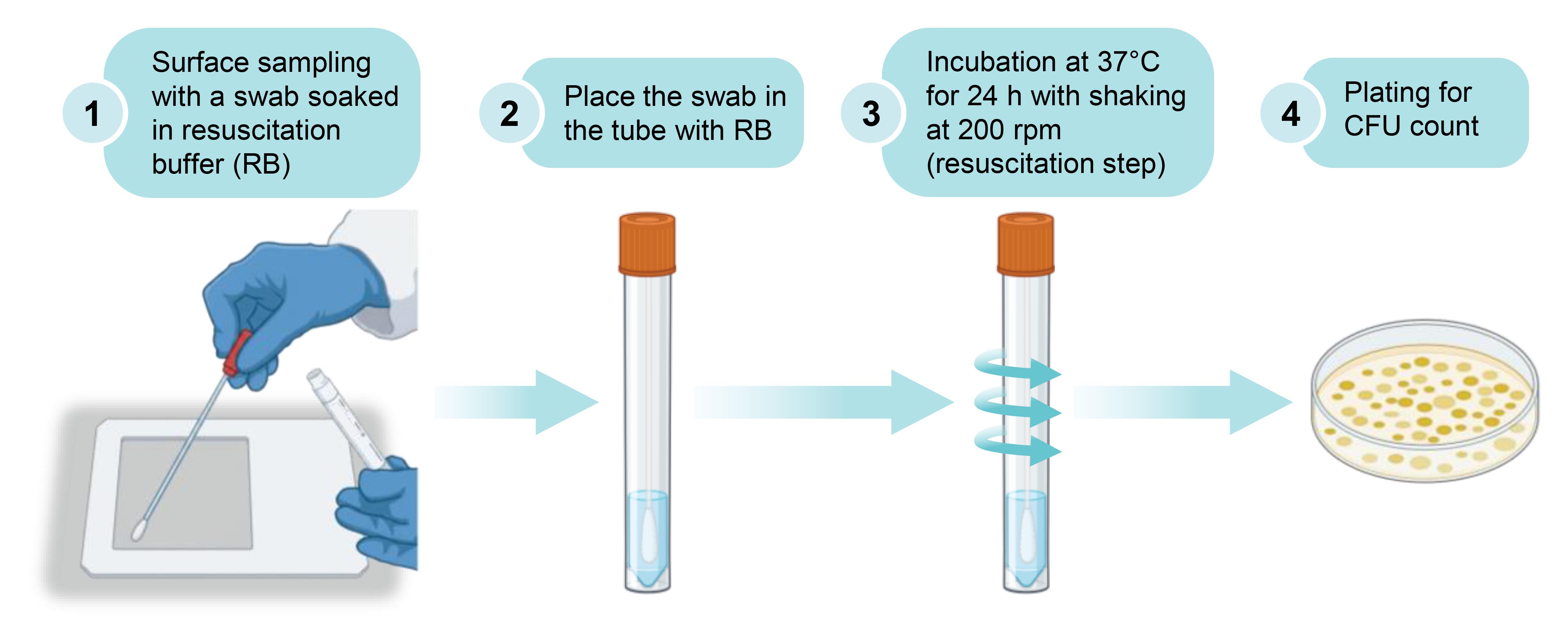
Schematic overview of the MORECOVERY surface sampling protocol, including the resuscitation step
Background
Difficult-to-treat (DTR) infections caused by some Gram-negative bacteria, including Acinetobacter baumannii, Pseudomonas aeruginosa, Klebsiella pneumoniae, and other antibiotic-resistant Enterobacterales, are a major challenge in healthcare settings due to multidrug resistance [1]. Although the introduction of newer antimicrobial agents has broadened treatment options and increased their use in recent years, clinical outcomes have remained largely unchanged, with no significant reduction in mortality observed for most DTR Gram-negative infections, underscoring the need for complementary prevention and control strategies [2].
Hospital surfaces and medical devices constitute well-established reservoirs for healthcare-associated microorganisms and play a central role in their transmission [3]. In this context, environmental surveillance represents a critical component of effective infection prevention strategies [4]. However, standard biocontamination control methods rely predominantly on culture-based techniques, which may fail to detect bacteria capable of entering a viable but non-culturable (VBNC) state [5].
The VBNC state is a reversible physiological condition in which bacterial cells remain viable and metabolically active but lose the ability to grow in conventional culture media. It can be induced by environmental stressors commonly encountered in hospital settings, including nutrient limitation, exposure to disinfectants and antimicrobial agents, and prolonged desiccation. Among these, long-term desiccation represents a particularly critical challenge, as sustained water loss can severely impair survival and trigger a range of biochemical, metabolic, and physiological adaptations [6]. Evidence indicates that DTR Gram-negative pathogens exposed to desiccation on abiotic hospital surfaces can enter the VBNC state, thereby losing their ability to generate colonies on standard microbiological media. Culturability can, however, be restored upon rehydration in a carbon-free isotonic buffer, a process referred to as resuscitation [7]. Notably, VBNC cells may retain pathogenic potential despite being undetectable by standard culture-based methods [5,8], representing a hidden reservoir for infection transmission to susceptible patients.
Hereafter, the MORECOVERY protocol for the detection and quantification of Gram-negative bacteria in the VBNC state on abiotic surfaces is described. The protocol is adapted from and validated in [7]. According to the BS EN 17141:2020 standard for “Cleanrooms and associated controlled environments—Biocontamination control,” environmental sampling can be performed using different devices, including contact plates, sponges, and swabs, with swabs being the most frequently used in healthcare settings [9,10]. Within this framework, the MORECOVERY protocol differs from conventional surface sampling protocols only by the inclusion of an additional step, consisting of a 24-h incubation at 37 °C in a resuscitation buffer (RB) before plating. RB is a buffer devoid of any carbon sources to prevent bacterial growth during the resuscitation step, thereby ensuring that the increase in CFU is attributable to VBNC resuscitation rather than proliferation of culturable cells. Despite minimal modifications compared with standard protocols [9,10], the resuscitation step allows the recovery of VBNC cells by restoring their cultivability, ultimately enabling a more accurate estimation of the bacterial load on sampled surfaces. Owing to its simplicity and compatibility with existing workflows, this approach can readily be implemented on a large scale in routine environmental monitoring and may contribute to improved infection control strategies.
Materials and reagents
Reagents
1. Tryptic soy agar (TSA) (Becton, Dickinson and Company, catalog number: 212305)
2. M9, minimal salts, 5× (Merck, catalog number: M6030)
3. Calcium chloride (CaCl2) (Merck, catalog number: C7902)
4. Magnesium sulfate (MgSO4) (Merck, catalog number: M2643)
Solutions
1. Tryptic soy agar (TSA) (see Recipes)
2. Resuscitation buffer (RB) (see Recipes)
Recipes
1. TSA
To prepare 1 L of TSA, suspend 40 g of dehydrated TSA culture medium in 1 L of distilled (type II) water in a suitable flask or beaker under magnetic stirring and heat with agitation until completely dissolved. Sterilize by autoclaving at 121 °C for 15 min. After cooling to ~45–50 °C, aseptically pour into sterile Petri dishes (25 mL per plate) and allow to solidify.
Note: Commercially prepared TSA plates, purchased by many companies, may be used as an alternative.
2. Resuscitation buffer (RB)
| Reagent | Final concentration | Quantity or volume |
|---|---|---|
| 5× M9, minimal salts (56.4 g/L) | 11.28 g/L | 200 mL |
| 500× CaCl2 (10 g/L) | 0.02 g/L | 2 mL |
| 500× MgSO4 (60 g/L) | 0.12 g/L | 2 mL |
| Double-distilled (type I) water (ddH2O) | - | 796 mL |
Before preparing the RB, stock solutions of 5× M9 minimal salts solution, CaCl2 (500×, 10 g/L), and MgSO4 (500×, 60 g/L) should be made in advance. To prepare 1 L of M9 minimal salts solution (5×), dissolve 56.4 g of dehydrated powder in approximately 800 mL of ddH2O in a suitable flask or beaker under magnetic stirring until completely dissolved. Adjust the final volume to 1 L with ddH2O and sterilize by autoclaving at 121 °C for 15 min. The M9 basal salts solution contains KH2PO4 (15 g/L), NaCl (2.5 g/L), Na2HPO4 (33.9 g/L), and NH4Cl (5 g/L) [11]. The sterile M9 minimal salts solution (5×) can be stored at 4 °C for up to 6 months. Before use, inspect for precipitation or signs of contamination. The pre-mixed M9 minimal salts are also available from Merck (see Reagents).
For the CaCl2 stock solution (500×, 10 g/L), dissolve 0.20 g of CaCl2 in 20 mL of ddH2O in a 50 mL tube and sterilize by 0.2 μm filtration or by autoclaving. Similarly, prepare the MgSO4 stock solution (500×, 60 g/L) by dissolving 1.20 g of MgSO4 in 20 mL of ddH2O in a 50 mL tube, followed by sterilization through 0.2 μm filtration or by autoclaving. The sterile CaCl2 and MgSO4 stock solutions are stable and can be stored for extended periods at room temperature, when properly sealed; however, storage at 4 °C is recommended for long-term use.
The required volume of RB depends on the number of surfaces to be sampled. Considering that each swab contains 2 mL of RB, 1 L of RB is sufficient for 500 swabs. For RB preparation, combine the appropriate volumes of sterile stock solutions in a sterile container under aseptic conditions. As an example, to prepare 200 mL of RB, mix 40 mL of 5× M9 minimal salts solution, 400 μL of 500× CaCl2 stock solution, and 400 μL of 500× MgSO4 stock solution, then adjust the final volume to 200 mL with sterile ddH2O. Mix thoroughly to ensure homogeneity.
When needed, fresh preparation of RB from sterile stock solutions under aseptic conditions is recommended to avoid potential contamination. However, RB remains stable for up to five weeks at room temperature, and it must be used at room temperature to ensure optimal handling conditions and to avoid any potential thermal shock to stressed bacterial cells.
Laboratory supplies
1. Sterile nylon swabs (FLOQSwabs, Copan, catalog number: 519C)
2. Sampling templates (10 × 10 cm) (Copan, catalog number: T2905)
3. Sampling templates (5 × 4 cm) (Copan, catalog number: T2906)
4. Sterile Petri dishes (Sarstedt, catalog number: 82.1472)
5. Sterile disposable spreader (Sarstedt, catalog number: 86.1569.005)
6. Disposable sterile 10 mL syringe (Biosigma, catalog number: 050840)
7. Syringe filters 0.2 μm (Sarstedt, catalog number: 83.1826.001)
8. 1.5 mL tubes (Sarstedt, catalog number: 72.690.001)
9. 15 mL conical tubes (Sarstedt, catalog number: 62.554.001)
10. 50 mL conical tubes (Sarstedt, catalog number: 62.547.255)
11. 200 μL pipette tips (Sarstedt, catalog number: 70.3030)
Equipment
1. Vertical laminar flow hood (Bioair, model: TopSafe 1.2)
2. Vortex (Merck, catalog number: Z258423)
3. Microbiological incubator (Merck, catalog number: CLS6754)
4. Magnetic stirrer with heating plate (Carl Roth, catalog number: XT23.1)
5. Pipette P200 (Gilson, catalog number: FA10005M)
6. Refrigerator (4 °C) (Miele, catalog number: KFN 7795C)
7. Analytical balance (Merck, catalog number: Z742879)
8. Autoclave (ASAL, catalog number: 29960018)
9. Nitrile gloves (Merck, catalog number: Z677272)
10. Felt marker, waterproof (Sarstedt, catalog number: 95.954)
Procedure
A. Preparation of sampling materials
1. Prepare the RB.
2. Dispense 2 mL of RB into the sterile swab tube under aseptic conditions to preserve its sterility.
3. Keep nylon swabs sterile until use.
B. Surface sampling and incubation of the swab
1. Label all swab tubes with sample ID and date.
2. Select the area to be sampled.
3. If the area of interest is flat, a sampling template should be used. The sampling template may be either 100 cm2 (10 × 10 cm) or 20 cm2 (5 × 4 cm) (Figure 1), depending on the surface to be sampled. The sampled area should be representative of the surface being sampled. For irregular surfaces, the sampled area should be approximated by decomposing it into simple geometric shapes in order to estimate its surface area as accurately as possible.
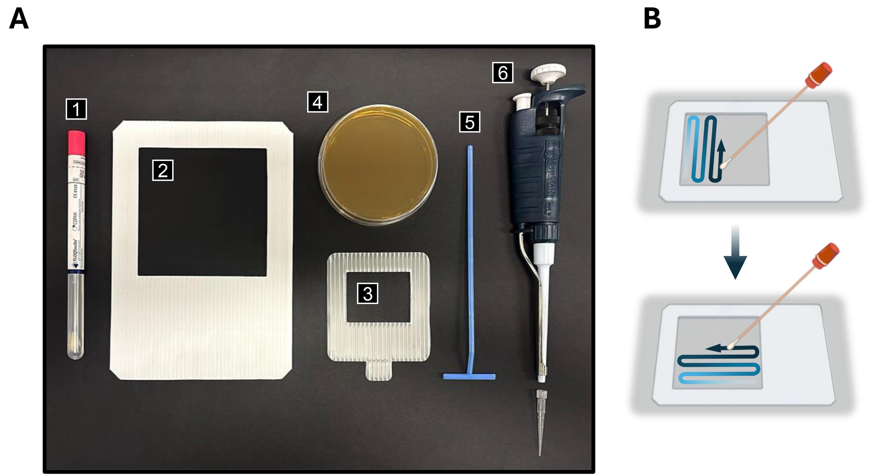
Figure 1. Materials and equipment for the MORECOVERY surface sampling protocol and method for surface sampling. (A) Materials and equipment: (1) Sterile plastic tube containing the nylon swab soaked with RB; (2–3) 10 × 10 cm and 5 × 4 cm sampling templates; (4) TSA plate; (5) disposable spreader for inoculation; and (6) P200 pipette with disposable tip. (B) Surface sampling procedure. For flat surfaces, swab the sampling area using a horizontal zig-zag pattern followed by a vertical zig-zag pattern, performing at least 10 strokes in each direction, to ensure sampling of the whole surface.
4. Keep the swab tube upright and check that the RB is at the bottom; if necessary, gently shake the tube downward so that all the liquid gathers at the bottom of the tube.
5. Gently press the swab tip against the inner wall of the tube to eliminate any excess RB, ensuring the swab remains damp and uniformly soaked with the solution.
6. Hold the swab handle between the thumb and forefinger. For flat surfaces, perform sampling by swabbing horizontally and vertically, carrying out at least 10 strokes in each direction. For small or hard-to-reach areas, ensure that the entire defined location is sampled, including crevices, gaps, joints, and other surface connections.
7. Transfer the swab back into the tube containing RB. Make sure the tube containing the swab is properly closed so that the swab remains submerged in RB until analysis.
8. If appropriate, include additional swabs to be used as field negative controls (i.e., swabs that are not used for surface sampling but are processed alongside the samples to monitor potential contamination).
Note: Samples should be transported to the laboratory at room temperature within 8 h. Ensure that storage and transport temperatures do not exceed 37 °C. Refrigerated conditions are not required, as the RB does not contain carbon sources that could support bacterial growth and potentially affect the accurate assessment of contamination.
C. Resuscitation of VBNC bacterial cells and plating
1. Incubate the tube containing the swab at 37 °C for 24 h with shaking at 200 rpm.
Critical: This step corresponds to the resuscitation phase, during which VBNC cells may regain culturability. Continuous shaking at 200 rpm during the resuscitation step is recommended to ensure adequate oxygenation and homogeneous contact between the cells and the RB. During incubation, swab tubes should preferably be positioned at a 45° angle on the shaker to facilitate mixing and aeration.
2. Following incubation, vortex the tube for 1 min prior to plating.
Critical: Vortexing for 1 min is essential to promote the release of bacterial cells from the swab fibers into the RB. Pipette 100 μL of the sample (presumptive bacterial cell suspension in RB) onto TSA agar plates and spread evenly using a sterile disposable spreader. Operate under a laminar flow hood to minimize the risk of contamination.
Note: Alternative culture media may be used according to analytical requirements. When the aim is the detection of a specific pathogen, selective media appropriate for the isolation of the target microorganism may also be employed, in addition to TSA.
3. Incubate plates at 37 °C for up to 72 h.
Note: Since this method can be applied to the detection of specific microorganisms, incubation time and temperature may be adjusted according to the growth requirements of the target organism.
D. Colony-forming unit (CFU) quantification and data analysis
1. Count the number of CFUs on each plate.
2. Calculate the bacterial load on the sampled surface, expressed as CFU/cm2 (Ns), using the following formula:
where:
N = number of CFUs on the plate.
A = sampled surface area (cm2).
D = the inverse of the plated fraction (e.g., if 100 μL out of 2 mL are plated, this corresponds to 1/20 of the sample, so D = 20).
Example results obtained with the MORECOVERY protocol, which integrates the resuscitation step into the sampling procedure, are shown in Figure 2. In particular, Figure 2A shows the CFU counts of representative Gram-negative bacteria (A. baumannii, Enterobacter cloacae, K. pneumoniae, and P. aeruginosa) before and after the resuscitation step in a simulated contamination experiment. The increase in bacterial recovery ranges approximately from 10 to 104, depending on the test species. Figure 2B shows the results of a field test in which different surfaces (laboratory bench, classroom desks, and washbasin) were sampled, demonstrating that the resuscitation step can markedly enhance bacterial detection, with up to a one-million-fold increase of CFU recovery in some cases.
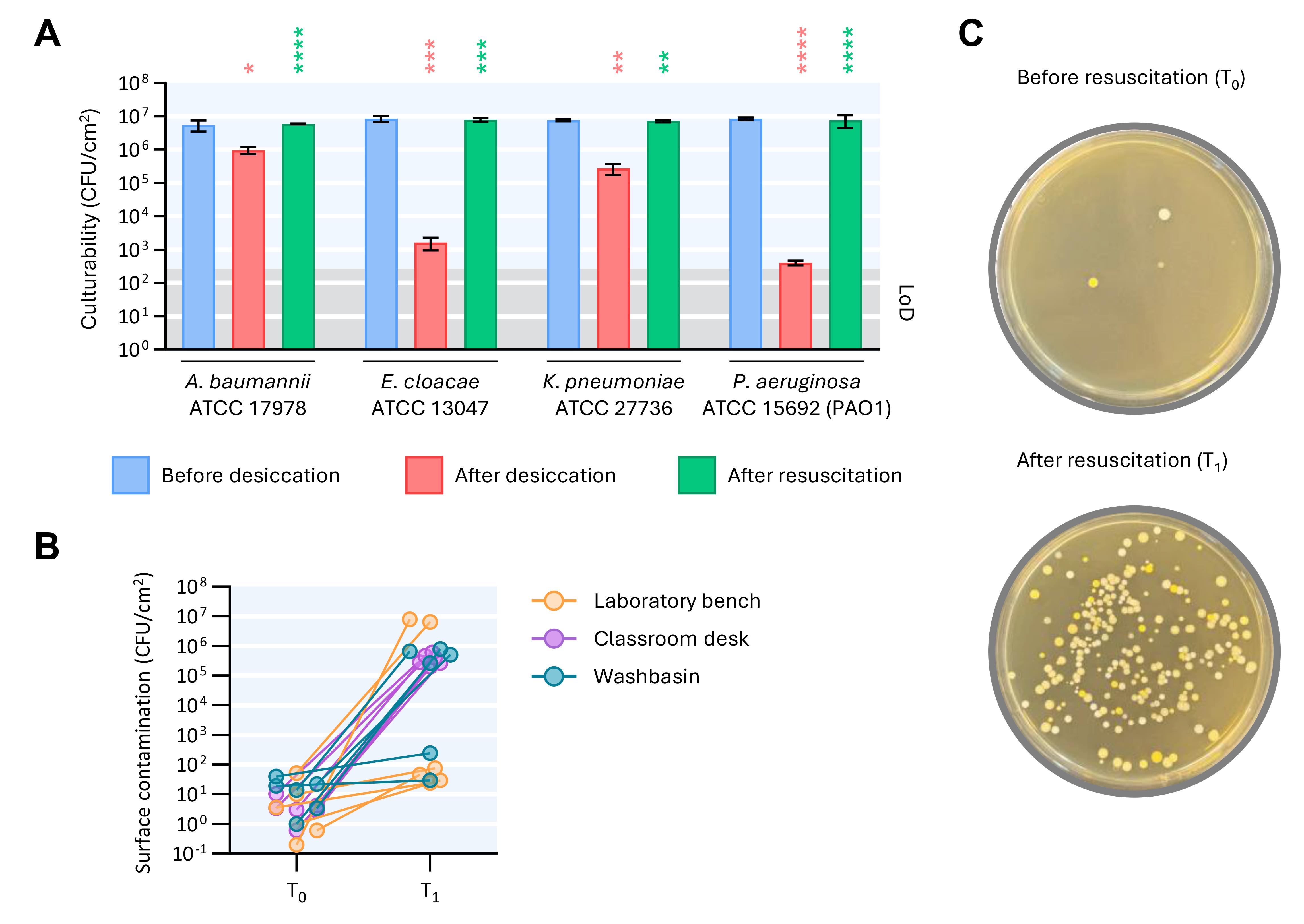
Figure 2. Resuscitation of viable but non-culturable (VBNC) cells by the MORECOVERY protocol improves bacterial detection [colony-forming unit (CFU) counts] on environmental surfaces under controlled laboratory conditions and in field tests. (A) Representative strains of Gram-negative bacteria were air-dried for 1 week on a glass surface. After desiccation, cells were suspended in RB and incubated at 37 °C under shaking for 24 h for resuscitation. Culturability was expressed as CFU recovery, as determined before desiccation, after 1-week desiccation, and after 24-h resuscitation in RB at 37 °C. The grey area indicates the limit of detection (LoD), corresponding to 2.7 × 10² CFU. Data are the mean ± standard deviation (error bars) of three independent experiments. Red asterisks indicate statistically significant differences between samples before desiccation and after desiccation. Green asterisks indicate statistically significant differences between samples after desiccation and after resuscitation. Statistically significant differences (*P < 0.05; **P < 0.01; ***P < 0.001; ****P < 0.0001) were determined using the unpaired t-test. (B) Microbial contamination of laboratory benches, classroom desks, and washbasins, determined before (T0) and after 24-h resuscitation in RB at 37 °C (T1). Six samples were collected and analyzed for each surface type (modified from [7]). (C) Representative photographs of Petri dishes inoculated with samples before (T0) and after resuscitation (T1).
E. Test report
Write a test report including the following information: date, identification of the sampling location, culture media and incubation conditions used, and the results obtained, expressed in relation to the sampled surface (including its size or designation). The report should also include details of any incidents or relevant deviations that may have influenced the test results.
Validation of protocol
This protocol has been used and validated in the following research article:
• Visaggio et al. [7]. ESKAPE Gram-negative bacteria escape culture-based detection upon desiccation on abiotic surfaces. Microbiol Spectrum. 14(3): e03357–25. https://doi.org/10.1128/spectrum.03357-25 (Figures 1 and 3)
General notes and troubleshooting
General notes
1. This protocol markedly increases the detection of Gram-negative bacteria, as it enables the recovery of both culturable cells and bacteria in the VBNC state. By contrast, the detection of some Gram-positive bacteria is not or only marginally improved by the resuscitation step [7].
2. This protocol can be adapted to different applications wherever monitoring of microbial contamination is required. When applied in settings where potentially pathogenic microorganisms may be present, appropriate institutional biosafety procedures should be followed, including BSL-2 practices when applicable.
Troubleshooting
Problem 1: Bacterial contamination below the limit of detection.
Possible cause: The swab is immersed in 2 mL of RB, from which 100 μL are plated for CFU counting. This results in a limit of detection (LoD) of 20 CFU per swab, corresponding to 10 CFU/mL, since only one-twentieth of the total sample volume is plated. Thus, contamination levels below the threshold of 20 CFU per sampled area cannot be detected.
Solution: For surfaces with expectedly low microbial contamination, or when detection of single bacterial cells is critical, sensitivity can be maximized by centrifuging the sample and plating the whole pellet originating from the 2-mL sample.
Problem 2: Plate overgrowth due to high bacterial contamination.
Possible cause: The sampled surface has a high bacterial load, leading to overgrowth and coalescence of colonies on the agar plate, which prevents accurate colony counting and reliable CFU quantification.
Solution: Perform serial ten-fold dilutions of RB prior to plating to obtain countable colonies on agar plates and ensure accurate quantification of the bacterial load.
Acknowledgments
Conceptualization, C.S., D.V., F.I., M.L., P.V.; Investigation, C.S., D.V.; Writing—Original Draft, C.S., D.V., M.L.; M.B.; Writing—Review & Editing, F.I., P.V.; Funding acquisition, D.V., P.V.; Supervision, P.V., F.I.
This work was supported by the Excellence Departments grant (art. 1, comma 314-337 Legge 232/2016) from the Italian Ministry of University and Research (MUR) to the Department of Science, Roma Tre University; the PRIN 2022 grant (CUP F53D23000860001) from MUR to D.V.; the Rome Technopole, PNRR grant (M4-C2-Inv. 1.5 CUP F83B22000040006).
The protocol was adapted from and validated as described in [7]. Early evidence of the ability to resuscitate the Gram-negative bacterium A. baumannii after incubation in RB was reported in [8]. The graphical overview was created using BioRender.
Competing interests
A patent application entitled “Metodo per la quantificazione di microrganismi patogeni su superfici abiotiche” was filed on June 19, 2025 (application number 102025000014596), and extended as a European patent (application number 26185781.7) on June 17, 2026. The authors have no additional conflicts of interest.
Ethical considerations
No ethical considerations apply to this study.
References
- GBD 2021 Antimicrobial Resistance Collaborators. (2024). Global burden of bacterial antimicrobial resistance 1990-2021: a systematic analysis with forecasts to 2050. Lancet. 04(10459): 1199–1226. https://doi.org/10.1016/S0140-6736(24)01867-1
- Walker, M. K., Yek, C., Sarzynski, S., Warner, S., Harris, A. D., Baghdadi, J. D., Goodman, K. E., Powers, J. H., Klompas, M., Rhee, C., et al. (2026). Survival trends in patients with difficult-to-treat, antibiotic-resistant, Gram-negative infections in the era of next-generation antibiotics in the USA: a retrospective cohort study. Lancet Infect Dis. 26(7): 659–670. https://doi.org/10.1016/s1473-3099(26)00020-4
- Mitchell, B. G., McDonagh, J., Dancer, S. J., Ford, S., Sim, J., Thottiyil Sultanmuhammed Abdul Khadar, B., Russo, P. L., Maillard, J. Y., Rawson, H., Browne, K., et al. (2023). Risk of organism acquisition from prior room occupants: An updated systematic review. Infect Dis Health. 28(4): 290–297. https://doi.org/10.1016/j.idh.2023.06.001
- Gastaldi, S., Accorgi, D. and D'Ancona, F. (2025). Tools and strategies for monitoring hospital environmental hygiene services. J Hosp Infect. 159: 52–61. https://doi.org/10.1016/j.jhin.2025.01.011
- Liu, J., Yang, L., Kjellerup, B. V. and Xu, Z. (2023). Viable but nonculturable (VBNC) state, an underestimated and controversial microbial survival strategy. Trends Microbiol. 31(10): 1013–1023. https://doi.org/10.1016/j.tim.2023.04.009
- Lebre, P. H., De Maayer, P. and Cowan, D. A. (2017). Xerotolerant bacteria: surviving through a dry spell. Nat Rev Microbiol. 15(5): 285–296. https://doi.org/10.1038/nrmicro.2017.16
- Visaggio, D., Lucidi, M., Spagnoli, C., Ciccone, I., Imperi, F. and Visca, P. (2026). ESKAPE Gram-negative bacteria escape culture-based detection upon desiccation on abiotic surfaces. Microbiol Spectrum. 14(3): e03357–25. https://doi.org/10.1128/spectrum.03357-25
- Lucidi, M., Capecchi, G., Spagnoli, C., Basile, A., Artuso, I., Persichetti, L., Fardelli, E., Capellini, G., Visaggio, D., Imperi, F., et al. (2025). The response to desiccation in Acinetobacter baumannii. Virulence. 16(1): e2490209. https://doi.org/10.1080/21505594.2025.2490209
- Rawlinson, S., Ciric, L. and Cloutman-Green, E. (2019). How to carry out microbiological sampling of healthcare environment surfaces? A review of current evidence. J Hosp Infect. 103(4): 363–374. https://doi.org/10.1016/j.jhin.2019.07.015
- Chen, F., Li, Y., Wang, W., Li, J., Wang, D., Sun, X., Peng, Y. and Deng, J. (2024). Comparative performance of contact plate metod and swab method for surface microbial contamination on medical fabrics. BMC Infect Dis. 24(1): 530. https://doi.org/10.1186/s12879-024-09416-8
- Sambrook, J., Fritsch, E. R. and Maniatis, T. (1989). Molecular Cloning: A Laboratory Manual (2nd ed.). Cold Spring Harbor, NY: Cold Spring Harbor Laboratory Press.
Article Information
Publication history
Received: May 17, 2026
Accepted: Jul 8, 2026
Available online: Jul 16, 2026
Published: Aug 20, 2026
Copyright
© 2026 The Author(s); This is an open access article under the CC BY-NC license (https://creativecommons.org/licenses/by-nc/4.0/).
How to cite
Visaggio, D., Spagnoli, C., Lucidi, M., Beccarini, M., Imperi, F. and Visca, P. (2026). MORECOVERY: A Swab-Based Surface Sampling Protocol Incorporating a Nutrient-Free Resuscitation Step for the Detection of Clinically Relevant Gram-Negative Pathogens in the Viable but Non-Culturable State. Bio-protocol 16(16): e5782. DOI: 10.21769/BioProtoc.5782.

 Download Citation in RIS Format
Category
Microbiology > Microbial cell biology > Cell viability
Microbiology > Microbial cell biology > Cell isolation and culture
Environmental science > Bacterium > Isolation
Do you have any questions about this protocol?
Post your question to gather feedback from the community. We will also invite the authors of this article to respond.
 Post a Question


