


- Protocols

- Articles and Issues
- For Authors
- About
- Become a Reviewer


Optimized Buffer for Preservation of Hepatitis E Virus During Freeze-Thaw Cycles

Published: Vol 16, Iss 16, Aug 20, 2026 DOI: 10.21769/BioProtoc.5781 Views: 220
Reviewed by: Luis Alberto Sánchez VargasHepzibah Bacilio OcampoLuis Martínez Robles
 PDF
PDF  Ask a question
Advertisement


Protocol Collections
Comprehensive collections of detailed, peer-reviewed protocols focusing on specific topics




Abstract
Hepatitis E virus (HEV) is a zoonotic pathogen responsible for approximately 20 million infections annually worldwide. The lack of robust cell culture systems and the absence of approved antiviral therapies have hindered HEV research and drug development. A major technical challenge is the rapid loss of viral infectivity during freeze–thaw cycles following virus purification. Here, we describe a simple and reproducible method to preserve HEV infectivity during storage. We systematically evaluated the effects of salt, serum, and sucrose on viral stability under freezing conditions. We identified an optimized buffer containing 2% fetal bovine serum (FBS), 150 mM NaCl, and 7% sucrose, which significantly maintained the infectivity of non-enveloped HEV (nHEV) and quasi-enveloped HEV (eHEV) following freeze–thaw cycles based on immunofluorescence. The buffer also demonstrated good stability across three independent repeat infection experiments. This protocol provides a practical and scalable approach for maintaining HEV infectivity and will facilitate HEV-related virological studies.
Key features
• Optimized buffer formulation for preserving HEV infectivity during freeze–thaw cycles.
• Compatibility with nHEV and eHEV.
• Simple and low-cost formulation using FBS, NaCl, and sucrose.
• Downstream validation using an infectivity assay.
Keywords: HEV Viral stability
Freeze–thaw
Storage buffer
Virus storage
Viral infectivity
Cryopreservation
Viral stability
Freeze–thaw
Storage buffer
Virus storage
Viral infectivity
Cryopreservation
Graphical overview
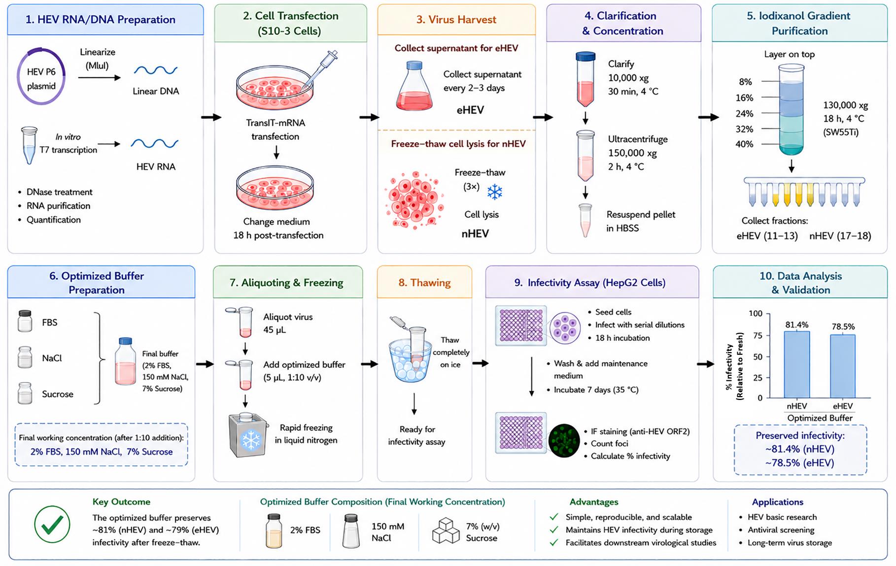
Overview of the optimized buffer for preservation of hepatitis E virus (HEV) during freeze–thaw cycles. Created with the assistance of ChatGPT (GPT-5.5) on July 13, 2026.
Background
Hepatitis E virus (HEV) is a main cause of acute hepatitis with clinical symptoms including jaundice, loss of appetite, nausea, vomiting, and abdominal pain. It is generally self-limiting with a case fatality rate of 0.5%–3% in young adults [1]. However, it can cause up to 30% mortality in pregnant women in the third trimester and can become chronic in immunocompromised people [2]. WHO estimates that hepatitis E caused approximately 44,000 deaths in 2015 (accounting for 3.3% of the mortality due to viral hepatitis) [3–5]. To date, no specific drugs have been approved for the treatment of HEV infections [6]. There is a critical medical need for developing novel anti-HEV treatment strategies.
HEV exists in two infectious forms: non-enveloped HEV (nHEV), which is mainly present in the bile and feces, and quasi-enveloped HEV (eHEV), which circulates in the bloodstream and is completely cloaked in a host-derived lipid membrane [7, 8]. The lack of an efficient cell culture system for virus cultivation has severely hindered the development of new antiviral drugs. Although cell culture systems for HEV infection have improved in recent years, they remain relatively cumbersome and time-consuming compared to those for many other viruses [9].
Viral infectivity declines rapidly following exposure to ambient temperatures and freeze-thaw cycles [10]. Previous studies have used stabilizers such as salt buffers, serum, or sucrose to mitigate these effects [11–13]. Sodium chloride provides a suitable storage environment for viruses by maintaining the solution’s osmotic pressure and ionic strength. For example, high-salt-dependent viruses such as HHPV-3 require at least 3 M NaCl to remain stable; a decrease in salinity leads to the dissociation of viral particles [14]. A certain proportion of serum is added to viral preservation solutions, particularly for viruses that are sensitive to freeze-thaw cycles, have low titers, or are prone to inactivation. This is because the albumin and other proteins present can reduce viral adsorption to tube walls, mitigate freeze–thaw damage, and, to some extent, stabilize the envelope or capsid structure [11]. Sucrose reduces damage to viral structures caused by ice crystals by inhibiting their formation and helps maintain the integrity of viral particles; for example, high concentrations of sucrose (such as 5%) can stabilize equine poxvirus particles and preserve their infectivity [15].
However, no optimized storage conditions have been established for HEV. Therefore, this protocol aims to provide a practical solution by evaluating commonly used stabilizing additives and defining an optimized buffer that preserves the infectivity of HEV particles during freezing and thawing.
Materials and reagents
1. HepG2 cells (ATCC, catalog number: CRL-10741)
2. Huh7 (S10-3) cells (a kind gift from Suzanne Emerson, NIH, available from the authors upon reasonable request) [16]
3. HEV Kernow-C1 p6 plasmid (a kind gift from Suzanne Emerson, NIH, available from the authors upon reasonable request) [16]
4. Anti-HEV capsid polyclonal antibody (produced in-house by immunizing rabbits with purified recombinant HEV p6 ORF2 p239 protein containing amino acids 422–660)
5. mMACHINE® T7 ULTRA Transcription kit (Thermo Fisher, catalog number: AM1345)
6. MluI (NEB, catalog number: R3198L)
7. TransIT®-mRNA Transfection kit (MirusBio, catalog number: MIR 2250)
8. Opti-MEM (Thermo Fisher, catalog number: 51985034)
9. Opti-PrepTM (60% iodixanol) (Sigma, catalog number: D1556-250ML)
10. Hank’s balanced salt solution (HBSS) (Gibco, catalog number: 14025-092)
11. 8%, 16%, 24% and 40% iodixanol gradient solutions (Sigma, catalog number: D1556-250ML; Gibco, catalog number: 14025-092)
12. Ultracentrifuge tubes [Beckman Coulter, catalog numbers: 326823 (36 mL); 344057 (5mL)]
13. Sodium chloride (Fisher Scientific, catalog number: BP358-1)
14. Sucrose (Fisher Scientific, catalog number: BP220-212)
15. Fetal bovine serum (FBS) (Atlasbio, catalog number: F0500-DR)
16. Optimized formulation buffer (Fisher Scientific, catalog number: BP358-1, BP220-212; Atlasbio, catalog number: F0500-DR)
17. NucleoSpin® Gel and PCR Clean-up (TaKaRa, catalog number: 740609.250)
18. Goat anti-rabbit IgG (H+L) cross-adsorbed secondary antibody, Alexa FluorTM 488 (Fisher Scientific, catalog number: A-11008)
19. RNeasy Mini kit (QIAGEN, catalog number: 74104)
20. NuncTM biobanking and cell culture cryogenic tubes (Fisher Scientific, catalog number: 12-565-167N)
21. Dulbecco’s phosphate buffered saline (DPBS) (Thermo Fisher, catalog number: 14040133)
Solutions
1. Iodixanol gradient solutions (see Recipes)
2. Optimized formulation buffer (see Recipes)
Recipes
1. Iodixanol gradient solutions
| Concentration gradient | Volume of 60% Opti-Prep stock (mL) | Volume of HBSS (mL) | Volume of total solution (mL) |
|---|---|---|---|
| 8% | 4 | 26 | 30 |
| 16% | 8 | 22 | 30 |
| 24% | 12 | 18 | 30 |
| 40% | 20 | 10 | 30 |
2. Optimized formulation buffer
a. Dilute 2 mL of 100% FBS with 8 mL of water to a final concentration of 20% (v/v).
b. Prepare a 10 mL solution containing 1,500 mM NaCl and 70% (w/v) sucrose by dissolving 0.877 g of NaCl and 7.0 g of sucrose in the above solution and adjusting the final volume to 10 mL.
c. Filter the solution, aliquot, and store at -20 °C.
d. For using, add the buffer to the virus stock at a 1:10 (v/v) ratio, followed by rapid freezing in liquid nitrogen.
e. Final working concentration after mixing: 2% FBS, 150 mM NaCl, and 7% sucrose.
Equipment
1. NanoDropTM One Microvolume UV-Vis Spectrophotometer (Thermo Scientific, catalog number: 13-400-518)
2. -80 °C deep freezer
3. 5% CO2 incubator
4. Beckman Coulter Optima XPN-80 Ultracentrifuge (Beckman Coulter, catalog number: 34015)
5. SW 32 Ti Swinging-Bucket Rotor (Beckman Coulter, catalog number: 369650)
6. SW 55 Ti Swinging-Bucket Rotor (Beckman Coulter, catalog number: 342194)
7. EVOS fluorescence microscopes (Thermo Scientific, model: EVOS M5000)
8. EVOSTM Light Cube Starter kit, DAPI, GFP, Texas Red (Thermo Scientific, catalog number: AMEP5016)
9. Liquid nitrogen tank
10. Eppendorf tabletop centrifuge 5810R, rotor A-4-81, F-34-6-38
Software and datasets
1. GraphPad Prism 10 (GraphPad, Version 10.6.0)
2. EVOS M5000 Imaging System (Thermo Scientific, EVOS M5000)
Procedure
A. eHEV and nHEV virus stocks production
Note: To produce HEV virus stocks, we drew on Debing’s methods [9] with some optimizations.
1. Linearize the HEV P6 plasmid with MluI at 37 °C for 3 h and purify the DNA with the NucleoSpin Gel and PCR Clean-Up kit.
2. Perform in vitro transcription using the T7 Transcription kit (Table 1). Incubate the reaction mixture at 37 °C for 2 h.
Table 1. In vitro transcription reaction
| Component | Volume (μL) |
|---|---|
| 2× NTP/CAP | 10 |
| 10× T7 reaction buffer | 2 |
| GTP | 1 |
| Linear template DNA | 5 |
| T7 enzyme mix | 2 |
3. Add 1 μL of TURBO DNase, mix well, and incubate for 15 min at 37 °C.
4. Recover RNA using the RNA Clean-up protocol from the Qiagen RNA Mini-prep kit. Measure RNA concentration by Nanodrop. The expected RNA yield is approximately 50 μg.
5. Seed the S10-3 cells on 10 cm plates (1 × 10⁶).
6. After 24 h, perform RNA transfection using the Mirus TransIT mRNA Transfection kit (Table 2). At the time of transfection, the cell confluency is typically 70%–80%, and the transfection efficiency is expected to be 85%–95%.
Table 2. RNA transfection reaction
| Opti-MEM | RNA | Boost | RNA transfection reagent |
|---|---|---|---|
| 1,500 μL | 15 μg | 30 μL | 30 μL |
7. Add the mixture to the medium.
8. Change the medium 18 h post-transfection.
9. When confluency reaches 100%, split the transfected cells into T175 flasks.
10. Collect the culture supernatant every 2–3 days.
11. Around 21 days post-transfection, wash the cells with DPBS twice and then add 10 mL of DPBS to each flask.
12. Place the flasks at -80 °C for at least 2 h and thaw them at 37 °C for 30 min. Repeat at least three times.
13. Transfer the cell lysates and culture supernatant to 50 mL Falcon tubes and spin at 10,000× g for 30 min at 4 °C.
14. Transfer the supernatant into ultracentrifuge tubes and spin at 150,000× g for 2 h at 4 °C.
15. Decant the supernatant. Let the pellet sit in a minimal volume of supernatant for 18 h at 4 °C.
16. Resuspend the pellet in 0.5 mL of HBSS.
17. Prepare 8%, 16%, 24%, 32%, and 40% iodixanol solutions from the 60% Opti-Prep stock solution with HBSS. Slowly add them (0.9 mL of each, starting from the highest concentration) into a 5 mL ultracentrifuge tube.
18. Load 0.5 mL of the resuspended pellet (step A16) on the top of the gradient. Balance the tubes and spin at 130,000× g for 18 h at 4 °C. Make sure the brake is off.
19. Collect fractions (250 μL each) from the top.
20. Collect fractions 11, 12, and 13 from the culture supernatant and store them as the eHEV stock; collect fractions 17 and 18 from the cell lysate and store them as the nHEV stock.
B. Virus storage and a single freeze-thaw cycle
1. Divide the virus stock into 45 μL each in cryopreservation tubes.
2. Add 5 μL of the optimized storage buffer (20% FBS with 1,500 mM NaCl and 70% sucrose) to each aliquot.
3. Store the aliquoted virus stock in liquid nitrogen for long-term preservation.
4. Take out the tubes and thaw completely on ice.
C. Virus infectivity titration
1. Seed HepG2 cells in a 96-well plate at 2 × 10⁴ cells per well.
2. Incubate the plate in a 5% CO2 incubator at 37 °C for 24 h.
3. Dilute the 1 μL HEV stock in 150 μL of medium. Inoculate the cells with the virus.
4. Incubate the plate in a 5% CO2 incubator at 35 °C for 18 h.
5. Remove the inoculum and wash the cells three times with 0.2 mL of DPBS.
6. Add 150 µL of medium to each well.
7. Incubate the plate in a 5% CO2 incubator at 35 °C for 7 days.
8. Perform the immunofluorescence assay (IFA) using a rabbit anti-HEV capsid polyclonal antibody. In brief, wash the cells twice with DPBS, followed by fixation and permeabilization. Add the anti-HEV capsid antibody (1:300 dilution) and incubate the plate at 37 °C for 1 h, followed by three times with DPBS for 5 min each. Add the Alexa FluorTM 488-conjugated goat anti-rabbit IgG (H+L) cross-adsorbed secondary antibody (1:1,000 dilution) and incubate at 37 °C for 1 h, followed by three washes with DPBS for 5 min each.
9. Acquire fluorescence images using microscope exposure settings optimized to achieve a clear positive signal with minimal background fluorescence. For quantitative analysis, count positive fluorescence signals across the entire infected cell well. Representative images are obtained by randomly selecting fields of view from each well under the optimized imaging conditions.
Data analysis
One freeze-thaw cycle reduced HEV infectivity by approximately 70%; the optimized buffer preserved approximately 81.4% (nHEV) and 78.5% (eHEV) infectivity compared to fresh viruses (Figures 1 and 2).
Statistical significance between groups is determined by a two-way ANOVA using GraphPad Prism 10. Data are shown as means ± SD from three independent experiments, each run in duplicate. *P < 0.05; ****P < 0.0001.
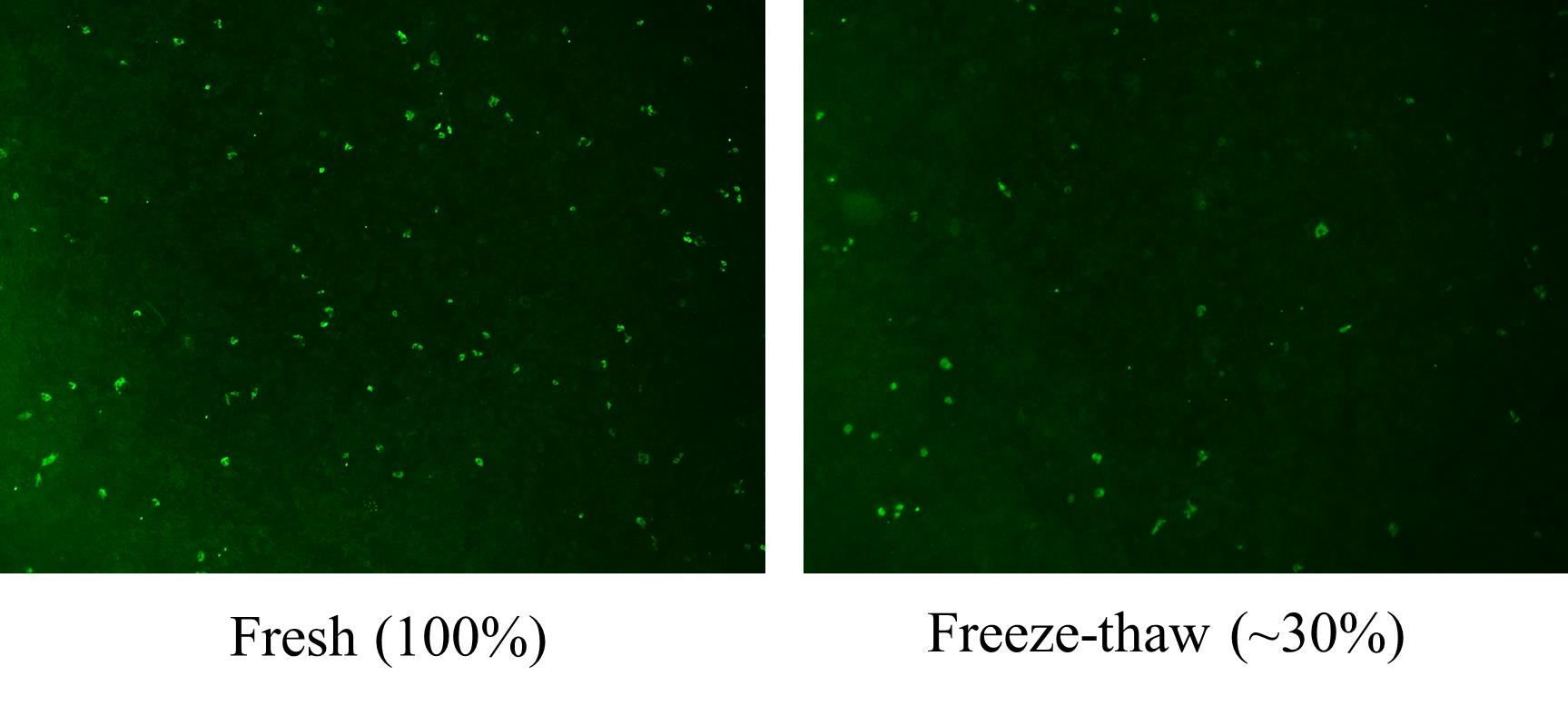
Figure 1. One freeze-thaw cycle reduced hepatitis E virus (HEV) infectivity by approximately 70%. After production, non-enveloped HEV (nHEV) virus stocks—including those that had not undergone freeze–thaw cycles (Fresh) and had undergone one freeze–thaw cycle (Freeze-thaw)—were used to infect HepG2 cells, inoculated with an equal amount of virus (1,200 gene copies/cell). Seven days after infection, the HEV viral load was quantified using the immunofluorescence assay (IFA) method. The green signal in the image indicates HEV infection. The photo was taken using a 4× objective lens. The numbers represent the percentage of HEV-positive foci relative to the Fresh group.
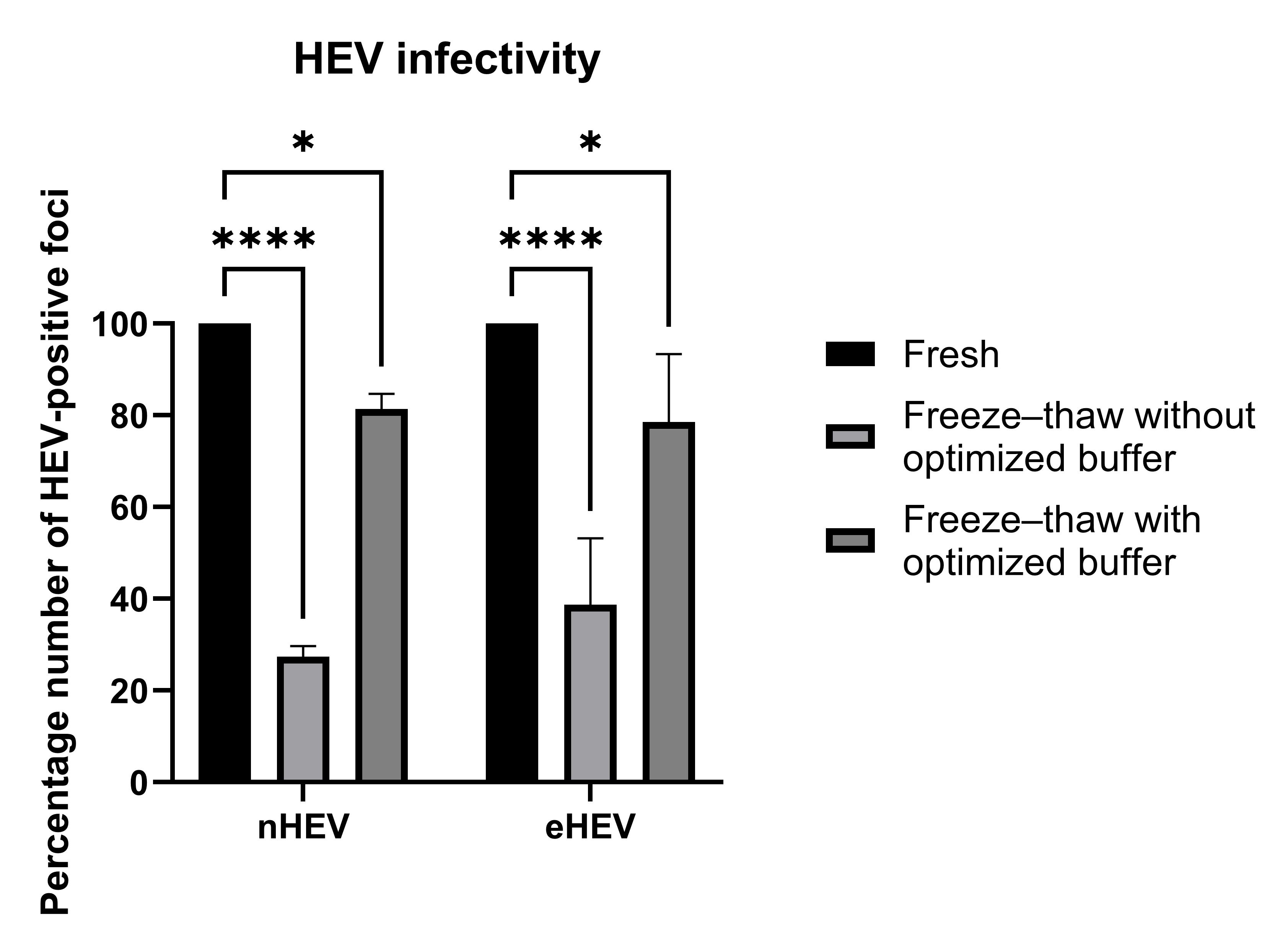
Figure 2. Higher infectivity is maintained by the optimized buffer. The numbers represent the relative infectivity, as determined by the number of hepatitis E virus (HEV)-positive foci, to the Fresh group. Data are shown as mean ± SD from three independent experiments, each run in duplicate. Statistical significance between groups was determined by a two-way ANOVA using GraphPad Prism 10. *P < 0.05; ****P < 0.0001.
Validation of protocol
We tested various combinations of different components (NaCl, sucrose, FBS, etc.) and found that the storage solution described in this protocol performed the best. We conducted three independent tests using this storage solution, and the results are shown in Table 3. The results demonstrate high reproducibility in all cases.
Table 3. HEV infectivity after freeze-thaw cycles
| nHEV | eHEV | ||||||
| Test 1 | Test 2 | Test 3 | Test 1 | Test 2 | Test 3 | ||
| Fresh | 31 | 190 | 220 | 20 | 47 | 37 | |
| Freeze-thaw | 8 | 57 | 58 | 11 | 16 | 10 | |
| Freeze-thaw with optimized buffer | 26 | 157 | 171 | 18 | 29 | 31 | |
The numbers indicate the number of positive foci in the HEV infectivity titration test.
General notes and troubleshooting
1. In a storage solution containing 2% FBS, 150 mM NaCl, and 7% sucrose, both nHEV and eHEV remained relatively stable after one freeze-thaw cycle.
2. Virus stocks should be aliquoted immediately after preparation and stored in liquid nitrogen (they can also be stored at -80 °C for short periods). For optimal infectivity, use within 3–6 months. Longer storage may gradually reduce viral infectivity and should be validated experimentally.
3. Prepare single-use aliquots whenever possible. Repeated freeze–thaw cycles can significantly reduce virus infectivity due to virion structural damage. Avoid more than one freeze–thaw cycle.
4. Infectivity after purification or concentration may vary depending on the method used. Recovery should be evaluated by infectivity assays. Typical recovery efficiencies should be established for each laboratory protocol.
5. Troubleshooting for low infectivity: If virus infectivity is lower than expected, consider the following: excessive freeze–thaw cycles; prolonged storage or inappropriate storage temperature; inaccurate virus quantification; loss of virus during purification or concentration; reduced permissiveness or poor health of target cells; incorrect viral input; and contamination or unsuitable culture conditions.
Acknowledgments
Authors’ contribution
Conceptualization, Z.J.; Investigation, Z.J.; Writing—Original Draft, Z.J.; Writing—Review & Editing, Z.J. and Z.F.; Funding acquisition, Z.F.; Supervision, Z.F.
This work was supported by the National Institutes of Health under the grant numbers R01AI174180, R01AI175800, and R01AI194642 (Z.F.).
Competing interests
The authors declare that they have no competing interests.
References
- Kamar, N., Dalton, H. R., Abravanel, F. and Izopet, J. (2014). Hepatitis E virus infection. Clin Microbiol Rev. 27(1): 116–138. https://doi.org/10.1128/cmr.00057-13
- Rein, D. B., Stevens, G. A., Theaker, J., Wittenborn, J. S. and Wiersma, S. T. (2012). The global burden of hepatitis E virus genotypes 1 and 2 in 2005. Hepatology. 55(4): 988–997. https://doi.org/10.1002/hep.25505
- Patra, S., Kumar, A., Trivedi, S. S., Puri, M. and Sarin, S. K. (2007). Maternal and Fetal Outcomes in Pregnant Women with Acute Hepatitis E Virus Infection. Ann Intern Med. 147(1): 28–33. https://doi.org/10.7326/0003-4819-147-1-200707030-00005
- Kamar, N., Selves, J., Mansuy, J. M., Ouezzani, L., Péron, J. M., Guitard, J., Cointault, O., Esposito, L., Abravanel, F., Danjoux, M., et al. (2008). Hepatitis E Virus and Chronic Hepatitis in Organ-Transplant Recipients. N Engl J Med. 358(8): 811–817. https://doi.org/10.1056/nejmoa0706992
- World Health Organization. (2015). Hepatitis E fact sheet. Updated reports.
- Todt, D., Gisa, A., Radonic, A., Nitsche, A., Behrendt, P., Suneetha, P. V., Pischke, S., Bremer, B., Brown, R. J. P., Manns, M. P., et al. (2016). In vivo evidence for ribavirin-induced mutagenesis of the hepatitis E virus genome. Gut. 65(10): 1733–1743. https://doi.org/10.1136/gutjnl-2015-311000
- Feng, Z., Hensley, L., McKnight, K. L., Hu, F., Madden, V., Ping, L., Jeong, S. H., Walker, C., Lanford, R. E., Lemon, S. M., et al. (2013). A pathogenic picornavirus acquires an envelope by hijacking cellular membranes. Nature. 496(7445): 367–371. https://doi.org/10.1038/nature12029
- Yin, X., Ambardekar, C., Lu, Y. and Feng, Z. (2016). Distinct Entry Mechanisms for Nonenveloped and Quasi-Enveloped Hepatitis E Viruses. J Virol. 90(8): 4232–4242. https://doi.org/10.1128/jvi.02804-15
- Debing, Y., Dallmeier, K. and Neyts, J. (2014). Infectious Virus Yield Assay for Hepatitis E Virus. Bio Protoc. 4(15): e1195. https://doi.org/10.21769/bioprotoc.1195
- Okamoto, H. (2012). Culture systems for hepatitis E virus. J Gastroenterol. 48(2): 147–158. https://doi.org/10.1007/s00535-012-0682-0
- Gould, E. A. (1995). Virus cryopreservation and storage. Methods Mol Biol. 38: 7–20. https://doi.org/10.1385/0-89603-296-5:7
- Wallis, C. and Melnick, J. L. (1968). Stabilization of viruses by proteins. J Bacteriol. 96(3): 713–719.
- Hubálek, Z. (2003). Protectants used in the cryopreservation of microorganisms. Cryobiology. 46(3): 205–229. https://doi.org/10.1016/s0011-2240(03)00046-4
- Demina, T. A., Atanasova, N. S., Pietilä, M. K., Oksanen, H. M. and Bamford, D. H. (2016). Vesicle-like virion of Haloarcula hispanica pleomorphic virus 3 preserves high infectivity in saturated salt. Virology. 499: 40–51. https://doi.org/10.1016/j.virol.2016.09.002
- Joklik, W. (1962). The purification of four strains of poxvirus. Virology. 18(1): 9–18. https://doi.org/10.1016/0042-6822(62)90172-1
- Shukla, P., Nguyen, H. T., Faulk, K., Mather, K., Torian, U., Engle, R. E. and Emerson, S. U. (2012). Adaptation of a Genotype 3 Hepatitis E Virus to Efficient Growth in Cell Culture Depends on an Inserted Human Gene Segment Acquired by Recombination. J Virol. 86(10): 5697–5707. https://doi.org/10.1128/jvi.00146-12
Article Information
Publication history
Received: Jun 9, 2026
Accepted: Jul 7, 2026
Available online: Jul 22, 2026
Published: Aug 20, 2026
Copyright
© 2026 The Author(s); This is an open access article under the CC BY-NC license (https://creativecommons.org/licenses/by-nc/4.0/).
How to cite
Jiao, Z. and Feng, Z. (2026). Optimized Buffer for Preservation of Hepatitis E Virus During Freeze-Thaw Cycles. Bio-protocol 16(16): e5781. DOI: 10.21769/BioProtoc.5781.

 Download Citation in RIS Format
Category
Microbiology > Microbial cell biology > Virus propagation
Biochemistry > Virus > Isolation and purification
Do you have any questions about this protocol?
Post your question to gather feedback from the community. We will also invite the authors of this article to respond.
 Post a Question


