


- Protocols

- Articles and Issues
- For Authors
- About
- Become a Reviewer


In Vivo and In Vitro SUMOylation Assays in Arabidopsis

Published: Vol 16, Iss 15, Aug 5, 2026 DOI: 10.21769/BioProtoc.5769 Views: 195
Reviewed by: Tasleem JavaidAnonymous reviewer(s)
 PDF
PDF  Ask a question
Original research article
The authors used this protocol in:
Feb 2026

Protocol Collections
Comprehensive collections of detailed, peer-reviewed protocols focusing on specific topics




Abstract
Small ubiquitin-like modification (SUMOylation) is a crucial post-translational modification that modulates protein stability, localization, and interaction dynamics. Despite the identification of thousands of putative small ubiquitin-like modifier (SUMO) substrates, functional validation remains challenging due to the low abundance and highly dynamic nature of SUMOylated proteins. Here, we present a protocol for detecting protein SUMOylation, integrating bioinformatic site prediction, and rapid substrate screening via in vivo tobacco transient expression and in vitro E. coli assay, followed by precise validation using transgenic Arabidopsis lines. However, detection of low-abundance SUMOylated proteins may require coupling with mass spectrometry, and the in vitro system does not fully recapitulate the complex regulatory network in vivo. This workflow provides a useful tool for studying SUMOylation in plants.
Key features
• Integrates bioinformatic prediction, in vitro validation, and in vivo confirmation for SUMOylation analysis.
• E. coli co-expression system enables rapid SUMOylation detection without protein purification.
• Arabidopsis transgenic line system confirms SUMOylation under physiological conditions.
• The protocol is applicable to most Arabidopsis proteins.
Keywords: SUMOylation Post-translational modification
In vitro SUMOylation assay
In vivo SUMOylation assay
Arabidopsis
Post-translational modification
In vitro SUMOylation assay
In vivo SUMOylation assay
Arabidopsis
Graphical overview
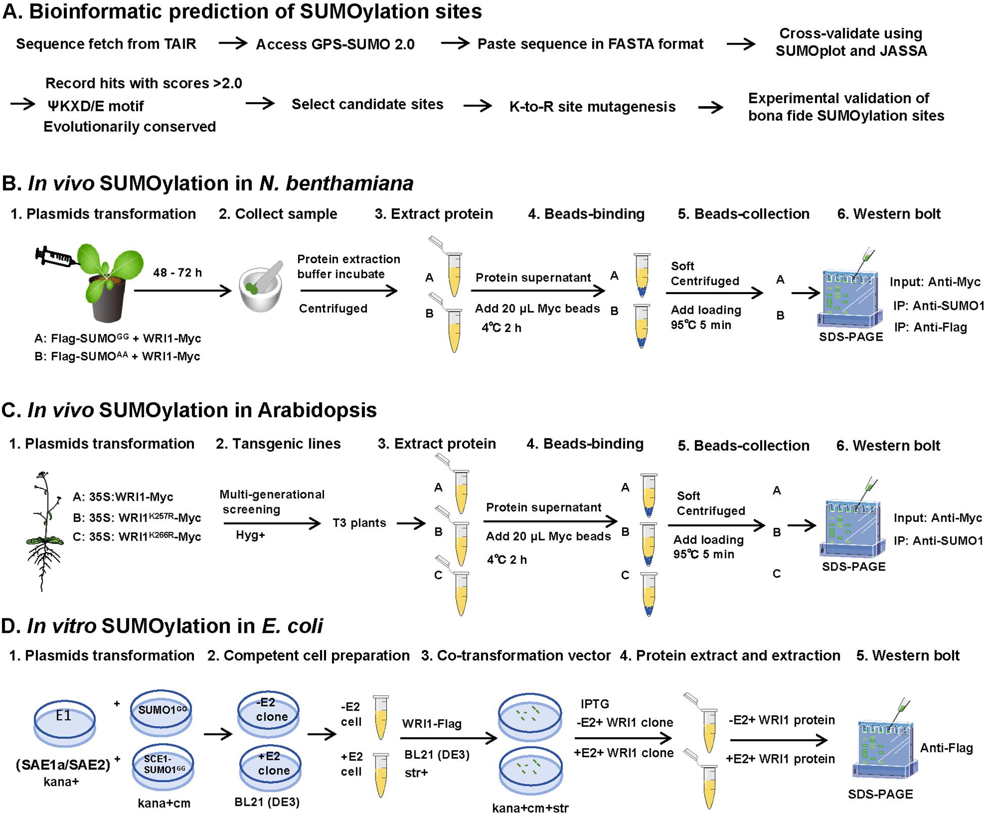
In vivo and in vitro SUMOylation. (A) Bioinformatic prediction of SUMOylation sites (dry-lab; expected time: 1–2 days). The target protein sequence is retrieved from TAIR and analyzed using GPS-SUMO 2.0, SUMOplot, and JASSA. GPS-SUMO 2.0 is suitable for large-scale prediction with customizable thresholds; SUMOplot focuses on the canonical ΨKXD/E motif; JASSA provides additional features such as secondary structure analysis and database hit searching. (B) In vivo SUMOylation assay in N. benthamiana (wet-lab; expected time: 3–4 days). Plasmids encoding Flag-SUMOGG or Flag-SUMOAA together with WRI1-Myc were transformed into N. benthamiana leaves. (C) In vivo SUMOylation assay in Arabidopsis (wet-lab; expected time: 9–10 months). Transgenic lines of 35S:WRI1-Myc, 35S:WRI1K257R-Myc, or 35S:WRI1K266R-Myc were generated through multi-generational screening. (D) In vitro SUMOylation in E. coli (wet-lab; expected time: 15–20 days). The competent cells of BL21 (DE3) containing pET28a-AtSAE1a-His-SAE2 + pACYCDuet-AtSUMO1GG (-E2) or pET28a-AtSAE1a-His-SAE2 + pACYCDuet-1-His-AtSCE1-Myc-AtSUMO1GG (+E2), respectively. The pCDFDuet-WRI1-Flag, WRI1K266R-Flag, WRI1K257R-Flag, and WRI12KR-Flag were transformed into -E2 and +E2 competent cells.
Background
Small ubiquitin-like modification (SUMOylation) is a reversible post-translational modification that plays essential roles in plant development, stress responses, and metabolic regulation [1]. The modification process involves a cascade of enzymes: small ubiquitin-like modifier (SUMO) activating enzyme E1, SUMO conjugating enzyme E2, and, usually, a SUMO ligase E3, which together conjugate SUMO molecules to lysine residues of target proteins [2]. Despite the identification of thousands of potential SUMO substrates through proteomic screens [3,4], functional validation of specific SUMOylation events remains technically challenging due to the low abundance of SUMOylated proteins and the presence of active SUMO proteases that rapidly remove SUMO conjugates.
Several methods have been developed to detect protein SUMOylation in plants. Qu et al. (2020) comprehensively described classic approaches, including in vitro assays using purified recombinant proteins and in vivo assays using tobacco transient expression, Arabidopsis protoplasts, and transgenic plants [5]. Similarly, a protocol for detecting SUMOylated phytochromes in plants was established to address the specific challenges of monitoring phytochrome SUMOylation [6]. Huang et al. (2022) established an efficient in vitro SUMOylation detection system in E. coli [7]. Lai et al. (2023) further expanded this system for high-throughput substrate screening, validating SUMOylation of 95% of candidate proteins from a maize cDNA library [8]
For large-scale identification of SUMO substrates, Miller et al. (2010) generated transgenic Arabidopsis lines expressing a His-tagged SUMO1 mutant H89R and identified 357 SUMOylated proteins by affinity purification coupled with mass spectrometry [9]. To improve detection efficiency, Hendriks et al. (2014) introduced a His-tagged SUMO2 mutant (KO-Q87R) and developed a two-step IMAC enrichment strategy, identifying over 4,300 SUMOylation sites and 1,600 target proteins [10]. A key technical breakthrough came with the discovery of the α-lytic protease WaLP from Lysobacter enzymogenesis, which specifically cleaves SUMO-modified peptides to generate KGG-containing peptides, enabling enrichment with anti-KGG antibodies [11]. Using this approach, Lumpkin et al. (2017) identified 1,209 endogenous SUMOylation sites [12]. Recently, Sang et al. (2024) employed a lysine-null SUMO1 in the sumo1 sumo2 mutant background, combined with a two-step lysine-null SUMO enrichment method, to identify 2,235 SUMOylation sites across 1,300 substrates [13]. Despite the identification of numerous SUMOylated proteins, in vivo experimental validation of these substrates remains challenging.
Building upon these established methods, we present an integrated protocol that combines bioinformatic site prediction, the E. coli co-expression system for in vitro validation, and both tobacco transient expression and transgenic plants for in vivo confirmation. Using the master regulator of seed oil synthesis WRINKLED1 (WRI1) as a case study, we provide detailed step-by-step instructions from SUMOylation site prediction to functional validation. The protocol is applicable to most target proteins in Arabidopsis and can be adapted for use in other plant species.
Materials and reagents
Biological materials
1. Escherichia coli DH5α (store at -80 °C)
2. Escherichia coli BL21 (DE3) (store at -80 °C)
3. Agrobacterium tumefaciens GV3101 (store at -80 °C)
4. Arabidopsis thaliana Columbia-0 (Col-0) seeds (store at -20 °C)
5. Nicotiana benthamiana seeds (store at -20 °C)
Reagents
1. Tris (Sigma-Aldrich, catalog number: T1503)
2. SDS (Sigma-Aldrich, catalog number: 436143)
3. NaCl (Sigma-Aldrich, catalog number: S3014-500G)
4. EDTA (Sigma-Aldrich, catalog number: E9884)
5. Triton X-100 (Sangon Biotech, catalog number: 73049-73-7)
6. Na2HPO4·12H2O (Sigma-Aldrich, catalog number: 71649)
7. KH2PO4 (Sigma-Aldrich, catalog number: P5655)
8. KCl (Sigma-Aldrich, catalog number: P9541)
9. Glycine (MACKLIN, catalog number: 50-01-1)
10. Coomassie Brilliant Blue R-250 (Sigma-Aldrich, catalog number: B1047)
11. Tween-20 (Biosharp, catalog number: BS100-500)
12. MES (Sangon Biotech, catalog number: A610214)
13. Acetosyringone (AS) (Sigma-Aldrich, catalog number: D134406)
14. MgCl2·6H2O (Sigma-Aldrich, catalog number: M2670)
15. Kanamycin sulfate (Sangon Biotech, catalog number: A506636-0100); 50 μg/mL
16. Chloramphenicol (Sangon Biotech, catalog number: A600118-0050); 50 μg/mL
17. Streptomycin sulfate (Sangon Biotech, catalog number: A610494); 50 μg/mL
18. Rifampicin (Sigma-Aldrich, catalog number: R3510); 50 μg/mL
19. Methanol (Thermo Fisher, catalog number: A412-4)
20. Glacial acetic acid (Thermo Fisher, catalog number: A38C212)
21. Protease inhibitor cocktail (Roche, catalog number: 04693159001)
22. Isopropyl-β-D-thiogalactopyranoside (IPTG) (Aladdin, catalog number: I108498)
23. BSA (Sangon Biotech, catalog number: A500023)
24. Coomassie Brilliant Blue R-250 (Sigma-Aldrich, catalog number: B1047)
25. Ponceau S staining solution (Affinibody, catalog number: AIWB-013)
26. ColorMixed Protein Marker 180 (10–180 kDa) (Abclone, catalog number: RM19001)
27. Anti-Myc agarose beads (Abmart, catalog number: M20012M)
28. Anti-FLAG agarose beads (Abmart, catalog number: M20008L)
29. Anti-Myc antibody (Abmart, catalog number: M20002L, 1:5,000)
30. Anti-FLAG antibody (Abmart, catalog number: M20008L, 1:5,000)
31. Anti-SUMO1 antibody (Abcam, catalog number: ab5316, 1:5,000)
32. Anti-actin antibody (Abmart, catalog number: M20009L, 1:5,000)
33. Goat anti-mouse IgG-HRP (Abmart, catalog number: M21001, 1:5,000–1:10,000)
34. Goat anti-rabbit IgG-HRP (Abmart, catalog number: M21002,1:5,000–1:10,000)
35. Competent Cell Preparation kit (Sangon Biotech, catalog number: B529305)
36. One-step PAGE Gel Preparation kit 10% (Affinibody, catalog number: NSF100)
37. BCA Protein Assay kit (Sangon Biotech, catalog number: C503031-1000)
38. High-sensitivity ECL chemiluminescence reagent (Affinibody, catalog number: AIWB-006)
39. Non-fat milk (Sangon Biotech, catalog number: A600669-0250)
Solutions
1. LB liquid medium (see Recipes)
2. Plant protein extract buffer (see Recipes)
3. 10× SDS-PAGE running buffer (see Recipes)
4. 10× PBS buffer (see Recipes)
5. 10× TBS buffer (see Recipes)
6. 10× Transfer buffer (see Recipes)
7. Coomassie blue staining solution (R-250 buffer) (see Recipes)
8. Destaining buffer (see Recipes)
9. 5% non-fat milk (10 mL)
10. 3% BSA (10 mL)
Recipes
1. LB liquid medium (1 L)
Tryptone (10 g)
Yeast extract (5 g)
NaCl (10 g)
LB solid medium: same as LB liquid medium with the addition of 15 g of agar; autoclave at 121 °C for 20 min.
Store at room temperature for up to 6 months. After addition of antibiotics, store at 4 °C and use within 2 weeks.
2. Plant protein extract buffer (1 L)
50 mM Tris-HCl pH 7.4 (7.88 g)
150 mM NaCl (8.77 g)
1 mM EDTA pH 8.0 (0.37 g)
0.5% (v/v) Triton X-100 (5 mL)
1× protease inhibitor cocktail (100 µL)
Store at -20 °C for 6 months. Add protease inhibitor cocktail fresh before use; after adding inhibitor, use within 24 h.
3. 10× SDS-PAGE running buffer (1 L)
250 mM Tris base (30 g)
1.92 M glycine (144 g)
1% (w/v) SDS (10 g)
Prepare 1× SDS-PAGE running buffer fresh by diluting 10× SDS-PAGE buffer 1:10 with ddH2O.
Store at room temperature for up to 6 months.
4. 10× PBS buffer (1 L)
100 mM Na2HPO4·12H2O (35.8 g)
18 mM KH2PO4 (2.4 g)
1.37 M NaCl (80 g)
27 mM KCl (2 g)
Prepare 1× PBS buffer fresh by diluting 10× PBS buffer 1:10 with ddH2O. Add 1× protease inhibitor cocktail to the 1× PBS buffer before use.
Store at room temperature for up to 6 months.
5. 10× TBS buffer (1 L)
200 mM Tris-HCl pH 8.0 (3.03 g)
1.5 M NaCl (87.66 g)
Prepare 1× TBST buffer fresh by diluting 10× TBS buffer 1:10 with ddH2O and add 1 mL of Tween-20.
Store at room temperature for up to 6 months.
6. 10× transfer buffer (1 L)
250 mM Tris (30.03 g)
1.92 M glycine (144 g)
1× Transfer buffer: Add 80 mL of 10× transfer buffer, 200 mL of methanol, and 720 mL of ddH2O to a total volume of 1 L. Prepare the 1× transfer buffer fresh on the day before use. Store 10× transfer buffer at room temperature for up to 6 months.
7. Coomassie blue staining solution (R-250 buffer) (1 L)
0.1% (w/v) Coomassie Brilliant Blue R-250 (1 g)
45% (v/v) methanol (450 mL)
10% (v/v) glacial acetic acid (100 mL)
45% (v/v) ddH2O (450 mL)
Store at room temperature in a dark bottle.
8. Destaining buffer (1 L)
10% (v/v) methanol (100 mL)
10% (v/v) glacial acetic acid (100 mL)
ddH2O (800 mL)
Store at room temperature. Can be reused until saturated with dye.
9. 5% non-fat milk (10 mL)
Non-fat milk (0.5 g)
1× TBST buffer (prepared fresh) (10 mL)
Prepare fresh and store at 4 °C for up to 1 day.
10. 3% BSA (10 mL)
3% BSA (0.3 g)
1× TBST buffer (prepared fresh) (10 mL)
Prepare fresh and store at 4 °C for up to 1 day.
Laboratory supplies
1. Microcentrifuge tubes, 1.5 mL (Pierce, catalog number: 69715)
2. Pipette tips 10, 200, 1000 μL (Axygen, catalog numbers: T-300, T-200-Y, T-1000-B)
3. Centrifuge tubes 15, 50 mL (Corning, catalog number: CLS430828-100EA)
4. PVDF membrane 0.45 μm (Merck Millipore, catalog number: IPVH00010)
5. Western blot filter papers (Bio-Rad, catalog number: 1703965)
6. Western blot sponge pads (Bio-Rad, catalog number: 1703932)
7. Syringe, 1 mL, needle-free (Welch Materials, catalog number: 00824-11121)
8. Tissue grinder or mortar and pestle (Aladdin, catalog number: 1245-160mm-1EA)
9. Liquid nitrogen (self-prepared)
Equipment
1. Ultrapure water system (Sartorius, model: Arium Pro Ultrapure Water System)
2. End-over-end rotator (Elmi, model: ROTAMIX RM1)
3. High-pressure cell disruptor (JNBIO, model: JN-10C)
4. Refrigerated centrifuge (Eppendorf, model: 5424R)
5. SDS-PAGE electrophoresis system (Bio-Rad, model: Mini-PROTEAN® Tetra)
6. Transfer system (Bio-Rad, model: Mini Trans-Blot®)
7. e-Blot for chemiluminescence imaging (Touch IMAGETM, Version 2.1)
Software and datasets
1. GPS-SUMO2.0 [1] (http://sumosp.biocuckoo.org/)
2. SUMOplot1.0 (http://www.abgent.com/sumoplot)
3. JASSA1.0 (http://www.jassa.fr/index.php?jassa)
4. GraphPad Prism9.0 (GraphPad Software, Version 9.0)
5. ImageJ 1.54 (https://imagej.nih.gov/ij/)
Procedure
A. SUMOylation site predication
1. Retrieve the sequence from TAIR (https://www.arabidopsis.org/) and save the FASTA format sequence.
2. Access the GPS-SUMO2.0 prediction tool at http://sumosp.biocuckoo.org/. Paste the FASTA sequence, set the threshold to “Medium,” and click “Submit.” Record all lysine (K) residues with a score >2.0. Cross-validate predictions using additional tools such as SUMOplot (http://www.abgent.com/sumoplot) or JASSA1.0 (http://www.jassa.fr/).
3. Select candidate lysine residues for experimental validation by prioritizing sites that are predicted by multiple software tools, have high prediction scores, are conserved across multiple species, and contain the canonical ΨKXD/E motif.
Note: Focus on sites containing the canonical ΨKXD/E motif, where Ψ is a hydrophobic amino acid (I, L, V, F), K is the target lysine, X is any amino acid, and D/E is aspartic or glutamic acid.
B. Site-directed mutagenesis (K to R mutants)
1. Design mutagenic primers to change the target codon from AAA/AAG to AGA/CGG.
2. Introduce mutations using overlapping PCR.
3. Clone the mutated CDS into the entry vector and verify by sequencing.
Note: Mutating lysine (K) to arginine (R) maintains the positive charge but prevents SUMOylation, allowing validation of critical SUMOylation sites.
C. In vivo SUMOylation assay in N. benthamiana
Note: This method is suitable for rapid validation of SUMOylation within 3–4 days.
1. Vector construction
Table 1. Plasmids used for in vivo SUMOylation assay in N. benthamiana
| Plasmid | Description |
|---|---|
| 35S:WRI1-3 × Myc | Expresses the target protein WRI1 (Myc-tag) |
| Flag-SUMO1GG | Experimental group |
| Flag-SUMO1AA | Negative control |
| P19 | Enhances expression efficiency |
Note: Transform the plant expression vector into A. tumefaciens GV3101-pSOUP. Other common plant expression tags, such as Flag or GFP, are also recommended.
2. Sample preparation
a. Culture Agrobacterium strain 35S:WRI1-Myc with Flag-SUMO1GG or Flag-SUMO1AA and P19 (Table 1) to OD600 = 1.0 in LB liquid medium (Recipe 1) containing appropriate antibiotics (kanamycin 50 μg/mL, rifampicin 50 μg/mL).
b. (Critical) Prepare infiltration buffer containing 10 mM MgCl2, 150 μM AS, and 10 mM MES. Mix gene and P19 at a 1:1 ratio and incubate at room temperature for 3 h.
Note: Prepare two mixtures: 35S:WRI1-Myc + Flag-SUMO1GG + P19; 35S:WRI1-Myc + Flag-SUMO1AA + P19.
Caution: Acetosyringone (AS) is an irritant. Prepare the 100 mM stock solution in a fume hood and store at -20 °C. Avoid skin contact.
c. Infiltrate each bacterial mixture into 4-week-old N. benthamiana leaves. Using a 1 mL sterile syringe (without the needle), slowly infiltrate the activated bacterial suspension into the abaxial side of leaves until a water-soaked patch appears.
d. Mark the infiltrated areas and sample numbers on leaves with a permanent marker.
Note: After infiltration, keep plants in the dark for 12–16 h (overnight, as usual). Avoid strong light exposure during this period.
e. After 2–3 days post-infiltration, harvest the infiltrated leaf areas (avoiding major veins) into a 2 mL tube. Collect approximately 100–150 mg of leaf tissue per sample.
Pause point: Flash freeze in liquid nitrogen and store at -80 °C if not processing immediately.
3. In vivo SUMOylation assay
a. Add 800 μL of protein extraction buffer (Recipe 2) (add 1× protease inhibitor cocktail fresh before use) to the tobacco sample. Rotate at 4 °C for 30 min. Centrifuge at 12,000× g for 15 min at 4 °C and transfer the supernatant to a new tube.
b. Input sample preparation: take 80 μL of supernatant with 20 μL of 5× SDS sample loading buffer as the input sample. Boil at 95 °C for 5 min and store at -20 °C if needed.
Critical: Balance 20 μL of Myc-beads with 500 μL of protein extraction buffer three times (gently centrifuge at 1,000× g for 1 min at 4 °C each time).
Pause point: Boiled samples can be stored at -20 °C for future SDS-PAGE.
c. Add the 20 μL washed beads to 500 μL of supernatant and incubate at 4 °C for 2 h with end-over-end rotation.
d. Centrifuge at 1,000× g for 5 min at 4 °C and discard the supernatant. Gently wash the beads with 1 mL of protein extraction buffer by inverting the tube 5–10 times. Repeat three times.
e. Remove all supernatant and add 100 μL of 2× SDS sample loading buffer, boil at 95 °C for 5 min, and centrifuge at 12,000× g for 3 min. Collect the supernatant as the IP sample.
f. Prepare a 10% SDS-PAGE gel using a commercial gel preparation kit.
g. Load 10 μL of each reaction into a well, reserving a lane for 5 μL of protein marker.
h. Run SDS-PAGE at 150 V for approximately 75 min in 1× SDS-PAGE running buffer (Recipe 3), until the dye front reaches the bottom of the gel.
Critical (for optimization of induction conditions): If the goal is to optimize induction conditions (different induction temperatures, IPTG concentrations, or time points), Coomassie Brilliant Blue R-250 staining is recommended for rapid assessment of total protein expression before proceeding to western blotting. Follow these steps for Coomassie Blue R-250 staining:
i. After SDS-PAGE, carefully remove the gel and place it into a clean container with Coomassie Brilliant Blue R-250 staining solution (Recipe 7).
ii. Stain at room temperature for 1–2 h on a gentle shaker.
iii. Recycle the staining solution: Pour the staining solution into a fresh bottle for future use (can be reused 2–3 times).
iv. Add destaining buffer to cover the gel.
v. Destain at room temperature with gentle shaking, changing the destaining buffer (Recipe 8) 1–2 times (every 30–60 min), until the background is clear and induced protein bands are clearly visible.
vi. Once satisfactory bands are observed, the optimal induction conditions can be selected for large-scale protein expression and subsequent western blot analysis.
Note: If no clear induced bands are observed after Coomassie Blue staining, it is recommended to re-optimize the induction conditions rather than proceeding with western blotting (steps C3i–n). This saves time and reagents.
i. Gently separate gel plates, attach the gel tightly to a PVDF membrane (5.5 cm × 8.5 cm), assemble the transfer cassette as sponge > filter paper > gel > PVDF membrane > filter paper > sponge, place the cast into the wet transfer blot, cover with 1× transfer buffer (Recipe 6), and run at 16 V and 400 mA at 4 °C for 45 min.
Critical (for transfer): PVDF membrane activation: Soak the PVDF membrane in 100% methanol for 15 s and rinse briefly in 1× transfer buffer for 5 min before assembling the transfer sandwich.
Caution: Do not let the PVDF membrane dry out at any step after methanol activation. A dry membrane will not bind proteins efficiently and will result in weak or no signal.
Note: Everything should be pre-wet with transfer buffer. Make sure that there are no bubbles between the gel and the membrane.
Transfer verification (optional but recommended): After transfer, stain the PVDF with 0.1% Ponceau S for 15 min with gentle shaking. The prestained protein marker and major protein bands should be clearly visible on the membrane. A successful transfer is indicated by clear, evenly distributed bands. The Ponceau S stain is reversible and can be removed by washing the membrane in TBST (Recipe 5) for 5–10 min before blocking.
j. Block the PVDF membrane with 5% non-fat milk (Recipe 9) or 3% BSA in 1× TBST buffer (Recipe 10) for 30 min at RT with gentle shaking.
k. Detect IP with anti-Flag and anti-SUMO1 antibody (1:5,000 dilution in blocking buffer, incubate overnight at 4 °C or 2 h at room temperature); detect input with anti-Myc antibody (1:5,000 dilution).
l. Use appropriate HRP-conjugated secondary antibodies (1:5,000) and incubate for 1 h at room temperature. Wash membranes 3 × 10 min with TBST after each antibody incubation.
m. Mix ECL substrate A and B at a 1:1 ratio (e.g., 500 μL of A + 500 μL of B per membrane). Use a clean tube and prepare just before use. Protect from light. Place the membrane on a clean plastic box. Pipette the ECL working solution evenly over the membrane. Incubate for 1 min at room temperature.
Caution: Avoid overexposed (saturated) bands. If any bands are saturated, reduce exposure time and re-capture. Saturated bands cannot be used for gray value quantification.
n. Then, capture chemiluminescent signals using the eBlot imaging system. Adjust exposure time (1 s to 1 min) based on signal intensity. Save the image.
Note: Membranes can be stripped and re-probed with different antibodies if needed. Store the membrane in TBST at 4 °C (wrapped to prevent drying) for short-term storage.
Critical: For accurate gray value quantification using ImageJ, it is critical to use uncompressed TIFF files (8-bit or 16-bit TIFF).
o. Result interpretation: SUMOylation bands above the target protein should be detected only in samples co-expressed with Flag-SUMO1GG, but not in Flag-SUMO1AA samples, indicating that the target protein can be SUMOylated.
D. In vivo SUMOylation in Arabidopsis
Note: This method is suitable for transgenic lines, providing more accurate and reliable results, essential for validating in vivo SUMOylation.
1. Vector construction
2. Plant materials preparation
a. Transform plant expression vectors (Table 2) into A. tumefaciens GV3101 and transform Arabidopsis Col-0 plants using the floral dip method [15].
b. Harvest T0 seeds and select positive transformants. Continue selection of T1 plants to obtain homozygous T2 transgenic lines (at least three independent lines).
Note: For WRI1 SUMOylation detection, collect developing siliques at 10 days after flowering (DAF), which corresponds to the peak of WRI1 expression and oil synthesis.
c. Collect samples from homozygous lines at the developmental stage. Flash freeze in liquid nitrogen and store at -80 °C.
Table 2. Plasmids used for in vivo SUMOylation assay
| Plasmid | Description |
|---|---|
| 35S:WRI1-3 × Myc | Detection of in vivo SUMOylation of WRI1 |
| 35S:WRI1K257R-3 × Myc | Validation of K257 as a SUMOylation site |
| 35S:WRI1K266R-3 × Myc | Validation of K266 as a SUMOylation site |
Note: All lines are homozygous T3 generations. Generate at least three independent transgenic lines per construct.
3. In vivo SUMOylation assay
a. Grind frozen silique tissue (100–200 mg) into a fine powder. Add 600 μL of ice-cold protein extraction buffer (1× protease inhibitor cocktail added fresh). Rotate at 4 °C for 30 min.
b. Centrifuge at 12,000× g for 15 min at 4 °C and transfer the supernatant to a new tube.
c. Input sample preparation: Take 80 μL of supernatant with 20 μL of 5× SDS sample loading buffer as the input sample. Boil at 95 °C for 5 min.
Critical: Wash 20 μL of Myc-beads with 1 mL of protein extraction buffer, centrifuge at 1,000× g for 1 min at 4 °C, and repeat three times.
Pause point: The boiled input samples can be stored at -20 °C for future SDS-PAGE.
d. Add the beads to 500 μL of the remaining supernatant and incubate at 4 °C for 2 h with end-over-end rotation.
e. Centrifuge at 1,000× g for 2 min at 4 °C and discard the supernatant. Gently wash the beads with 1 mL of ice-cold protein extraction buffer. Repeat three times.
f. Remove all supernatant, add 80 μL of 2× SDS sample loading buffer, boil at 95 °C for 5 min, and centrifuge at 12,000× g for 3 min. Collect the supernatant as the IP sample.
Pause point: The boiled IP samples can be stored at -20 °C for future SDS-PAGE.
g. Prepare a 10% SDS-PAGE gel.
h. Load 10 μL of the completed reaction into a well in a 10% SDS-PAGE gel.
i. Transfer the proteins from the gel to a PVDF membrane.
j. Detect input with anti-Myc (1:5,000) and anti-actin (1:5,000) antibodies; detect IP with anti-SUMO1 antibody (1:5,000).
k. Result interpretation: A ladder of bands approximately 15 kDa above the unmodified target protein in IP samples indicates in vivo SUMOylation (Figure 1A–B). Reduced or absent bands in mutant lines indicate critical SUMOylation sites. Quantify band intensities using ImageJ (see Data analysis section).
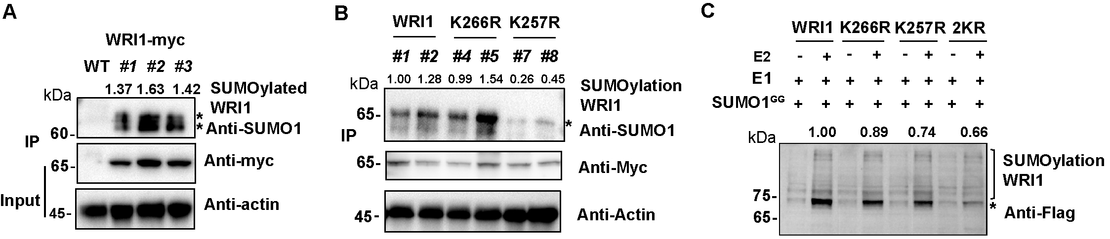
Figure 1. In vivo and in vitro SUMOylation of WRI1. (A) In vivo SUMOylation of WRI1. Total proteins from Myc-tag (WRI1-myc) transgenic lines were immunoprecipitated with Myc beads and detected using anti-SUMO1 antibodies. The vertical line indicates SUMOylated WRI1. Input samples were detected with anti-Myc and anti-actin antibodies. WT served as a negative control. Three independent transgenic lines (#1, #2, and #3) are shown. (B) In vivo SUMOylation assay indicating that K257 is an important SUMO site of WRI1. The K257R mutation significantly reduced the bands; the K266R mutation had a minor effect. (C) In vitro SUMOylation of WRI1. WRI1 protein was co-expressed in E. coli with E1, E2, and SUMO1GG; SUMOylation was detected by immunoblotting analysis using anti-Flag antibodies.
E. In vitro SUMOylation assay
1. Vector construction
2. Protein induction and extraction
a. Transform pCDF-Duet-1-WRI1-Flag and SUMOylation site mutation vectors into +E2 BL21 (DE3) and -E2 BL21 (DE3) competent cells (Table 3), respectively.
b. Pick a single colony (Str, kana, cm) and inoculate into 300 μL of LB medium for recovery. Transfer to 5 mL of LB medium at 37 °C and 220 rpm.
c. Inoculate the overnight culture into 50 mL of fresh LB medium with a 1:50 dilution at 37 °C and 220 rpm for 4–6 h until OD600 = 0.6–0.8.
d. Add 0.5 mM IPTG to induce protein expression. Incubate at 200 rpm at 28 °C for 4–6 h or overnight at 16 °C.
Critical: Induction at 28 °C for 4–6 h is suitable for a rapid detection of SUMOylation. The optimal induction temperature and duration should be determined empirically for each protein.
e. Centrifuge at 12,000× g for 5 min at 4 °C and discard the supernatant.
Pause point: The cell pellet can be stored at -20 or -80 °C.
f. Add 10 mL of ice-cold buffer 1× PBS (add 1× protease inhibitor cocktail fresh before use, Recipe 4).
g. Lyse cells using a high-pressure cell disruptor at 1,000 MPa or by sonication (on ice, 5 s on/5 s off, total 15–20 min).
Caution: For sonication, ensure the sample remains on ice throughout to prevent protein denaturation.
h. Centrifuge at 12,000× g for 15 min at 4 °C. Collect the supernatant as the induced protein extract.
Note: Determine protein concentration using a BCA Protein Assay kit. Use BSA standards (0–2 mg/mL) for calibration.
Critical: Adjust all samples to the same protein concentration using extraction buffer to facilitate comparison of SUMOylation status between +E2 samples and to assess the effect of point mutations on protein abundance.
i. Mix 80 μL of protein supernatant with 20 μL of 5× SDS sample loading buffer and heat at 95 °C for 5 min.
Table 3. Plasmids and competent cells used for in vitro SUMOylation assay
| Plasmid/competent cells | Source |
|---|---|
| pET28-AtSAE1a-His6-AtSAE2 | Huang et al., 2022 |
| pACYC-Duet-1-(His6-AtSCE1)-(MYC-AtSUMO1-GG) | Huang et al., 2022 |
| pACYC-Duet-1-SUMO1GG | Huang et al., 2022 |
| pCDF-Duet-1-target-Flag | Huang et al., 2022 |
pET28-AtSAE1a-His6-AtSAE2 + pACYC-Duet-1-(His6-AtSCE1)-(MYC-AtSUMO1-GG) | Liu et al., 2026 |
| pET28-AtSAE1a-His6-AtSAE2 + pACYC-Duet-1-SUMO1GG | Liu et al., 2026 |
| pCDF-Duet-1-WRI1-Flag | Liu et al., 2026 |
| pCDF-Duet-1-WRI1K257R-Flag | Liu et al., 2026 |
| pCDF-Duet-1-WRI1K266R-Flag | Liu et al., 2026 |
| pCDF-Duet-1-WRI1K257R/K266R-Flag | Liu et al., 2026 |
| pCDF-Duet-1-WRI1-Flag plasmid transform to +E2 competent cells | Liu et al., 2026 |
| pCDF-Duet-1-WRI1-Flag plasmid transform to -E2 competent cells | Liu et al., 2026 |
Note: Co-transform the above plasmid combinations into BL21(DE3) competent cells. Prepare competent cells using a commercial competent cell preparation kit (Sangon Biotech) and store at -80 °C until use. In this study, the pCDF-Duet-1 vectors were transformed into competent cells to obtain co-transformation cells.
3. In vitro SUMOylation assay
a. Prepare a 10% SDS-PAGE gel.
b. Load 10 μL of completed reaction to an equal volume of protein into wells. Include a marker lane.
c. Transfer the proteins from the gel to a PVDF membrane.
d. Use anti-Flag antibody (1:5,000) to detect the target protein and SUMOylated bands. Use HRP-conjugated secondary antibody and ECL detection.
e. A ladder of bands approximately 15 kDa above the unmodified target protein indicates SUMOylation. These bands should be present only in +E2 samples, not in -E2 controls (Figure 1C).
Note: For in vitro SUMOylation of WRI1, please refer to [16].
Data analysis
1. Western blot quantification
Use Image J software to quantify band intensities. Normalize SUMOylated band intensity to the intensity of the unmodified target protein (or to actin as loading control) to obtain relative SUMOylation levels.
a. Open the image: Click File > Open and select the saved western bolt image.
b. Convert to 8-bit: Click image > type > 8 bit to convert the image to grayscale. This ensures consistent measurement across all samples.
c. Invert background (critical for dark bands on light background): Click Edit > Invert. This converts the background from black to white and the bands from white to black, which is necessary for accurate peak detection by the software.
d. Select the lane: Use the Rectangle Selection Tool to draw a box around the first lane. Drag the rectangle to the next lane and repeat for all lanes to be quantified.
e. Obtain quantitative: Click Analyze > measure (or press Ctrl + M). A Results window will appear. For each measured band, record the “IntDen” (Integrated Density) value. IntDen represents the sum of pixel intensities within the selected area and is the most accurate value for comparing band intensities.
f. Normalization: Normalize the SUMOylated band IntDen value to the IntDen value of the unmodified target protein (or to actin as loading control) to obtain relative SUMOylation levels. Set the wildtype or negative control group as 1.0 and calculate fold changes for experimental groups. Perform at least three independent biological replicates for statistical analysis.
2. Statistical analysis
Perform at least three independent biological replicates. Data are presented as mean ± SD. Statistical significance is assessed using a two-tailed Student’s t-test (* p < 0.05, ** p < 0.01).
3. Result interpretation
a. In vitro assay (E. coli BL21 DE3): A ladder of bands 15 kDa above the target protein in the +E2 sample indicates SUMOylation; no bands in the -E2 sample confirms E2 dependence.
b. In vivo assay (Arabidopsis): A ladder of bands detected by anti-SUMO1 in IP samples indicates SUMOylation; reduced bands in mutant lines indicate critical SUMOylation sites.
c. In vivo assay (tobacco): SUMOylation bands detected only in Flag-SUMO1GG co-expressed samples, not in Flag-SUMO1AA samples.
Validation of protocol
This protocol has been used and validated in the following research article(s):
Liu, X. et al. (2026). SUMO-specific proteases SPF1 and SPF2 negatively regulate seed oil synthesis by mediating WRI1 deSUMOylation. Plant Communications.
Figure 4C: In vivo SUMOylation of WRI1 in transgenic Arabidopsis lines. Total proteins from Myc-tag (WRI1-myc) transgenic lines were immunoprecipitated with Myc beads and detected using anti-SUMO1 antibodies.
Figure 5A: In vitro SUMOylation of WRI1. Clear SUMOylated bands were detected above WRI1-Flag. The K257R mutation significantly reduced the bands, the K266R mutation had a minor effect, and the double mutation almost completely abolished SUMOylation, indicating that K257 is the major SUMOylation site of WRI1.
Figure 5B: In vivo SUMOylation of WRI1 in transgenic Arabidopsis lines. The 35S:WRI1-Myc lines showed clear SUMOylated bands, which were significantly reduced in 35S:WRI1K257R-Myc lines, confirming K257 as the major SUMOylation site.
General notes and troubleshooting
General notes
1. Optimize induction conditions for each protein. The optimal induction temperature and duration for protein expression in E. coli should be determined empirically. Soluble proteins are typically induced at 28 °C for 4–6 h, while proteins prone to form inclusion bodies may require 16 °C overnight or 37 °C for 4–6 h.
2. Protein concentration normalization: Adjust all protein samples to the same concentration before SDS-PAGE to ensure accurate comparison of SUMOylation levels between different samples.
3. Use negative controls in each experiment:
a. For in vitro assays: -E2 control (without SUMO E2) to confirm E2-dependent SUMOylation.
b. For tobacco assays: Flag-SUMO1AA control (SUMO1 mutant that cannot be conjugated).
c. For Arabidopsis assays: wild-type Col-0 plants as negative control.
4. Keep all samples on ice during protein extraction and immunoprecipitation to minimize protein degradation. Use prechilled buffers and centrifuge at 4 °C.
Troubleshooting
Problem 1: No SUMOylation signal in in vitro assay.
Possible cause: Low protein expression abundance.
Solutions: 1) Optimize induction conditions (IPTG concentration, temperature, time). Some proteins require an E3 ligase for efficient SUMOylation. 2) Co-express the target protein with AtSIZ1 or other relevant E3 ligase in the E. coli system. Alternatively, perform in vivo assays in plant systems where endogenous E3 is present. 3) Ensure Agrobacterium OD600 reaches exactly 1.0. Always co-infiltrate with P19 to suppress RNA silencing and enhance protein expression. Shade plants for 12–16 h after infiltration.
Problem 2: Protein degradation during extraction.
Possible cause: Lack of protease inhibitors.
Solutions: Ensure protease inhibitor cocktail is added fresh, keep samples frozen until extraction, and work quickly on ice. Use a stronger promoter (2×35S) or enrich the target protein by immunoprecipitation before detection.
Acknowledgments
This work was supported by the grants from the National Key Research and Development Plan of China (2021YFD2200103), the National Natural Science Foundation of China (32470339). The authors thank Prof. Chenwei Yang (South China Normal University) for providing plasmids for the in vitro SUMOylation assay.
Competing interests
The authors declare no conflicts of interest.
References
- Augustine, R. C. and Vierstra, R. D. (2018). SUMOylation: re-wiring the plant nucleus during stress and development. Curr Opin Plant Biol. 45: 143–154. https://doi.org/10.1016/j.pbi.2018.06.006
- Roy, D. and Sadanandom, A. (2021). SUMO mediated regulation of transcription factors as a mechanism for transducing environmental cues into cellular signaling in plants. Cell Mol Life Sci. 78(6): 2641–2664. https://doi.org/10.1007/s00018-020-03723-4
- Miller, M. J., Barrett-Wilt, G. A., Hua, Z. and Vierstra, R. D. (2010). Proteomic analyses identify a diverse array of nuclear processes affected by small ubiquitin-like modifier conjugation in Arabidopsis. Proc Natl Acad Sci USA. 107(38): 16512–16517. https://doi.org/10.1073/pnas.1004181107
- Rytz, T. C., Miller, M. J., McLoughlin, F., Augustine, R. C., Marshall, R. S., Juan, Y. t., Charng, Y. y., Scalf, M., Smith, L. M., Vierstra, R. D., et al. (2018). SUMOylome profiling reveals a diverse array of nuclear targets modified by the SUMO ligase SIZ1 during heat stress. Plant Cell. 30(5): 1077–1099. https://doi.org/10.1105/tpc.17.00993
- Qu, G. P., Li, H., Lin, X. L., Kong, X., Hu, Z. L., Jin, Y. H., Liu, Y., Song, H. L., Kim, D. H., Lin, R., et al. (2020). Reversible SUMOylation of FHY1 regulates phytochrome a signaling in Arabidopsis. Mol Plant. 13(6): 879–893. https://doi.org/10.1016/j.molp.2020.04.002
- Orosa, B. and Viczián, A. (2019). Detection of SUMOylated phytochromes in plants. Methods Mol Biol. 69–83. https://doi.org/10.1007/978-1-4939-9612-4_5
- Huang, J., Feng, Q., Zheng, K., Huang, J., Wang, L., Lai, R., Lai, J. and Yang, C. (2022). An effective in vitro SUMOylation detection system for plant proteins. Chin Bull Bot. 57(4): 490–499. https://doi.org/10.11983/CBB22080
- Lai, R., Li, W., Xu, Z., Liu, W., Zeng, Q., Lin, W., Jiang, J., Lai, J. and Yang, C. (2023). A robust method for identification of plant SUMOylation substrates in a library-based reconstitution system. Plant Commun. 4(4): 100573. https://doi.org/10.1016/j.xplc.2023.100573
- Miller, M. J., Barrett-Wilt, G. A., Hua, Z. and Vierstra, R. D. (2010). Proteomic analyses identify a diverse array of nuclear processes affected by small ubiquitin-like modifier conjugation in Arabidopsis. Proc Natl Acad Sci USA. 107(38): 16512–16517. https://doi.org/10.1073/pnas.1004181107
- Hendriks, I. A., D'Souza, R. C. J., Yang, B., Verlaan-de Vries, M., Mann, M. and Vertegaal, A. C. O. (2014). Uncovering global SUMOylation signaling networks in a site-specific manner. Nature Structural & Molecular Biology 21(10): 927–936. https://doi.org/10.1038/nsmb.2890
- Meyer, J. G., Kim, S., Maltby, D. A., Ghassemian, M., Bandeira, N. and Komives, E. A. (2014). Expanding proteome coverage with orthogonal-specificity α-Lytic proteases. Molecular & Cellular Proteomics 13(3): 823–835. https://doi.org/10.1074/mcp.m113.034710
- Lumpkin, R. J., Gu, H., Zhu, Y., Leonard, M., Ahmad, A. S., Clauser, K. R., Meyer, J. G., Bennett, E. J. and Komives, E. A. (2017). Site-specific identification and quantitation of endogenous SUMO modifications under native conditions. Nat Commun. 8(1): 1171. https://doi.org/10.1038/s41467-017-01271-3
- Sang, T., Xu, Y., Qin, G., Zhao, S., Hsu, C. C. and Wang, P. (2024). Highly sensitive site-specific SUMOylation proteomics in Arabidopsis. Nat Plants. 10(9): 1330–1342. https://doi.org/10.1038/s41477-024-01783-z
- Zhao, Q., Xie, Y., Zheng, Y., Jiang, S., Liu, W., Mu, W., Liu, Z., Zhao, Y., Xue, Y., Ren, J., et al. (2014). GPS-SUMO: a tool for the prediction of sumoylation sites and SUMO-interaction motifs. Nucleic Acids Res. 42: W325–W330. https://doi.org/10.1093/nar/gku383
- Clough, S. J. and Bent, A. F. (1998). Floral dip: a simplified method for Agrobacterium-mediated transformation of Arabidopsis thaliana. Plant J 16(6): 735-743. https://doi.org/10.1046/j.1365-313x.1998.00343.x
- Liu, X., Liu, L., Li, S., Zhao, J., Chen, Y., Ma, W., Tang, S., Guo, L., Guo, X., Fan, C., et al. (2026). SUMO-specific proteases SPF1 and SPF2 negatively regulate seed oil synthesis by mediating WRI1 deSUMOylation. Plant Commun. 7(2): 101672. https://doi.org/10.1016/j.xplc.2025.101672
Article Information
Publication history
Received: Apr 14, 2026
Accepted: Jun 17, 2026
Available online: Jul 3, 2026
Published: Aug 5, 2026
Copyright
© 2026 The Author(s); This is an open access article under the CC BY-NC license (https://creativecommons.org/licenses/by-nc/4.0/).
How to cite
Liu, X., Tang, S., Guo, X., Fan, C. and Hu, Z. (2026). In Vivo and In Vitro SUMOylation Assays in Arabidopsis. Bio-protocol 16(15): e5769. DOI: 10.21769/BioProtoc.5769.

 Download Citation in RIS Format
Category
Plant Science > Plant biochemistry > Protein > Modification
Biochemistry > Protein > Modification > Sumoylation
Do you have any questions about this protocol?
Post your question to gather feedback from the community. We will also invite the authors of this article to respond.
 Post a Question


