


- Protocols

- Articles and Issues
- For Authors
- About
- Become a Reviewer


Isolation of Mononucleated and Binucleated Hepatocytes by Flow Cytometry

Published: Vol 16, Iss 14, Jul 20, 2026 DOI: 10.21769/BioProtoc.5760 Views: 306
Reviewed by: Vinit SharmaAnonymous reviewer(s)
 PDF
PDF  Ask a question
Original research article
The authors used this protocol in:

Jul 2026

Protocol Collections
Comprehensive collections of detailed, peer-reviewed protocols focusing on specific topics




Abstract
Polyploid hepatocytes are one of the unique features of the liver. Some polyploid hepatocytes have chromosomes in a single nucleus (e.g., 1x4n, 1x8n), while others separate their chromosomes into two nuclei (e.g., 2x2n, 2x4n). In ploidy research, hepatocytes are typically sorted according to their cellular ploidy, revealing their contribution to tumorigenesis and cellular senescence. However, the conventional sorting method fails to distinguish 1x4n from 2x2n, or 1x8n from 2x4n cells, leaving it unclear whether hepatocytes with the same cellular ploidy but different nuclear configurations are identical or phenotypically different. Here, we describe a detailed protocol for fractionating mononucleated and binucleated hepatocytes. First, we present the method for isolating primary mouse hepatocytes and staining them with the DNA dye Hoechst 33342. Flow cytometry is then used to detect fluorescence differences between mononucleated and binucleated hepatocytes. This protocol enables the discrimination of hepatocyte subpopulations with identical cellular ploidy, providing a useful tool to investigate the functional heterogeneity of polyploid hepatocytes.
Key features
• This protocol summarizes a method for isolating primary mouse hepatocytes.
• This protocol enables us to isolate mononucleated and binucleated subpopulations from mixed primary hepatocytes.
Keywords: Polyploid hepatocyte Binucleated hepatocyte
Liver perfusion
Flow cytometry
Doublet
Binucleated hepatocyte
Liver perfusion
Flow cytometry
Doublet
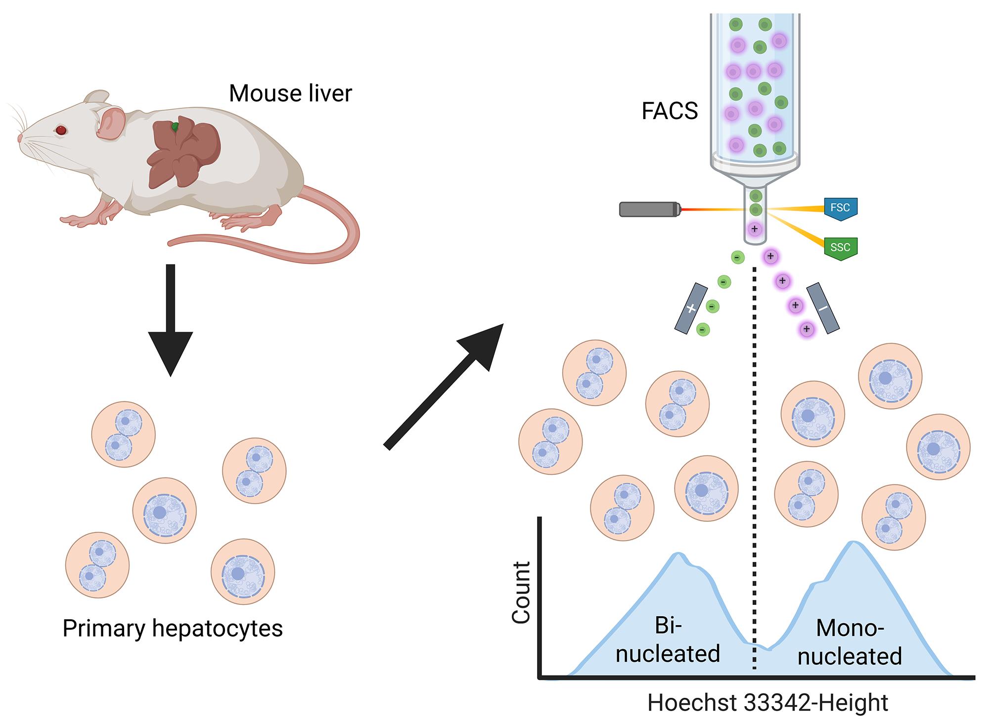
Graphical overview
Background
Hepatocytes, which comprise approximately 70%–80% of the liver cell population, exhibit a unique ploidy status [1,2]. While most other mammalian cell types possess two copies of the genome, a state known as diploidy (2n), hepatocytes often contain more than two copies. In the adult mouse liver, tetraploid (4n) hepatocytes are the most abundant population, followed by octoploid (8n) cells, whereas diploid (2n) cells account for only approximately 10% of the total [3,4]. Earlier studies have explored the differences among hepatocytes with different cellular ploidy levels, revealing that polyploid hepatocytes are more prone to senescence and more resistant to tumorigenesis caused by loss of heterozygosity [5–8]. In addition to the variation in cellular ploidy status, polyploid hepatocytes are further classified based on the number of nuclei [9–11]. Specifically, tetraploid hepatocytes exist either as mononucleated cells containing four genomic copies within a single nucleus (1x4n) or as binucleated cells containing two genomic copies in each of two nuclei (2x2n). Similarly, 8n hepatocytes can be classified into 1x8n and 2x4n hepatocytes. Since these hepatocytes have the same cellular ploidy levels, they have generally been treated as equivalent populations, and their differences remain poorly understood [12,13].
The major limitation for this has been the lack of a method to isolate these subpopulations. In conventional ploidy research, hepatocytes are commonly stained with a DNA-binding dye and distinguished according to total DNA content (2n, 4n, and 8n) in flow cytometry [6, 8–11,13,14]. Fluorescence signals are characterized by three parameters—width, height, and area—with area signals most commonly used due to their stability [14,15]. Height vs. area plots of the forward scatter (FSC) signal are widely applied for doublet discrimination, as doublet events typically exhibit lower height values than singlets. Based on this principle, our protocol leverages the height parameter to distinguish and isolate mononucleated and binucleated hepatocytes. Freshly isolated mouse hepatocytes are stained with the DNA-binding dye Hoechst 33342 and sorted by flow cytometry. Microscopy confirms the successful isolation of 1x4n mononucleated and 2x2n binucleated hepatocytes.
Materials and reagents
Biological materials
1. Adult male mice (purchased from CLEA Japan, bred, and housed in our laboratory)
Note: Female mice can also be used; however, all data in this study were obtained from male mice.
Reagents
1. Sterile distilled water [purified by an Elix system and Milli-Q purification system (Merck Millipore) and sterilized by autoclaving prior to use]
2. Ethanol (Wako Pure Chemical Corporation, catalog number: 055-00457)
3. Isoflurane (VTRS, Viatris Healthcare)
4. HBSS (10×), no calcium, no magnesium, no phenol red [HBSS (-)] (Thermo Fisher Scientific, GibcoTM, catalog number: 14185052)
5. 10× PBS (-) (Wako Pure Chemical Corporation, catalog number: 163-25265)
6. Liver perfusion medium (Thermo Fisher Scientific, GibcoTM, catalog number: 17701038)
7. CaCl2 (Wako Pure Chemical Corporation, catalog number: 038-24985)
8. Collagenase (Wako Pure Chemical Corporation, catalog number: 032-22364)
9. D-MEM (high glucose) with L-glutamine and phenol red (Wako Pure Chemical Corporation, catalog number: 044-29765)
10. Fetal bovine serum (FBS) (BioWest, catalog number: S1810-500)
Note: FBS should be heat-inactivated at 56 °C for 30 min and kept at 4 °C before use.
11. Penicillin-streptomycin-amphotericin B suspension (100×) (PSA) (Wako Pure Chemical Corporation, catalog number: 161-23181)
12. DNase I (Roche, catalog number: 10104159001)
13. PercollTM density gradient media (Cytiva, catalog number: 17089101)
14. Trypan Blue solution, 0.4% (Thermo Fisher Scientific, GibcoTM, catalog number: 15250061)
15. Bisbenzimide H33342 trihydrochloride (Hoechst 33342) (Wako Pure Chemical Corporation, catalog number: 080-09981)
16. Reserpine (Wako Pure Chemical Corporation, catalog number: 184-00691)
17. DMSO (Wako Pure Chemical Corporation, catalog number: 031-24051)
18. HEPES (Sigma-Aldrich, catalog number: H3375)
19. Magnesium chloride (MgCl2) (Wako Pure Chemical Corporation, catalog number: 136-03995)
20. D-(+)-glucose (Sigma-Aldrich, catalog number: G7528)
21. MEM non-essential amino acids solution (100×) (NEAA) (Wako Pure Chemical Corporation, catalog number: 139-15651)
22. GlutaMAXTM supplement (Thermo Fisher Scientific, GibcoTM, catalog number: 35050061)
23. Sodium pyruvate solution (100×) 100 mmol/L (Wako Pure Chemical Corporation, catalog number: 190-14881)
24. Sodium hydroxide solution (NaOH) 5 mol/L (Wako Pure Chemical Corporation, catalog number: 196-05375)
25. Monomeric cyanine nucleic acid stains TO-PRO-3 iodide (Thermo Fisher Scientific, InvitrogenTM, catalog number: T3605)
Solutions
1. HBSS (-) (1×) (see Recipes)
2. PBS (-) (1×) (see Recipes)
3. Collagenase stock solution (see Recipes)
4. CaCl2 stock solution (see Recipes)
5. Collagenase solution (see Recipes)
6. Wash medium (see Recipes)
7. DNase stock solution (see Recipes)
8. Complete Percoll solution (see Recipes)
9. Staining medium (see Recipes)
10. Hoechst stock solution (see Recipes)
11. Reserpine stock solution (see Recipes)
12. HEPES stock solution (see Recipes)
13. MgCl2 stock solution (see Recipes)
14. Glucose stock solution (see Recipes)
15. Flow buffer (see Recipes)
16. Collection buffer (see Recipes)
17. Plating medium (see Recipes)
Recipes
1. HBSS (-) (1×)
| Reagent | Final concentration | Quantity or volume |
|---|---|---|
| HBSS (-) (10×) | 1× | 50 mL |
| Sterile distilled water | --- | 450 mL |
Dilute HBSS (-) (10×) with sterile distilled water under aseptic conditions.
2. PBS (-) (1×)
| Reagent | Final concentration | Quantity or volume |
|---|---|---|
| PBS (-) (10×) | 1× | 50 mL |
| Sterile distilled water | --- | 450 mL |
Dilute PBS (-) (10×) with sterile distilled water under aseptic conditions.
3. Collagenase stock solution
| Reagent | Final concentration | Quantity or volume |
|---|---|---|
| PBS (-) (1×) | 1× | 25 mL |
| Collagenase | 20 mg/mL | 500 mg |
Dissolve 500 mg of collagenase in 25 mL of PBS (-) (1×). Filter with a 0.22 μm filter, aliquot into 1 mL portions under aseptic conditions, and store at -20 °C until use.
Note: Avoid freeze-thaw cycles and use one vial for each mouse dissection.
4. CaCl2 stock solution
| Reagent | Final concentration | Quantity or volume |
|---|---|---|
| CaCl2 | 1 M | 2.22 g |
| Sterile distilled water | 20 mL |
Dissolve 2.22 g of CaCl2 in 20 mL of sterile distilled water. Filter with a 0.22 μm filter under aseptic conditions and store at room temperature before use.
5. Collagenase solution
| Reagent | Final concentration | Quantity or volume |
|---|---|---|
| HBSS (-) (1×) | 1× | 40 mL |
| CaCl2 stock solution | 5 mM | 200 μL |
| Collagenase stock solution | 500 μg/mL | 1 mL |
Add 200 μL of CaCl2 stock solution into 40 mL of HBSS (-) (1×). This solution can be stored at room temperature for a long period. When the volume of liver perfusion medium is reduced to approximately half during liver perfusion, add 1 mL of collagenase stock solution into collagenase solution and invert gently several times to make a homogeneous solution.
6. Wash medium
| Reagent | Final concentration | Quantity or volume |
|---|---|---|
| D-MEM | --- | 500 mL |
| FBS | 5% | 26.3 mL |
D-MEM is supplemented with 5% FBS by adding 26.3 mL of FBS into 500 mL of D-MEM under aseptic conditions.
7. DNase stock solution
| Reagent | Final concentration | Quantity or volume |
|---|---|---|
| DNase I | 1 mg/mL | 100 mg |
| Sterile distilled water | --- | 100 mL |
Dissolve 100 mg of DNase I in 100 mL of sterile distilled water. Filter with a 0.22 μm filter under aseptic conditions, aliquot into 1 mL portions, and store at -20 °C before use.
8. Complete Percoll solution
| Reagent | Final concentration | Quantity or Volume |
|---|---|---|
| Wash medium | 50% | 12 mL |
| HBSS (-) (10×) | 0.5× | 1.2 mL |
| PercollTM density gradient media | 45% | 10.8 mL |
Prepare complete Percoll solution in a 50 mL conical tube just before use by mixing 12 mL of wash medium, 1.2 mL of HBSS (-) (10×), and 10.8 mL of PercollTM density gradient media under aseptic conditions.
9. Staining medium
| Reagent | Final concentration | Quantity or volume |
|---|---|---|
| D-MEM | --- | 500 mL |
| FBS | 5% | 26.6 mL |
| PSA (100×) | 1× | 5.3 mL |
D-MEM is supplemented with 5% FBS and 1× PSA by adding 26.6 mL of FBS and 5.3 mL of PSA into 500 mL of D-MEM under aseptic conditions.
10. Hoechst stock solution
| Reagent | Final concentration | Quantity or volume |
|---|---|---|
| Hoechst 33342 | 1 mg/mL | 10 mg |
| Sterile distilled water | --- | 10 mL |
Dissolve 10 mg of Hoechst 33342 in 10 mL of sterile distilled water. Filter with a 0.22 μm filter and aliquot into 1 mL portions under aseptic conditions. Store at -20 °C before use.
11. Reserpine stock solution
| Reagent | Final concentration | Quantity or volume |
|---|---|---|
| Reserpine | 50 mM | 60.9 mg |
| DMSO | --- | 2.0 mL |
Dissolve 60.9 mg of reserpine in 2.0 mL of DMSO. Filter with a 0.20 μm PTFE filter and aliquot into 200 μL portions under aseptic conditions. Store at -20 °C before use.
12. HEPES stock solution
| Reagent | Final concentration | Quantity or volume |
|---|---|---|
| HEPES | 1 M | 4.77 g |
| Sterile distilled water | --- | 20 mL |
Dissolve 4.77 g of HEPES in 20 mL of sterile distilled water. Filter with a 0.22 μm filter under aseptic conditions and store at 4 °C before use.
13. MgCl2 stock solution
| Reagent | Final concentration | Quantity or volume |
|---|---|---|
| MgCl2 | 1 M | 4.06 g |
| Sterile distilled water | --- | 20 mL |
Dissolve 4.06 g of MgCl2 in 20 mL of sterile distilled water. Filter with a 0.22 μm filter under aseptic conditions and store at room temperature before use.
14. Glucose stock solution
| Reagent | Final concentration | Quantity or volume |
|---|---|---|
| D-(+)-glucose | 200 mg/mL | 6.00 g |
| HBSS (-) (1×) | 1× | 30 mL |
Dissolve 6.00 g of D-(+)-glucose in 30 mL of HBSS (-) (1×). Filter with a 0.22 μm filter under aseptic conditions and store at 4 °C before use.
15. Flow buffer
| Reagent | Final concentration | Quantity or volume |
|---|---|---|
| HBSS (-) (1×) | 1× | 86.5 mL |
| HEPES stock solution | 25 mM | 2.5 mL |
| MgCl2 stock solution | 5 mM | 0.5 mL |
| Glucose stock solution | 3 mg/mL | 1.5 mL |
| PSA (100×) | 1× | 1 mL |
| NEAA (100×) | 1× | 1 mL |
| GlutaMAX supplement | 1× | 1 mL |
| Sodium pyruvate solution (100×) | 1× | 1 mL |
| FBS | 5% | 5 mL |
| NaOH (5 mol/L) | (for pH adjustment) | Approximately 200 μL |
Add 2.5 mL of HEPES stock solution, 0.5 mL of MgCl2 stock solution, 1.5 mL of glucose stock solution, 1 mL of PSA (100×), 1 mL of NEAA (100×), 1 mL of GlutaMAX, 1 mL of 100 mM sodium pyruvate solution, and 5 mL of FBS into 86.5 mL of HBSS (-) (1×). Then, add 200 μL of 5 mol/L NaOH and adjust pH to 7.4 ± 0.1. Additional NaOH may be added if required. Filter with Sartolab RF 150 under aseptic conditions and store at 4 °C before use.
16. Collection buffer
| Reagent | Final concentration | Quantity or volume |
|---|---|---|
| D-MEM | 50% | --- |
| FBS | 50% | --- |
Prepare collection buffer in a 15 mL conical tube before use by mixing equal volumes of D-MEM and FBS under aseptic conditions, adjusting the total volume according to the number of samples.
17. Plating medium
| Reagent | Final concentration | Quantity or volume |
|---|---|---|
| D-MEM | --- | 500 mL |
| FBS | 10% | 56.2 mL |
| PSA (100×) | 1× | 5.6 mL |
D-MEM is supplemented with 10% FBS and 1× PSA by adding 56.2 mL of FBS and 5.6 mL of PSA (100×) into 500 mL of D-MEM under aseptic conditions.
Laboratory supplies
1. Cotton gauze (Iwatsuki, catalog number: 001-10510)
2. Needle for injection (Japan Becton Dickinson, catalog number: 388412)
3. Needle for fixation (Terumo, catalog number: NN-2525R)
4. 96-well microplate for tissue culture (Iwaki, catalog number: 3860-096)
5. 100 mm non-treated dish (Iwaki, catalog number: 1020-100)
6. 5 mL disposable pipette (Violamo, catalog number: 1-2247-03)
7. 10 mL disposable pipette (Violamo, catalog number: 1-2247-14)
8. 25 mL disposable pipette (Violamo, catalog number: 1-2247-05)
9. 50 mL disposable pipette (Violamo, catalog number: 1-2247-06)
10. NuncTM 50 mL conical sterile polypropylene centrifuge tubes (Thermo Fisher Scientific, Thermo ScientificTM, catalog number: 339652)
11. NuncTM 15 mL conical sterile polypropylene centrifuge tubes (Thermo Fisher Scientific, Thermo ScientificTM, catalog number: 339650)
12. pluriStrainer 40 μm, sterile (pluriSelect, catalog number: 43-50040-51)
13. pluriStrainer 70 μm, sterile (pluriSelect, catalog number: 43-50070-51)
14. Millex-GV syringe filter unit, 0.22 μm, PVDF, 33 mm, gamma sterilized (Merck, Millipore, catalog number: SLGVR33RS)
15. Millex-LG syringe filter unit, 0.20 μm, PTFE, 25 mm (Merck, Millipore, catalog number: SLLG025SS)
16. Sartolab RF 150 (Sartorius, catalog number: 180E02)
17. Sorting Chip-130 µm for SH800 and MA900 (Sony, catalog number: LE-C3210)
18. Automatic setup beads (Sony, catalog number: LE-B3001)
19. IsoFlow Sheath Fluid CE (Beckman Coulter, catalog number: 8546859)
Equipment
1. Surgical scissors (3 cm blade length, 12 cm overall length)
2. Surgical forceps (KFI, catalog number: K-4MM)
3. Perfusion tray (a metal tray 72 cm × 30 cm × 2 cm and a polystyrene foam tray 28 cm × 23 cm × 4 cm)
4. Anesthesia machine (Natsume, model: KN-1071 NARCOBIT-E II type)
5. Anesthesia chamber (27 cm × 14 cm × 14 cm)
6. Variable-speed peristaltic pump (VWR, model: 13-876-2)
7. Water bath for perfusion (Taitec, model: Personal-11)
8. Water bath for staining (Taitec, model: SdminiN, catalog number: 0068750-000)
9. Refrigerated centrifuge (Beckman Coulter, model: Allegra X-30R Centrifuge)
10. CountessTM 3 Automated Cell Counter (Thermo Fisher Scientific, model: AMQAX2000)
11. CountessTM Reusable Slide and Holder (Thermo Fisher Scientific, catalog number: A25750)
12. Cell sorter (Sony, model: SH800S)
13. Fluorescence microscope (Keyence, model: BZ-X810)
14. CO2 incubator (Panasonic Healthcare, catalog number: MCO-170AICUVH-PJ)
15. Compact Tabletop Refrigerated Centrifuge (KUBOTA, model: 2800)
16. Hemocytometers (Improved Neubauer) (Sunlead Glass, catalog number: A126)
17. Micropipette (Gilson, catalog numbers: F123600 for P20; F123601 for P200; F123602 for P1000)
18. Unit water bath Thermominder SDminiN (TEITEC, model: SdminiN, catalog number: 0068750-000)
Software and datasets
1. Cell Sorter Software (SONY, version: 2.1.7)
Procedure
A. Isolation of primary mouse hepatocytes
Notes:
1. Before starting, place 40 mL of HBSS (-) (1×), liver perfusion medium, and collagenase solution without collagenase at 38 °C.
2. Thaw 1 mL of collagenase stock solution and DNase stock solution at room temperature or 4 °C so that the reagents can be readily used.
3. Before starting, set the perfusion rate at approximately 10 mL/min, regardless of mice age or liver size.
1. Assemble the experimental setup as shown in Figure 1.
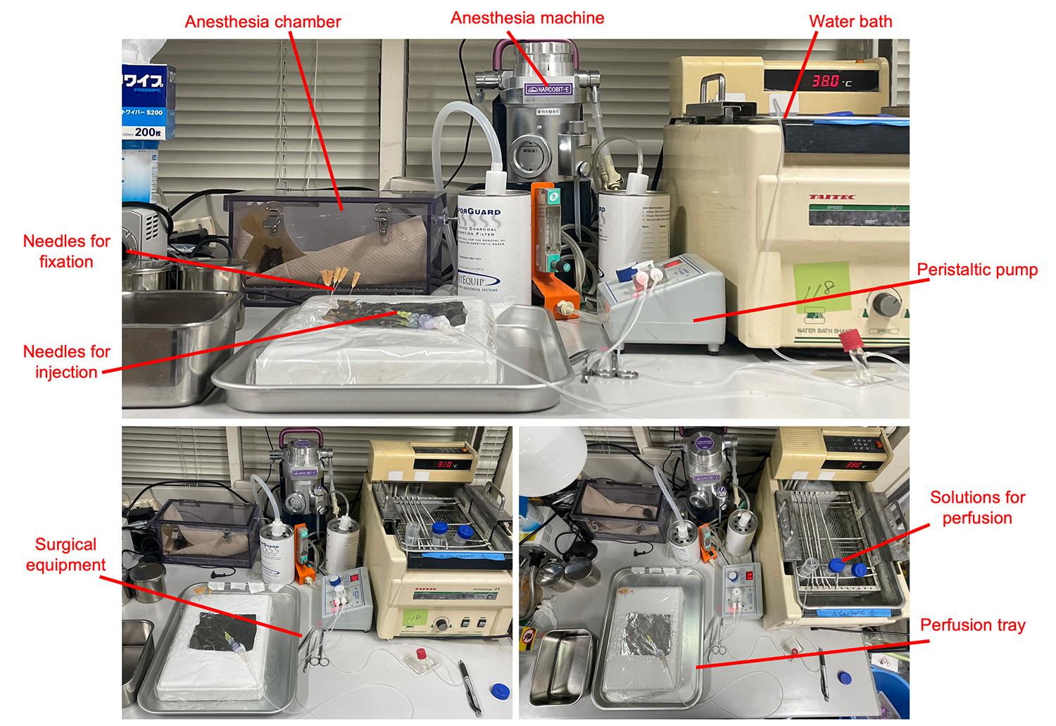
Figure 1. Equipment setup for liver perfusion
2. Euthanize a mouse by inhaling an overdose of isoflurane. Lay the mouse on the tray (Figure 2A). Sterilize the mouse by wiping its fur with 70% ethanol.
3. Open the abdomen using surgical scissors (Figure 2B). Move the mouse's small intestine to the right side and fold the left lobe toward the head of the mouse to expose the portal vein (Figure 2C).
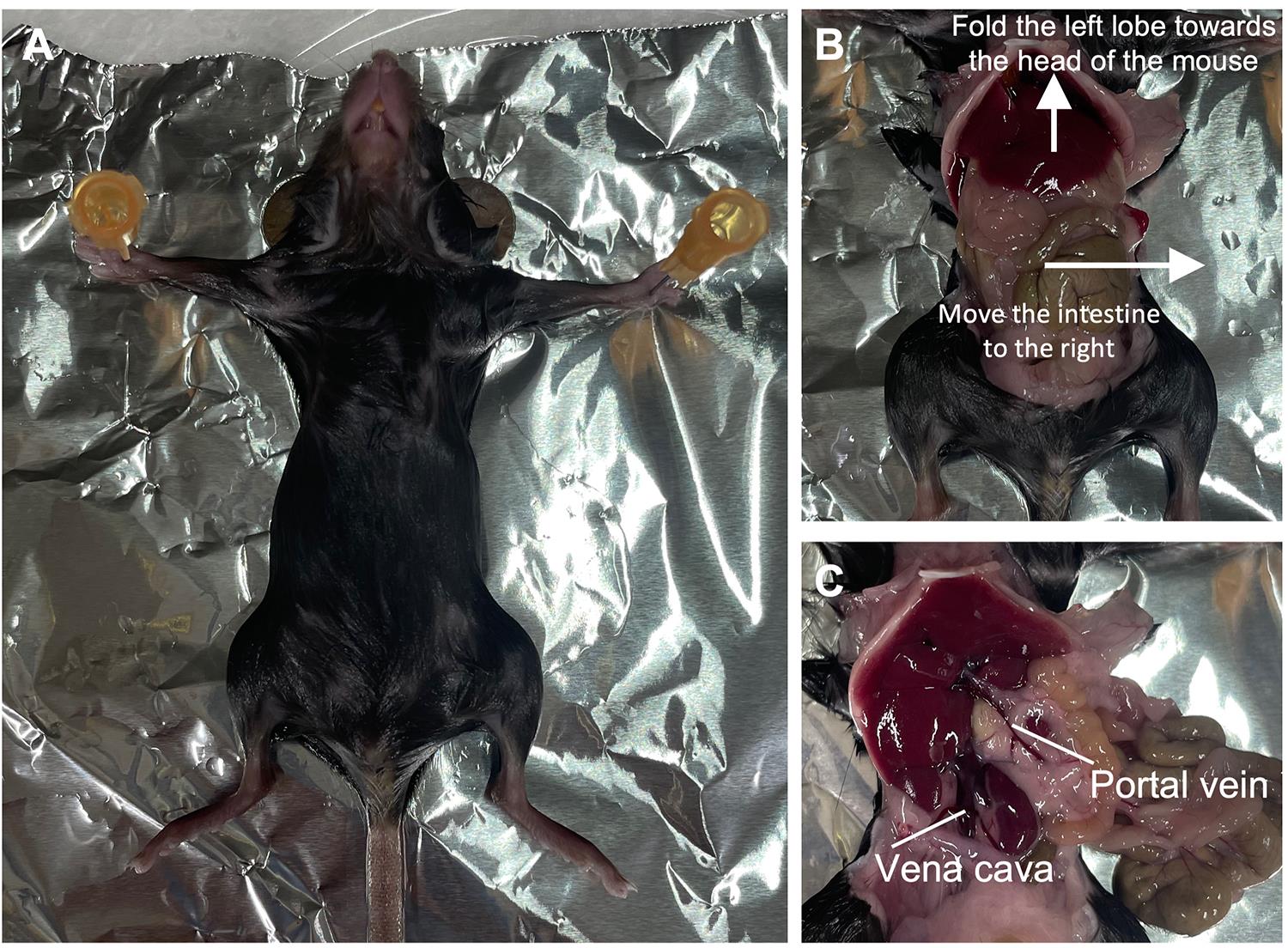
Figure 2. Laparotomy procedure in a mouse. (A) The anesthetized mouse is fixed in a supine position on the surgical platform. (B) The abdomen is opened; the liver is retracted cranially, and the small intestine is moved to the right. (C) The portal vein and inferior vena cava are visualized.
4. Flush a small volume of HBSS (-) (1×) through the perfusion pump, remove any air bubbles from the tubing, and turn off the pump.
5. Gently grasp the underside of the portal vein with forceps and apply tension to straighten the vessel (Figure 3A–C). Insert the needle into the portal vein and advance the needle sheath further in to prevent the needle tip from causing additional damage to the vessel (Figure 3D, E).
Notes:
1. Applying sufficient tension is critical. If the portal vein is displaced rather than penetrated by the needle, the tension applied with the forceps is insufficient.
2. If the needle sheath is pushed back, the needle was not placed properly in the blood vessel. In such a case, slightly draw back the needle and reinsert it into the vessel.
6. Start perfusion of HBSS (-). Immediately after confirming that the color of the liver changes from reddish brown to yellowish, cut the inferior vena cava (Figure 3F–I).
7. After perfusing 40 mL of HBSS (-), transfer the inlet tube to liver perfusion medium and perfuse 40 mL.
Note: Be careful not to allow air bubbles to enter the liver.
8. Clamp the upstream portion of the inferior vena cava with forceps and inflate the liver until it stands upright. Then, release the forceps and allow the liver to return to its original size (Figure 3J, K). Repeat this procedure three times.
Note: Excessive pumping may damage hepatocytes and lead to low viability. We usually release the outlet immediately after the last lobe (typically the middle-left lobe) starts to stand upright.
9. During this step, prepare collagenase solution by adding collagenase stock solution to HBSS (-) (1×) with 5 mM CaCl2 (see Recipes).
10. After perfusing 40 mL of liver perfusion medium, transfer the inlet tube to a freshly prepared collagenase solution. Leave it undisturbed until 40 mL of the solution has been delivered.
11. Stop perfusion and remove the needle from the portal vein. Cut the liver from the abdominal cavity and transfer it to a 100-mm sterile plastic dish. Remove surrounding tissues attached to the liver using scissors and forceps, such as the diaphragm and pancreas.
Note: From this step forward, perform all procedures on a clean bench.
12. Add 10 mL of wash medium and 400 μL of DNase stock solution. Dissociate the tissue using surgical scissors and forceps, followed by further dissociation by pipetting with a 10 mL pipette.
13. Filter the dissociated tissue with a 70 μm cell strainer.
Note: At this step, the filtered cells can be kept on ice for up to 2 h to pause the procedure. In our experience, this storage does not affect any subsequent treatments, allowing us to harvest liver cells from multiple mice and proceed to the downstream steps in parallel.
14. Centrifuge the tube at 69× g for 5 min at 4 °C.
15. Remove supernatant. Loosen the cell pellet by gently tapping the tube. Add 24 mL of complete Percoll solution and resuspend the cell pellet by gently pipetting several times. Centrifuge the tube at 69× g for 10 min at 4 °C.
16. Remove supernatant. Loosen the cell pellet by gently tapping the tube. Add 10 mL of staining medium and resuspend the cell pellet by gently pipetting several times.
17. Dilute 20 μL of cell suspension with 180 μL of staining medium to make a 1:10 dilution. Then, mix the diluted suspension and Trypan Blue solution at a 1:1 ratio. Count the cells using a cell counter and a disposable hemocytometer.
Notes:
1. We typically achieve a total of 1×10⁸ hepatocytes and cell viability of >90%.
2. Based on our experience, at this step, the sample can be kept on ice to pause the procedure for up to 6 h without affecting subsequent treatments.
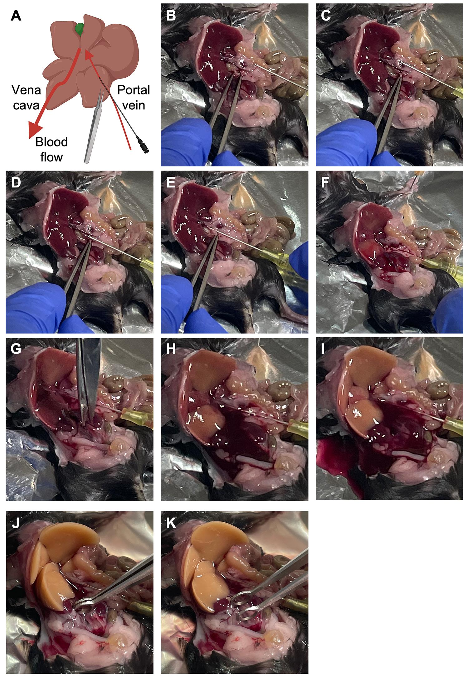
Figure 3. Cannulation of the portal vein. (A) Schematic representation. (B, C) The underside of the portal vein is gently grasped with forceps, and slight but sufficient tension is applied to straighten the vessel and prevent the needle from being pushed back. (D, E) The needle is inserted into the portal vein, and the needle sheath is advanced to minimize damage to the vessel. (F) Perfusion is initiated with HBSS (-). (G–I) The inferior vena cava is cut immediately after the color of the liver changes from reddish brown to yellowish. (J, K) The upstream portion of the inferior vena cava is clamped with forceps to inflate the liver, followed by release to allow it to return to its original size. Panel (A) was created in https://BioRender.com.
B. Staining for flow cytometry
1. Adjust the cell density to 2 × 10⁶ cells/mL by adding staining medium.
2. Add 750 μL of Hoechst stock solution and 5 μL of reserpine stock solution per 50 mL of cell suspension, resulting in a final concentration of 15 μg/mL Hoechst and 5 μM reserpine.
3. Incubate the cell tube at 37 °C in a water bath for 30 min.
Note: Hepatocytes tend to sediment even during incubation with agitation. For clear separation in flow cytometry, inverting the tube every 10 min is both essential and sufficient.
4. Place and cool the tube on ice for a few minutes. Centrifuge the tube at 69× g for 5 min at 4 °C. Remove supernatant.
5. Resuspend the cell pellet with flow buffer at a concentration of 1 × 10⁷ cells/mL.
6. Add 750 μL of Hoechst stock solution, 5 μL of TO-PRO-3 iodide, and 2 mL of DNase stock solution per 50 mL of cell suspension, resulting in a final concentration of 15 μg/mL of Hoechst, 1 nM of TO-PRO-3 iodide, and 40 μg/mL of DNase. Invert the tube several times.
7. Filter the cell suspension with a 40 μm filter and keep it on ice before the subsequent analysis.
C. Flow cytometry and FACS
1. Set up the cell sorter with a 130 μm sorting chip. Select "With 405 nm laser" for filter setting. Perform auto-calibration with automatic setup beads. Set the threshold to 3.00% on the FSC channel.
Note: Fluorescence compensation may improve the sorting accuracy, although the authors did not apply compensation.
2. Perform flow cytometry. Events contributing to noise in the results are removed from the analysis in the following order (Figure 4A):
a. Non-hepatocyte live cells are removed by back scatter area (BSC-A) vs. forward scatter area (FSC-A).
b. Doublet events are removed by forward scatter height (FSC-H) vs. FSC-A.
c. Dead cells are removed by the fluorescence of TO-PRO-3 iodide.
Note: Back scatter of Sony's flow cytometer corresponds to the side scatter of other companies' products.
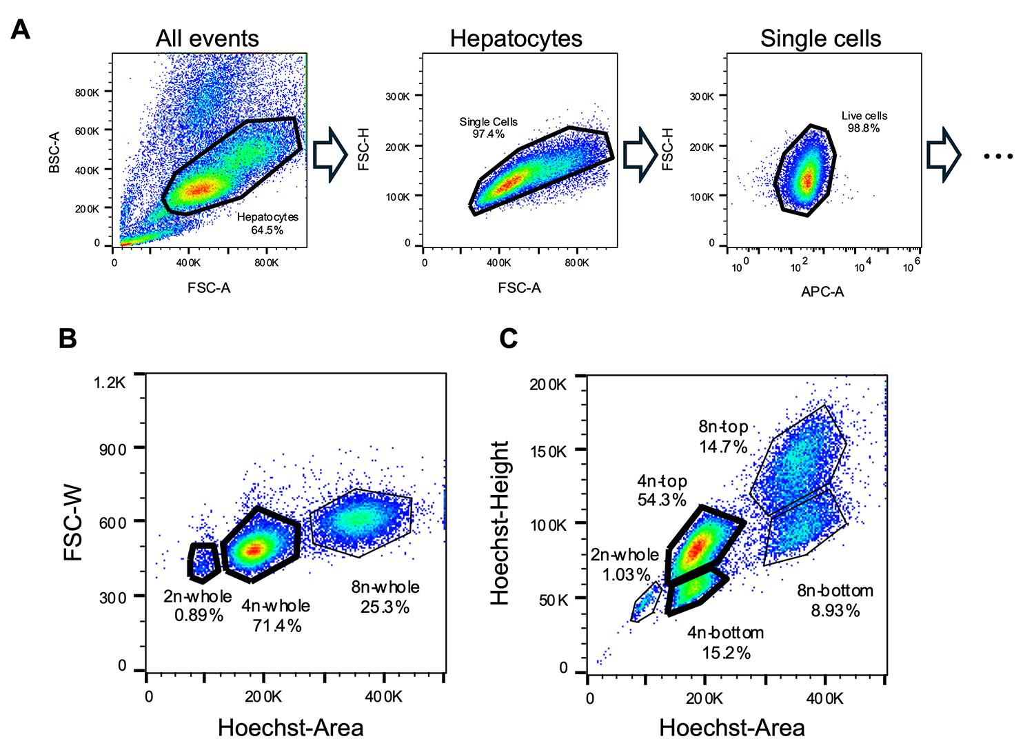
Figure 4. Flow cytometry gating strategy. (A) Live hepatocytes are gated by sequentially excluding small cell events, doublets, and dead cells. (B) The conventional method for distinguishing 2n, 4n, and 8n hepatocytes based on cellular ploidy. (C) Two distinct populations are observed within the 4n and 8n fractions based on Hoechst-height signals. FSC-A, forward scatter area; BSC-A, back scatter area; FSC-H, forward scatter height; APC, allophycocyanin; FSC-W, forward scatter width.
3. Then, analyze the fluorescence of DNA-stained hepatocytes by forward scatter width (FSC-W) vs. Hoechst area (Figure 4B). Confirm that hepatocytes are fractionated into three populations according to the cellular ploidy: 2n, 4n, and 8n.
Note: From here on, we explain the case of 4n hepatocytes, which are the major population in adult mouse liver cells.
4. Prepare collection tubes by adding 1 mL of collection buffer into 15 mL conical tubes, followed by inverting them gently. Set the tubes in the collection area of the flow cytometer.
5. Analyze hepatocytes by plotting the Hoechst height vs. Hoechst area values (Figure 4C). Set gates for the populations with high (4n-top) and low (4n-bottom) values and sort them. Optionally, set a gate for all 4n hepatocytes by the FSC-W vs. Hoechst area parameters and sort them.
Note: We typically set the flow rate to a maximum of 4,000 events per second (eps) and the "purity" mode as the sorting accuracy of the machine.
6. After sorting the two or three populations, perform post-sort by analyzing hundreds to a thousand cells and check the sorting accuracy (Figure 5).
Notes:
1. We found that binucleated hepatocytes are randomly distributed between the 4n-top and 4n-bottom populations [16]. Therefore, their distribution between these fractions is not considered critical. Instead, sorting accuracy should be evaluated by confirming that both populations are indeed 4n, using the FSC-W vs. Hoechst-area plot.
2. We typically achieve a sorting accuracy of >90% and viability of >90% after sorting.
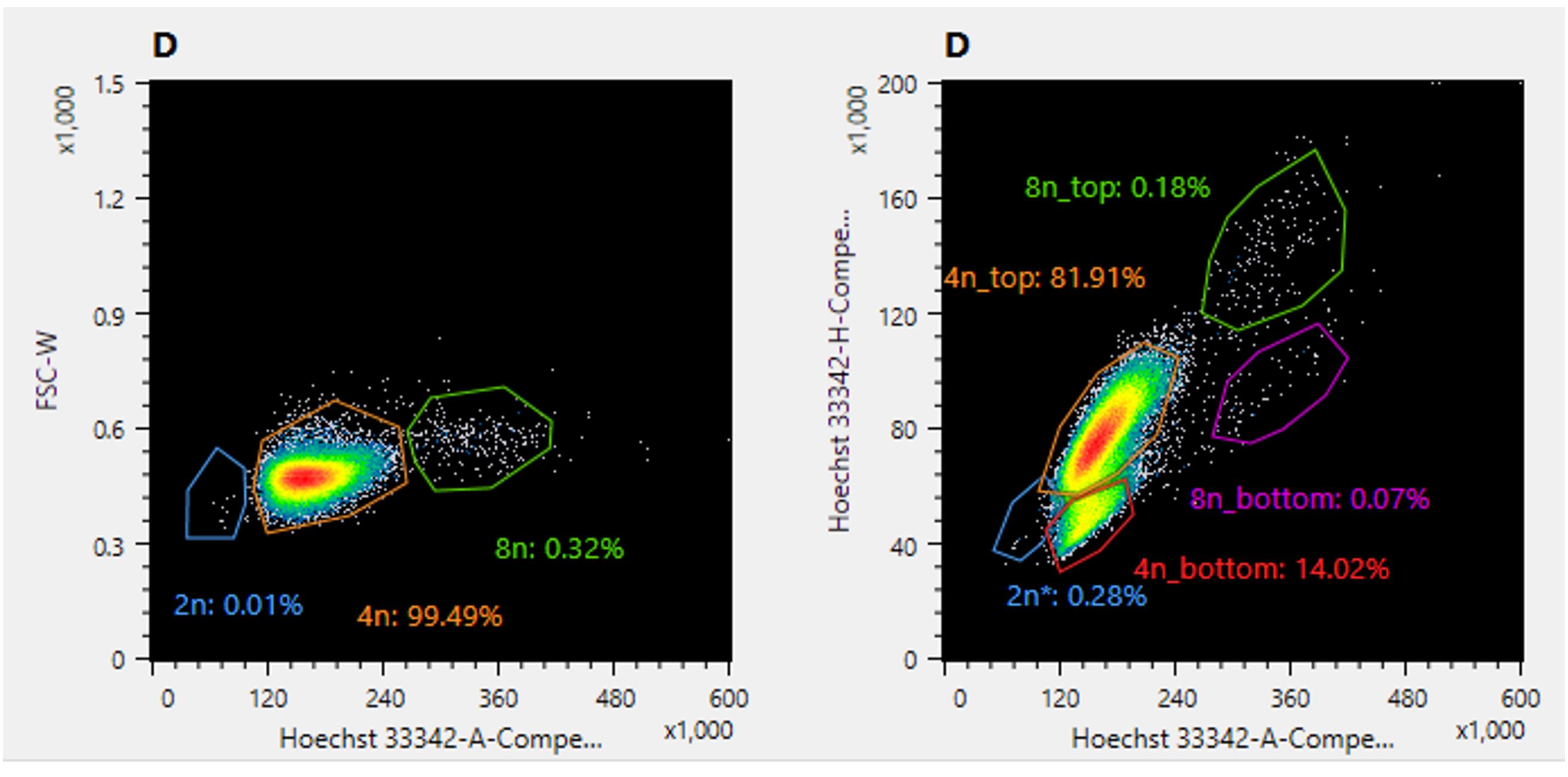
Figure 5. Post-sort analysis of the 4n-top population. (Left) Gate setting recommended for evaluating sorting accuracy. (Right) The 4n-top population is redistributed into both the 4n-top and 4n-bottom populations; therefore, this gate setting is not suitable for evaluation.
D. Evaluation of ploidy status of sorted hepatocytes
1. Centrifuge the samples at 69× g for 5 min at 4 °C.
Note: When treating 2n hepatocytes, we usually centrifuge them at 800× g for 5 min at 4 °C.
2. Carefully remove supernatant. Loosen the cell pellets by gently tapping the tubes. Add 1 mL of plating medium per 100,000 cells. Then, gently tap and invert the tubes.
3. Prepare a 96-well plate for evaluation of the proportion of binucleated hepatocytes. Prepare three wells per sample, each containing 90 μL of plating medium. Add 10 μL of the sample solution and 1.5 μL of Hoechst stock solution.
4. Shake the plate horizontally. After confirming that cells are evenly distributed in wells under a microscope, incubate the plate for 15 min to 1 h at 37 °C.
Note: You may proceed with subsequent experiments using the remaining cells.
5. Observe the cells by phase contrast and Hoechst fluorescence using a fluorescence microscope at 10× magnification (Figure 6). Phase-contrast illumination enables the distinction of cells.
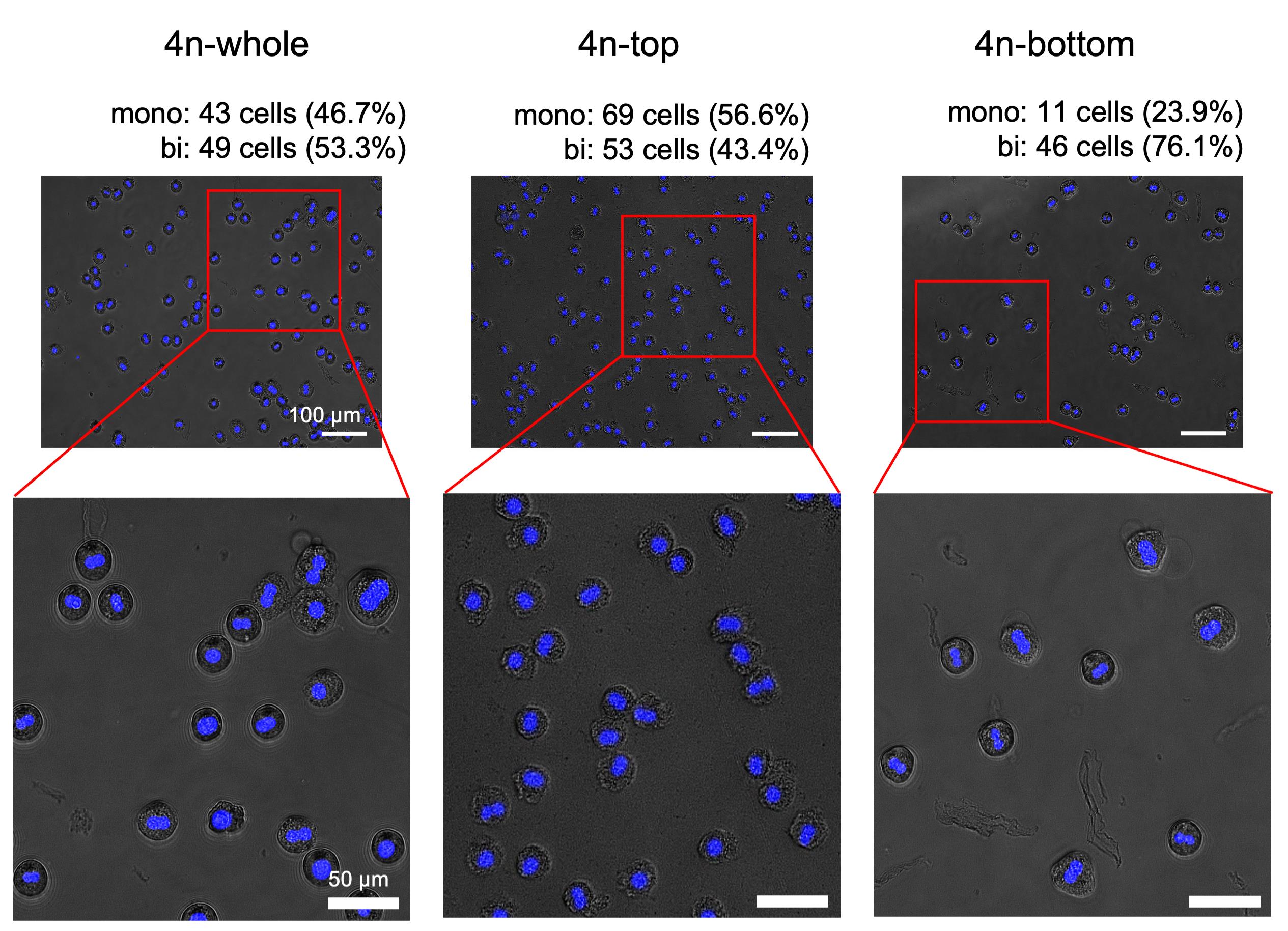
Figure 6. Representative images of plated hepatocytes used to assess the enrichment of mononucleated and binucleated fractions. 4n-whole (left), 4n-top (center), and 4n-bottom (right).
6. Take two or more pictures for each population and count mononucleated and binucleated cells.
Note: We typically count more than 200 cells in total for each population across two pictures to ensure statistical robustness.
7. Calculate the proportion of binucleated cells and assess the enrichment of 1x4n and 2x2n in the 4n-top and 4n-bottom population, respectively.
Data analysis
The proportion of binucleated cells was assessed using a Welch’s t-test to compare three 4n samples (Figure 7).
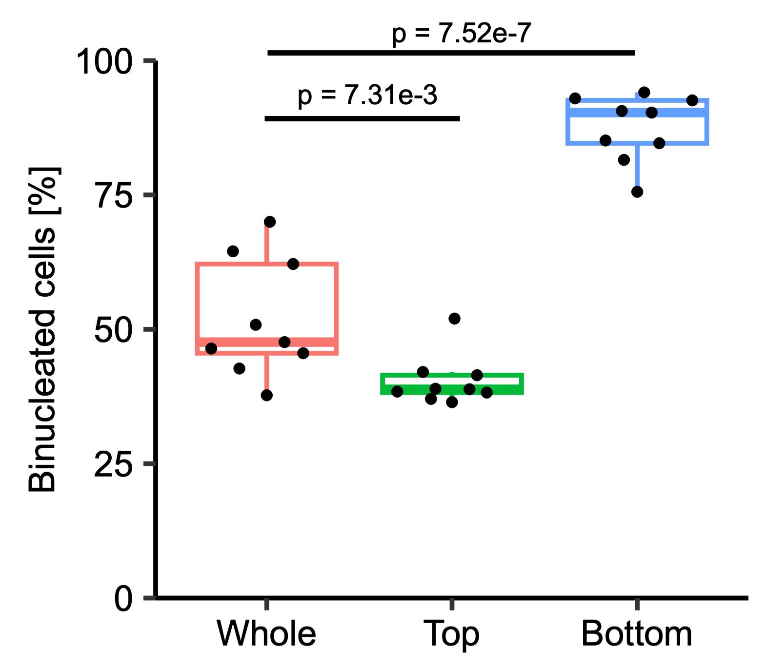
Figure 7. Proportions of binucleated cells in each of the sorted 4n populations. Boxes represent the interquartile range (IQR), with the center line indicating the median. Whiskers extend to the most extreme data points within 1.5 × IQR. Individual data points are overlaid. Significance was assessed using a Welch’s t-test (n = 9 in this study).
Validation of protocol
Data validation procedures are described in the Data analysis section.
This protocol (or parts of it) has been used and validated in the following research article(s):
• Watanabe et al. [16]. A novel sorting method uncovers metabolic heterogeneity between mononucleated and binucleated tetraploid hepatocytes. J Biol Chem.: 113324. https://doi.org/10.1016/j.jbc.2026.113324
General notes and troubleshooting
General notes
1. C57BL/6JJcl mice were purchased from CLEA, Japan. Mice were fed with a standard diet of CE-2 (10 kGy) (CLEA, Japan) and kept in a 12/12 h light/dark cycle. 7- to 15-week-old male mice were used.
2. Reagents stored at 4 °C were used within 3 months of preparation, while those stored at -20 °C were used within 1 year of preparation.
Troubleshooting
Problem 1: Low yield of live hepatocytes after Percoll treatment.
Possible cause: Hepatocytes might lose viability during the perfusion process.
Solutions: Ensure that the needle and tubing are filled with solutions to avoid introducing air bubbles. Avoid overinflation of the liver during perfusion. Confirm that the collagenase solution is sufficiently warmed to maximize enzymatic activity.
Problem 2: Inconsistencies in cell counts.
Possible cause: Hepatocytes are intrinsically large in size, which can make accurate cell counting more challenging compared to other cell types.
Solution: Repeat the cell count two or more times and calculate the average or median cell density.
Problem 3: Inadequate separation of 2n, 4n, and 8n hepatocytes during flow cytometry.
Possible cause: Insufficient Hoechst staining of hepatocytes.
Solution: Gently invert the tube every 10 min during staining to ensure uniform dye distribution.
Problem 4: Inadequate separation of 2n, 4n, and 8n hepatocytes during flow cytometry.
Possible cause: Inaccurate cell counting may lead to a discrepancy between the estimated and actual cell densities, resulting in an insufficient concentration of Hoechst stain.
Solutions: Repeat the cell count two or more times and re-estimate the cell density. Prepare a fresh staining suspension. Although repeating the incubation at 37 °C may reduce cell viability, dead cells can be excluded from the analysis based on TO-PRO-3 fluorescence, allowing live cells to be separated according to their ploidy status.
Problem 5: Poor sorting accuracy.
Possible cause: Sorting mode is "normal" or "yield".
Solution: Change the sorting mode to "purify" to achieve more than 90% sorting accuracy.
Acknowledgments
This work was supported by grants from the Uehara Memorial Foundation, the Nakatomi Foundation, Japan Society for the Promotion of Science (24K23176, 22H00595, and 21K19885), and Japan Agency for Medical Research and Development (1236023). This protocol was used in [16].
The following figures were created using BioRender: Graphical overview, https://app.biorender.com/illustrations/69ce9769859594f759c4459c?slideId=525ccdfc-b57c-434b-8ca9-f9cc24221688; Figure 3A, https://app.biorender.com/illustrations/69c7d0bf97553193d888594a?slideId=620d64ec-9caf-4454-835e-476a0787e39a.
Competing interests
The authors declare that they have no competing interests.
Ethical considerations
All animal procedures used in this study were conducted in accordance with the guidelines of the University of Tokyo and were approved by the University of Tokyo Animal Care and Use Committee (approval ID: A2024E011-05).
References
- Richter, M. L., Deligiannis, I. K., Yin, K., Danese, A., Lleshi, E., Coupland, P., Vallejos, C. A., Matchett, K. P., Henderson, N. C., Colome-Tatche, M., et al. (2021). Single-nucleus RNA-seq2 reveals functional crosstalk between liver zonation and ploidy. Nat Commun. 12(1): e1038/s41467–021–24543–5. https://doi.org/10.1038/s41467-021-24543-5
- Hildebrandt, F., Andersson, A., Saarenpää, S., Larsson, L., Van Hul, N., Kanatani, S., Masek, J., Ellis, E., Barragan, A., Mollbrink, A., et al. (2021). Spatial Transcriptomics to define transcriptional patterns of zonation and structural components in the mouse liver. Nat Commun. 12(1): e1038/s41467–021–27354–w. https://doi.org/10.1038/s41467-021-27354-w
- Donne, R., Saroul-Aïnama, M., Cordier, P., Celton-Morizur, S. and Desdouets, C. (2020). Polyploidy in liver development, homeostasis and disease. Nat Rev Gastroenterol Hepatol. 17(7): 391–405. https://doi.org/10.1038/s41575-020-0284-x
- Duncan, A. W. (2020). Single-Cell and Bulk Transcriptome Profiling Reveals Unique Features of Diploid and Polyploid Hepatocytes. CMGH Cell Mol Gastroenterol Hepatol. 9(1): 193–194. https://doi.org/10.1016/j.jcmgh.2019.09.008
- Zhang, S., Zhou, K., Luo, X., Li, L., Tu, H. C., Sehgal, A., Nguyen, L. H., Zhang, Y., Gopal, P., Tarlow, B. D., et al. (2018). The Polyploid State Plays a Tumor-Suppressive Role in the Liver. Dev Cell. 44(4): 447–459.e5. https://doi.org/10.1016/j.devcel.2018.01.010
- Nikopoulou, C., Kleinenkuhnen, N., Parekh, S., Sandoval, T., Ziegenhain, C., Schneider, F., Giavalisco, P., Donahue, K. F., Vesting, A. J., Kirchner, M., et al. (2023). Spatial and single-cell profiling of the metabolome, transcriptome and epigenome of the aging mouse liver. Nat Aging. 3(11): 1430–1445. https://doi.org/10.1038/s43587-023-00513-y
- Wang, M. J., Chen, F., Li, J. X., Liu, C. C., Zhang, H. B., Xia, Y., Yu, B., You, P., Xiang, D., Lu, L., et al. (2014). Reversal of Hepatocyte Senescence After Continuous In Vivo Cell Proliferation. Hepatology. 60(1): 349–361. https://doi.org/10.1002/hep.27094
- Pandit, S. K., Westendorp, B., Nantasanti, S., van Liere, E., Tooten, P. C. J., Cornelissen, P. W. A., Toussaint, M. J. M., Lamers, W. H. and de Bruin, A. (2012). E2F8 is essential for polyploidization in mammalian cells. Nat Cell Biol. 14(11): 1181–1191. https://doi.org/10.1038/ncb2585
- Duncan, A. W., Taylor, M. H., Hickey, R. D., Hanlon Newell, A. E., Lenzi, M. L., Olson, S. B., Finegold, M. J. and Grompe, M. (2010). The ploidy conveyor of mature hepatocytes as a source of genetic variation. Nature. 467(7316): 707–710. https://doi.org/10.1038/nature09414
- Wilkinson, P. D., Delgado, E. R., Alencastro, F., Leek, M. P., Roy, N., Weirich, M. P., Stahl, E. C., Otero, P. A., Chen, M. I., Brown, W. K., et al. (2019). The Polyploid State Restricts Hepatocyte Proliferation and Liver Regeneration in Mice. Hepatology. 69(3): 1242–1258. https://doi.org/10.1002/hep.30286
- Guidotti, J. E., Brégerie, O., Robert, A., Debey, P., Brechot, C. and Desdouets, C. (2003). Liver Cell Polyploidization: A Pivotal Role for Binuclear Hepatocytes. J Biol Chem. 278(21): 19095–19101. https://doi.org/10.1074/jbc.m300982200
- Matsumoto, T., Wakefield, L., Tarlow, B. D. and Grompe, M. (2020). In Vivo Lineage Tracing of Polyploid Hepatocytes Reveals Extensive Proliferation during Liver Regeneration. Cell Stem Cell. 26(1): 34–47.e3. https://doi.org/10.1016/j.stem.2019.11.014
- Katsuda, T., Hosaka, K., Matsuzaki, J., Usuba, W., Prieto-Vila, M., Yamaguchi, T., Tsuchiya, A., Terai, S. and Ochiya, T. (2020). Transcriptomic Dissection of Hepatocyte Heterogeneity: Linking Ploidy, Zonation, and Stem/Progenitor Cell Characteristics. CMGH Cell Mol Gastroenterol Hepatol. 9(1): 161–183. https://doi.org/10.1016/j.jcmgh.2019.08.011
- Rico, L. G., Bardina, J., Bistué‐Rovira, Ã., Salvia, R., Ward, M. D., Bradford, J. A. and Petriz, J. (2022). Accurate identification of cell doublet profiles: Comparison of light scattering with fluorescence measurement techniques. Cytometry Part A. 103(5): 447–454. https://doi.org/10.1002/cyto.a.24690
- Cosma, A. (2019). The Nightmare of a Single Cell: Being a Doublet. Cytometry Part A. 97(8): 768–771. https://doi.org/10.1002/cyto.a.23929
- Watanabe, Y. V., Sussman, J. H., Anami, T., Shimodaira, G., Hattori, M., Yamane, S., Nishikawa, M., Sakai, Y. and Katsuda, T. (2026). A novel sorting method uncovers metabolic heterogeneity between mononucleated and binucleated tetraploid hepatocytes. J Biol Chem.: 113324. https://doi.org/10.1016/j.jbc.2026.113324
Article Information
Publication history
Received: Apr 29, 2026
Accepted: Jun 10, 2026
Available online: Jun 24, 2026
Published: Jul 20, 2026
Copyright
© 2026 The Author(s); This is an open access article under the CC BY-NC license (https://creativecommons.org/licenses/by-nc/4.0/).
How to cite
Readers should cite both the Bio-protocol article and the original research article where this protocol was used:
- Watanabe, Y. V., Nishikawa, M., Sakai, Y. and Katsuda, T. (2026). Isolation of Mononucleated and Binucleated Hepatocytes by Flow Cytometry. Bio-protocol 16(14): e5760. DOI: 10.21769/BioProtoc.5760.
- Watanabe, Y. V., Sussman, J. H., Anami, T., Shimodaira, G., Hattori, M., Yamane, S., Nishikawa, M., Sakai, Y. and Katsuda, T. (2026). A novel sorting method uncovers metabolic heterogeneity between mononucleated and binucleated tetraploid hepatocytes. J Biol Chem.: 113324. https://doi.org/10.1016/j.jbc.2026.113324

 Download Citation in RIS Format
Category
Cell Biology > Cell isolation and culture > Cell isolation
Cell Biology > Single cell analysis > Flow cytometry
Cell Biology > Cell staining > Nucleic acid
Do you have any questions about this protocol?
Post your question to gather feedback from the community. We will also invite the authors of this article to respond.
 Post a Question


