- Protocols

- Articles and Issues
- For Authors
- About
- Become a Reviewer

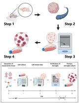
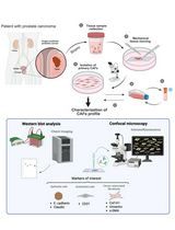
Isolation of Podocyte Cell Fractions From Mouse Kidney Using Magnetic Activated Cell Sorting (MACS)
(§Technical contact: scoobie@uw.edu) Published: Vol 15, Iss 13, Jul 5, 2025 DOI: 10.21769/BioProtoc.5364 Views: 2484
Reviewed by: Komuraiah MyakalaAnonymous reviewer(s)
Original research article
The authors used this protocol in:
Nov 2020
Advertisement


Protocol Collections
Comprehensive collections of detailed, peer-reviewed protocols focusing on specific topics


Related protocols
Abstract
Glomerular diseases characterized by injury to post-mitotic epithelial cells called podocytes are a leading cause of chronic kidney disease. Yet, isolating podocytes from the kidney for transcriptomic, proteomic, and metabolomic studies has been a major technical challenge. Protocols utilizing glomerular sieving and laser capture methods are of limited use because they are not podocyte-specific but instead capture all four glomerular cell types. Here, we present a magnetic-activated cell sorting (MACS) method where podocytes are isolated from digested whole kidneys using antibodies specific to extracellular antigens on podocytes. Using microbeaded secondary antibodies binding to the podocyte-specific primary antibodies allows sorting of the podocytes using a magnet. This podocyte-only cell fraction is a unique source of in vivo–derived cells for molecular and cellular experiments.
Key features
• The protocol isolates a podocyte-only cell population from kidneys that is readily available for molecular and cellular studies.
• The non-podocyte fraction serves as a matching negative control.
• High cell yields are obtained.
• The method can be applied to separately isolate podocytes from the outer cortex and juxtamedullary regions of the kidney.
Keywords: KidneyGraphical overview
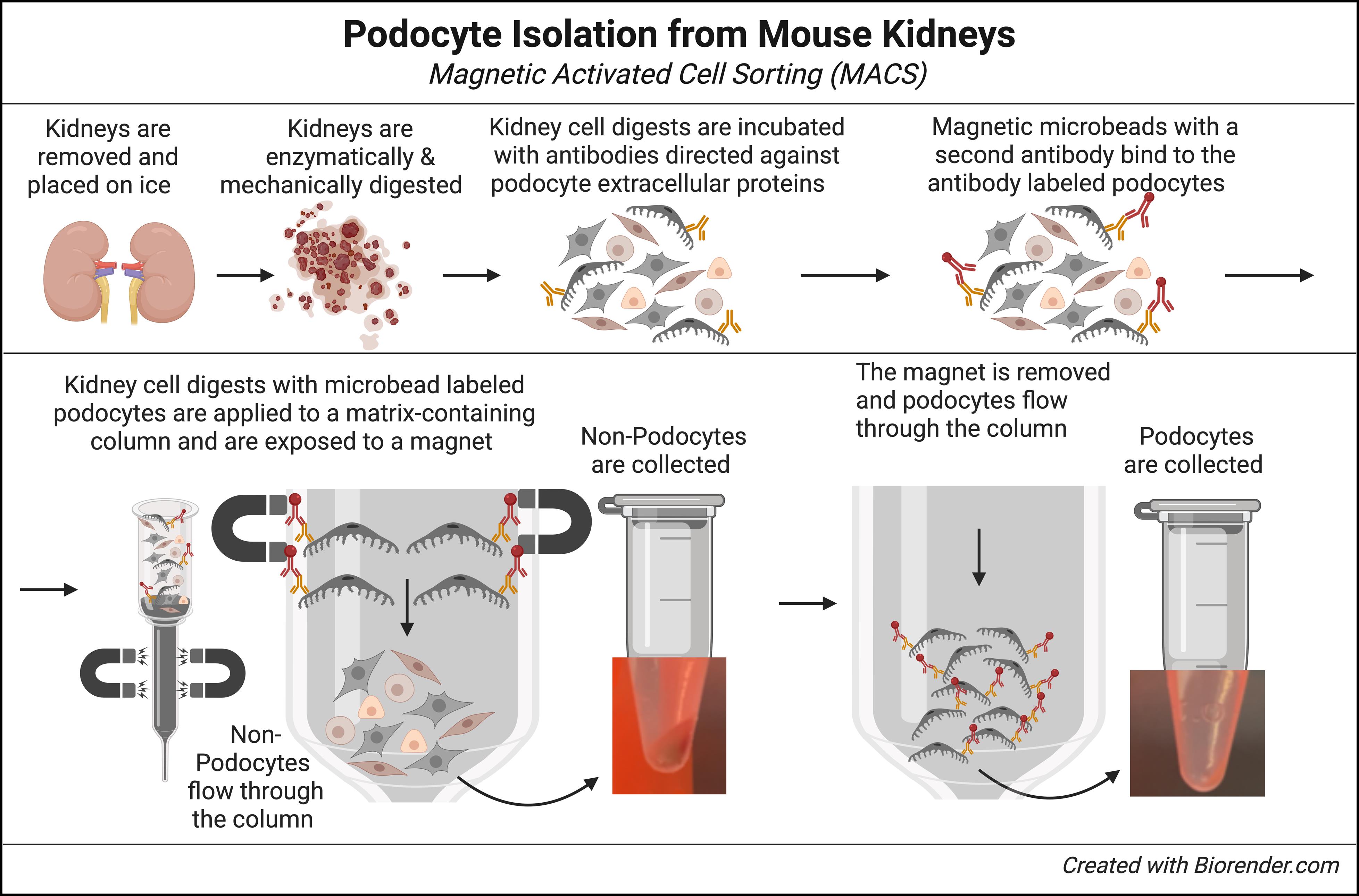
Mouse kidneys are enzymatically and mechanically digested into a single cell suspension. Surface antigens on podocytes are then labeled with a pool of specific antibodies that only bind to extracellular proteins expressed on podocytes. Magnetic microbead-conjugated anti-rabbit IgG antibodies bind to the podocyte-specific antibodies. Kidney cell suspensions containing microbead-conjugated podocytes are added to a column containing ferromagnetic spheres. Placement under a magnetic field retains podocytes in the column matrix, while unbound non-podocytes flow through and are collected. Finally, upon removal of the magnet, podocytes are flushed from the column and collected.
Background
Thanks to advances in biological technology and computing, the role of bioinformatics in biomedical research has exploded in recent years [1]. In nephrology, this has resulted in the collection of large datasets that include genome, transcriptome, proteome, and metabolome data from kidney patients, as well as from mice with experimental models of kidney disease [2–10].
However, a major challenge in collecting these data is the structural and cellular complexity of the kidney. The mouse kidney has been reported to contain >19 different cell types [11,12], and recent single-cell studies of the human kidney identified even up to 41 different cell types [11–13]. This makes it very difficult to collect data from any one specific kidney cell type. Podocytes are particularly very challenging, as these post-mitotic glomerular epithelial cells are tightly attached to the glomerular basement membrane and wrapped around the underlying vasculature.
One solution to this dilemma is the development of large-scale collections using single-cell and single-nuclear technology, which allows the extraction and analysis of individual cell types [14,15]. This methodology has some great advantages, such as the discovery of transcriptional similarities and differences within a given cell population of, e.g., healthy and diseased states, or the identification of rare cell populations that would otherwise go undetected in large pools of cells. Yet, isolating podocytes by single-cell/nuclear methods often produces low yields, and each single cell contains significantly less mRNA compared to a bulk sample of pooled cells. Thus, the detection of low-level expressed genes can be overlooked [14]. Microdissection or isolation of glomeruli and tubules can increase the granularity and depth in the data [16,17], but this approach then lacks cell type specificity.
In mice, one alternative to isolate specific kidney cell types is the use of cell-specific fluorescence reporter lines used for lineage tracing [18,19]. Performing fluorescence-activated cell sorting (FACS) on digested kidneys can obtain large pools of specific kidney cell types. Yet, limitations include the effects of long-term expression of exogenous reporters in these cells and, in the case of inducible reporters, toxic effects from the compounds used to induce expression, like tamoxifen [20], as well as the known toxicity of Cre recombinase [21–23]. Finally, a major limitation is that the vast majority of mutant and transgenic mice, and all human kidneys, lack fluorescence cell reporters.
In the protocol described herein, we provide a method for the isolation of large pools of mouse glomerular podocytes for use in bulk RNA sequencing as well as other bioinformatic applications that bypasses many of these issues.
Materials and reagents
Reagents
1. LiberaseTM TL research grade, low thermolysin (Sigma-Aldrich, catalog number: 5401020001, store at -20 °C)
2. DNase I recombinant, RNase-free (Sigma, catalog number: 4716728001, store at -20 °C)
3. Anti-nephrin, rabbit monoclonal antibody (G17-H); recognizes the epitope located between the Cys53-Cys101 extracellular domain (MyBiosource.com, catalog number: MBS684143, store at -80 °C)
4. Anti-nephrin, rabbit monoclonal antibody (Y17-R); recognizes the epitope located between the Cys465-Cys528 extracellular domain (MyBiosource.com, catalog number: MBS684100, store at -80 °C)
5. Anti-podoplanin (PDPN), rabbit monoclonal antibody; membranous mucin-type O-glycosylated glycoprotein, expressed on the surface of podocytes (MyBiosource.com, catalog number: MBS179563, store at -80 °C)
6. Anti-Rabbit IgG MicroBeads (Miltenyi Biotec, catalog number: 130-048-602, store at 4 °C)
7. Alexa Fluor® 594 AffiniPureTM donkey anti-rabbit IgG (Jackson ImmunoResearch Laboratories, Inc., catalog number: 711-585-152, store at 4 °C)
8. Dulbecco’s phosphate-buffered saline, 1× with calcium and magnesium (Corning, catalog number: 21-030-CM, store at 4 °C)
9. EDTA solution, 0.5 M pH 8.0 (Invitrogen, catalog number: AM9260G, store at room temperature)
10. DEPC-treated, DNase- and RNase-free, mol. biol. water (for RNA work) (Fisher Scientific, catalog number: BP5611, store at room temperature)
11. Bovine serum albumin (BSA), lyophilized powder (Sigma-Aldrich, catalog number: A2058, store at 4 °C)
12. HyCloneTM RPMI 1640 media, liquid (Cytiva, catalog number: SH30605.01, store at 4 °C)
13. HyCloneTM 100 mM sodium pyruvate solution (Cytiva, catalog number: SH3023901, store at 4 °C)
14. Corning® Nu-SerumTM IV growth medium supplement (optional); can be replaced with standard fetal bovine serum (Corning, catalog number: 355504, store at -20 °C)
15. Penicillin-streptomycin (10,000 U/mL) (optional); increases shelf life of the podocyte wash media (Thermo Fisher, catalog number: 15140122, store at 4 °C)
16. ProLongTM Gold antifade mountant (Thermo Fisher, catalog number: P10144, store at room temperature)
Solutions
1. Podocyte wash media (see Recipes)
2. MACS buffer (see Recipes)
3. Liberase stock solution (see Recipes)
4. DNase stock solution (see Recipes)
5. Digestion buffer (Liberase/DNase working solution) (see Recipes)
Recipes
1. Podocyte wash media
Filter sterilize. Store at 4 °C and use within 2 months.
| Reagent | Final concentration | Volume |
|---|---|---|
| RPMI 1640 | Classic liquid medium | 500 mL |
| Sodium pyruvate | 1 mM | 5 mL |
| Nu-SerumTM IV growth medium supplement | 9% | 50 mL |
| Penicillin-streptomycin (optional) | 100 U/mL | 5 mL |
| Total | 560 mL |
2. MACS buffer
Make fresh. Store at 4 °C and use within 1 week.
| Reagent | Final concentration | Quantity |
|---|---|---|
| Dulbecco’s phosphate-buffered saline | 1× | 50 mL |
| EDTA solution (0.5 M) | 2 mM | 0.2 mL |
| BSA | 0.5% | 0.25 g |
| Total | 50 mL |
3. Liberase stock solution
Freeze at -20 °C, avoid repeated freezing and thawing, and use within 1 year. 0.1 mL aliquots can be used to digest two mouse kidneys.
| Reagent | Final concentration | Quantity |
|---|---|---|
| LiberaseTM TL | 10 mg/mL | 5 mg/vial |
| Water (for RNA work) | 0.5 mL |
4. DNase stock solution
Freeze at -20 °C, avoid repeated freezing and thawing, and use within 1 year. 0.05 mL aliquots can be used to digest two mouse kidneys.
| Reagent | Final concentration | Quantity |
|---|---|---|
| DNase | 10 U/mL | 10,000 U/vial |
| Total |
5. Digestion buffer (Liberase/DNase working solution)
Make fresh on the day of digestion. Keep on ice. Can be used to digest two mouse kidneys.
| Reagent | Final concentration | Volume |
|---|---|---|
| Liberase stock solution (Recipe 3) | 0.2 mg/mL | 0.1 mL |
| DNase stock solution (Recipe 4) | 100 U/mL | 0.05 mL |
| RPMI 1640 | n/a | 5 mL |
| Total | 5 mL |
Laboratory supplies
1. FisherbrandTM 10 cm Petri dishes (Fisher Scientific, catalog number: FB0875713)
2. 15 mL conical tubes (FalconTM, catalog number: 352095)
3. 30 mL conical tubes (VWR, catalog number: 76521-346)
4. 50 mL conical tubes (FalconTM, catalog number: 352070)
5. 1.7 mL microfuge tubes (VWR, catalog number: 87003-294)
6. 10 × 75 test tubes (FalconTM, catalog number: 352054)
7. FisherbrandTM sterile 10cc syringes (Fisher Scientific, catalog number: 14-955-459)
8. BD PrecisionGlideTM needle 18 G × 1-1/2 in. (Becton Dickenson, catalog number: 305196)
9. Cell strainers, 100 μm (Sigma-Aldrich, catalog number: CLS431752)
10. Cell strainers, 40 μm (Sigma-Aldrich, catalog number: CLS431750)
11. LS columns (Miltenyi Biotec, catalog number: 130-042-401)
Equipment
1. Shaking water bath (e.g., Thermo Scientific, model: 2876)
2. Roto Shake Genie or tube rotator (e.g., Scientific Industries, model: SI-1100)
3. Centrifuge (Eppendorf, model: 5424)
4. MidiMACSTM separator (Miltenyi Biotec, catalog number: 130-042-302)
5. MACS® MultiStand (Miltenyi Biotec, catalog number: 130-042-303)
Procedure
A. Kidney dissociation (Figure 1)
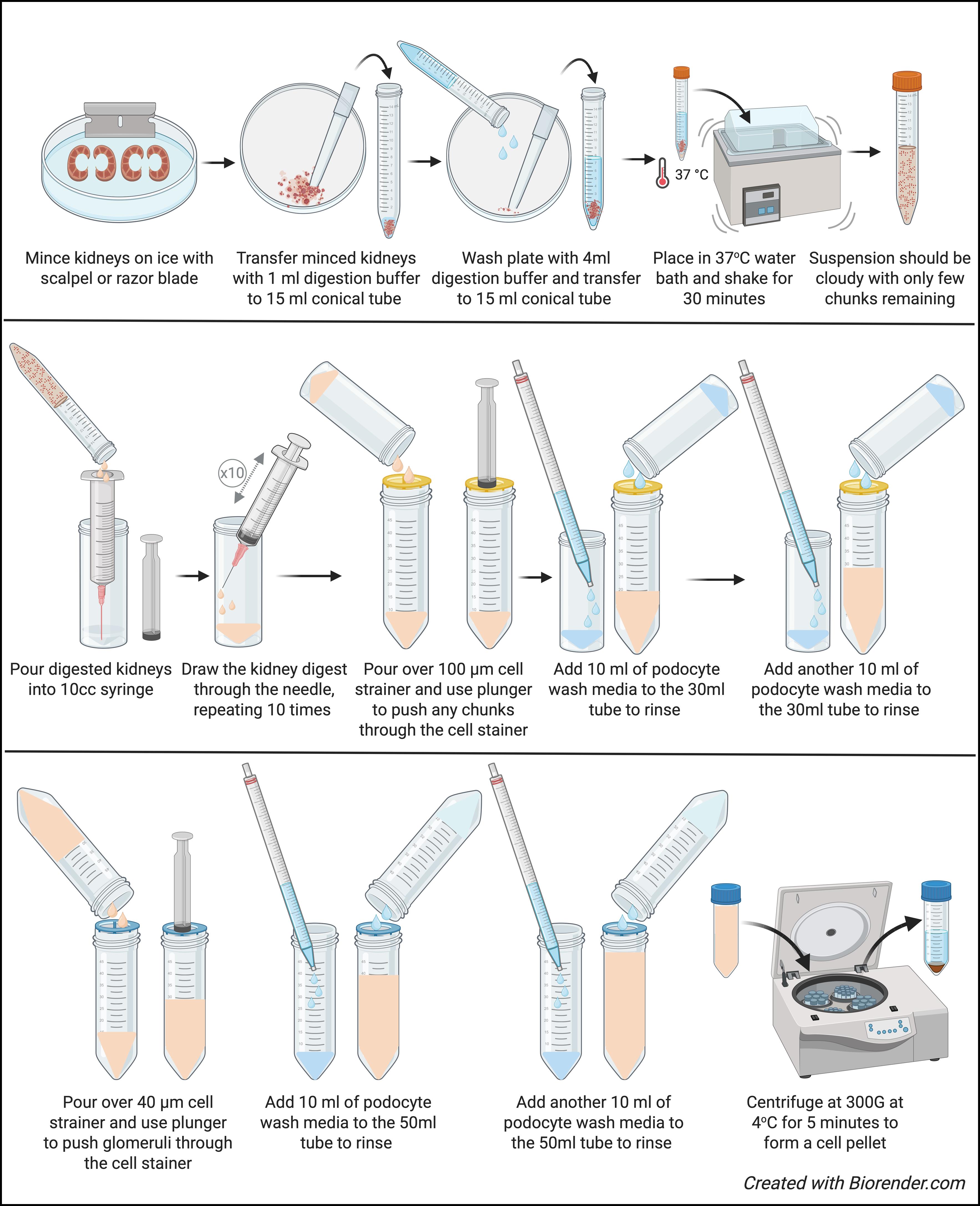
Figure 1. Kidney dissociation
1. Turn on the shaking water bath, fill with distilled water if needed, and set to 37 °C. Set the shaking speed to 125 cycles per minute (cpm).
2. Place 15 mL of RPMI 1640 media into a 10 cm Petri dish and place on ice to hold the extracted mouse kidneys.
3. Take out autoclaved forceps.
4. Take out sterile scalpel or razor blade.
5. Euthanize the mouse according to the IACUC-approved method, quickly extract kidneys, and place into a 10 cm Petri dish.
6. Place a 15 mL conical tube on ice. Remove Liberase stock solution (100 μL aliquot) and DNase stock solution (50 μL aliquot) from -20 °C and let thaw on ice.
7. Add 5 mL of RPMI 1640 media to the 15 mL tube.
8. Make the digestion solution by adding 100 μL of Liberase stock solution and 50 μL of DNAse stock solution to the 5 mL RPMI 1640 media.
9. Using forceps and a scalpel, remove the kidney capsule from the kidneys.
10. Slice the kidneys in half lengthwise. Using a scalpel or razor blade, trim off the cortex (red edge) containing the glomeruli and retain it. Discard the inner white part (medulla), which does not contain glomeruli. This dissection removes unwanted tissue and concentrates the podocytes; if the kidneys are very small, it is preferable to keep the whole kidney.
11. Pipette 0.5 mL of digestion buffer onto the kidneys.
12. With a scalpel or razor blade, very finely mince the kidneys, which were removed from the RPMI 1640 medium in which they were collected.
13. Cut off the tip of a 1,000 μL pipette tip so the minced kidney does not get trapped; apply 0.5 mL of digestion buffer to the minced kidney, pipette up and down, and transfer to a separate 15 mL conical tube (that has been kept on ice). Use the remaining 4 mL of digestion buffer to wash the Petri dish and transfer to the tube containing the minced kidneys; this tube now contains 2 minced kidneys and 5 mL of digestion buffer.
14. Tighten the cap and put parafilm around the cap to assure a good seal.
15. Place the 15 mL conical tube in a shaking water bath horizontally, making sure the tube is submerged.
16. Let the minced kidney shake (at 125 cpm) for 30 min. At the end of the digestion period, the suspension should be cloudy, with only a few chunks of tissue remaining.
17. Prepare the following materials: 18 G needle, 10 mL syringe, (1) 30 mL tube on ice, (2) 50 mL tubes on ice, (1) 1.7 mL microfuge tube on ice, 100 μm cell stainer on top of (1) 50 mL conical tube on ice, and 40 μm cell stainer on (1) 50 mL conical on ice.
18. Apply the 18 G needle to the 10 mL syringe and remove the plunger from the syringe.
19. With the needle and syringe inside the 30 mL conical tube, pour the 5 mL of kidney digest into the syringe.
20. Add 5 mL of RPMI 1640 media to the 15 mL conical tube used for digestion to rinse the tube and pour into the 10 mL syringe.
21. Replace the plunger and proceed to depress and draw the kidney digest through the needle, repeating 10 times to mechanically dissociate the kidney tissue. The suspension should now be cloudy and contain visibly fewer chunks of kidney tissue.
22. Slowly pour the 10 mL suspension over the 100 μm cell strainer, using the plunger from the syringe to gently work any remaining kidney chunks through the cell strainer.
23. Add 10 mL of podocyte wash media to the 30 mL tube to rinse the tube and pour over the 100 μm cell strainer into the 10 mL of kidney digest.
24. Add another 10 mL of podocyte wash media to the 30 mL tube to rinse the tube and pour over the 100 μm cell strainer into the 10 mL of kidney digest.
25. Slowly pour the 30 mL of suspension over the 40 μm cell strainer, using the plunger from the syringe to gently work any partially digested glomeruli through the cell strainer.
26. Add 10 mL of podocyte wash media to the 50 mL tube that contained the 100 μm cell strainer to rinse and pour over the 40 μm cell strainer.
27. Add another 10 mL of podocyte wash media to the 50 mL tube that contained the 100 μm cell strainer and pour over the 40 μm cell strainer.
28. You should now have 50 mL of kidney cell suspension.
29. Centrifuge at 300× g for 5 min at 4 °C.
30. Aspirate podocyte wash media and retain the cell pellet.
31. Resuspend the pellet with 10 mL of podocyte wash media for a second wash to remove any residual digestion reagents.
32. Centrifuge at 300× g for 5 min at 4 °C.
33. Resuspend the pellet with 1 mL of podocyte wash media and transfer the kidney cell suspension to a 1.7 mL microcentrifuge tube (kept on ice) for podocyte labeling.
B. Podocyte labeling (Figure 2)
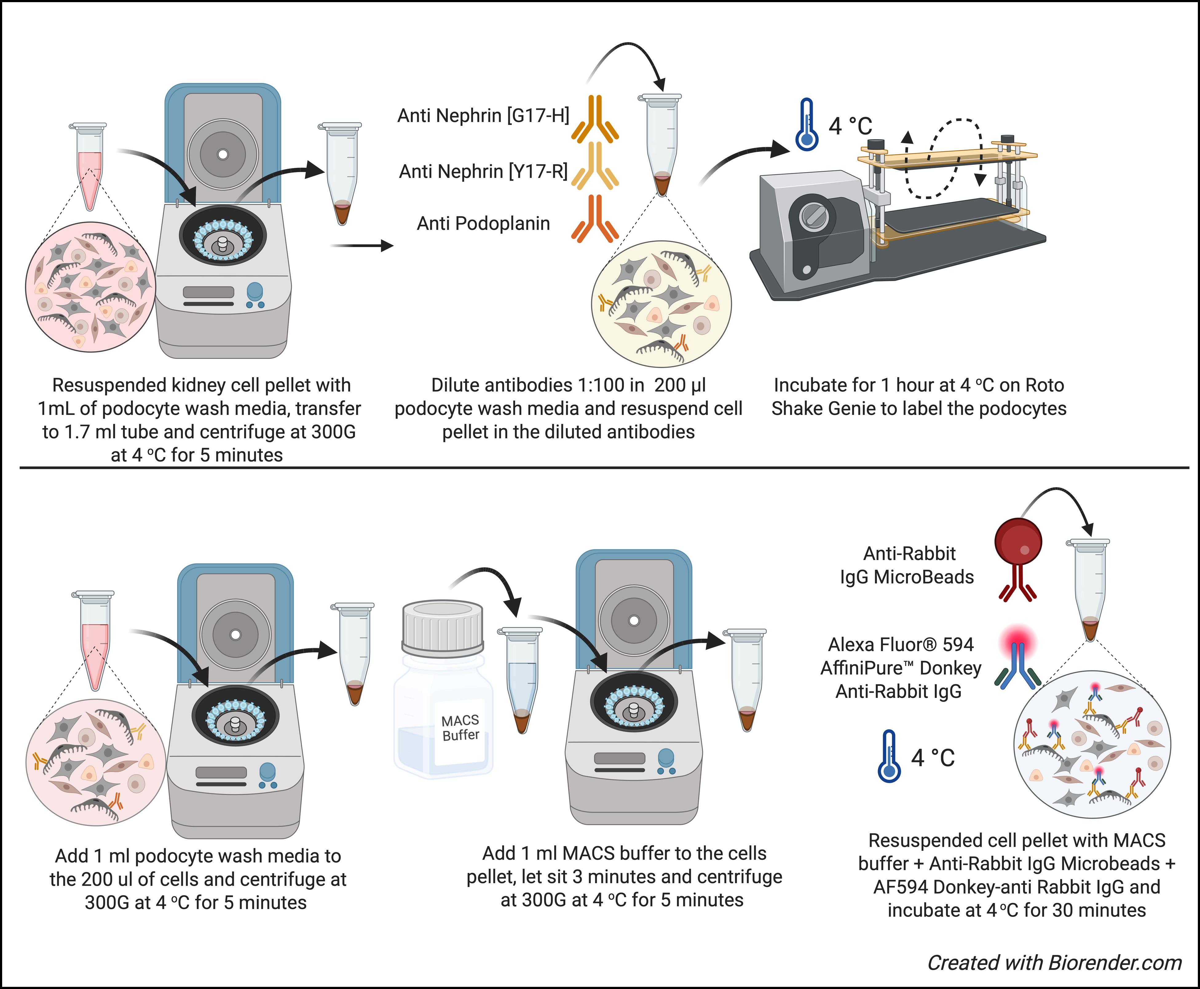
Figure 2. Podocyte labeling
All the subsequent steps should be performed in a cold room to decrease the possibility of RNA or protein degradation.
1. Centrifuge the 1.7 mL microfuge tube containing kidney digest at 300× g for 3 min at 4 °C.
2. Remove supernatant.
3. Dilute the nephrin and podoplanin antibodies 1:100 in a final volume of 200 μL in podocyte wash media and resuspend the cell pellet in the diluted antibodies.
4. Incubate for 1 h at 4 °C on Roto Shake Genie to label podocytes.
5. Following the incubation, add 1 mL of podocyte wash media to 200 μL of cells.
6. Centrifuge at 300× g for 3 min at 4 °C.
7. Remove supernatant.
8. Wash cells by adding 1 mL of MACS buffer and let it sit for 3 min.
9. Centrifuge at 300× g for 3 min at 4 °C.
10. Combine 80 μL of MACS buffer + 20 μL of anti-rabbit IgG Microbeads + 1 μL of Alexa Fluor® 594 AffiniPureTM donkey anti-rabbit IgG (1:100) with the resuspended cell pellet.
11. Incubate for 30 min at 4 °C without shaking on the Roto Shake.
12. Wash cells by adding 1 mL of MACS buffer and let it sit for 3 min.
13. Centrifuge at 300× g for 3 min at 4 °C.
14. Resuspend the cell pellet with 1 mL of MACS buffer.
Critical: MACS isolation of podocytes will not work if you do not use cell-specific antibodies that bind to the outside cell membranes of live intact podocytes. The antibodies used in this protocol have been carefully selected and validated to meet this requirement (see Figure 3). While other antibodies may be used, they must be selected with this requirement in mind and should be validated first. The antibodies listed have been tested on human kidney digests and do not work (data not shown). We are currently testing candidate antibodies for the isolation of human podocytes.
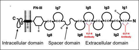
Figure 3. Nephrin/Nphs1 protein, showing the extracellular binding domains for the nephrin-specific antibodies [24]
C. MACS column podocyte isolation (Figure 4)
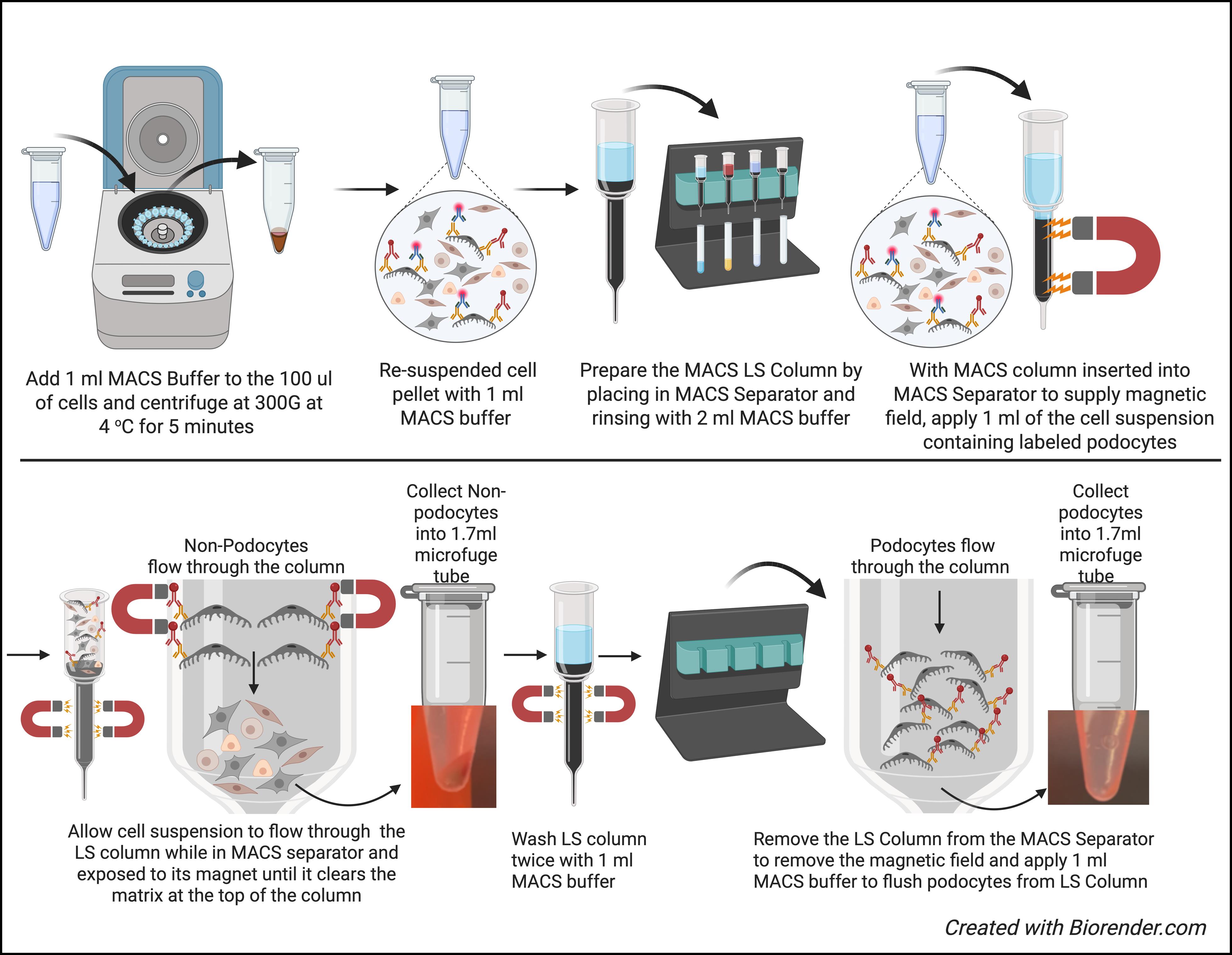
Figure 4. MACS column podocyte isolation
1. Prepare the LS column by rinsing with MACS buffer, apply 2 mL to the top of the LS column, and let the buffer run through into a collection tube (10 × 75 Test tubes).
2. Discard the effluent and change the collection tube.
3. Insert the MACS column into the MACS separator and apply 1 mL of cell suspension in MACS buffer to the top of the LS column.
4. Collect the pass-through of the LS column; podocytes will be retained in the LS column.
5. Reapply the pass-through containing the cells a second time to the LS column to ensure retention of most podocytes in the LS column.
6. Collect pass-through containing the unlabeled cells in a 1.7 mL microfuge tube labeled non-podocyte fraction.
7. With the LS column still in the MACS separator and wash the column twice with 1 mL of MACS buffer.
8. Remove the LS column from the MACS separator and place in a new 1.7 mL microfuge tube labeled podocyte fraction.
9. Apply 1 mL of MACS buffer to release the podocytes from the LS column.
10. Once the MACS buffer has passed through the column by gravity, use the plunger supplied with the LS column to flush out any remaining labeled podocytes from the LS column.
11. Centrifuge tubes with non-podocyte fraction and podocyte fraction at 300× g for 3 min at 4 °C.
12. Remove supernatant and snap-freeze cell pellets at -80 °C.
Note: Figure 5 shows a summary of the number of live cells at each step of the isolation protocol.
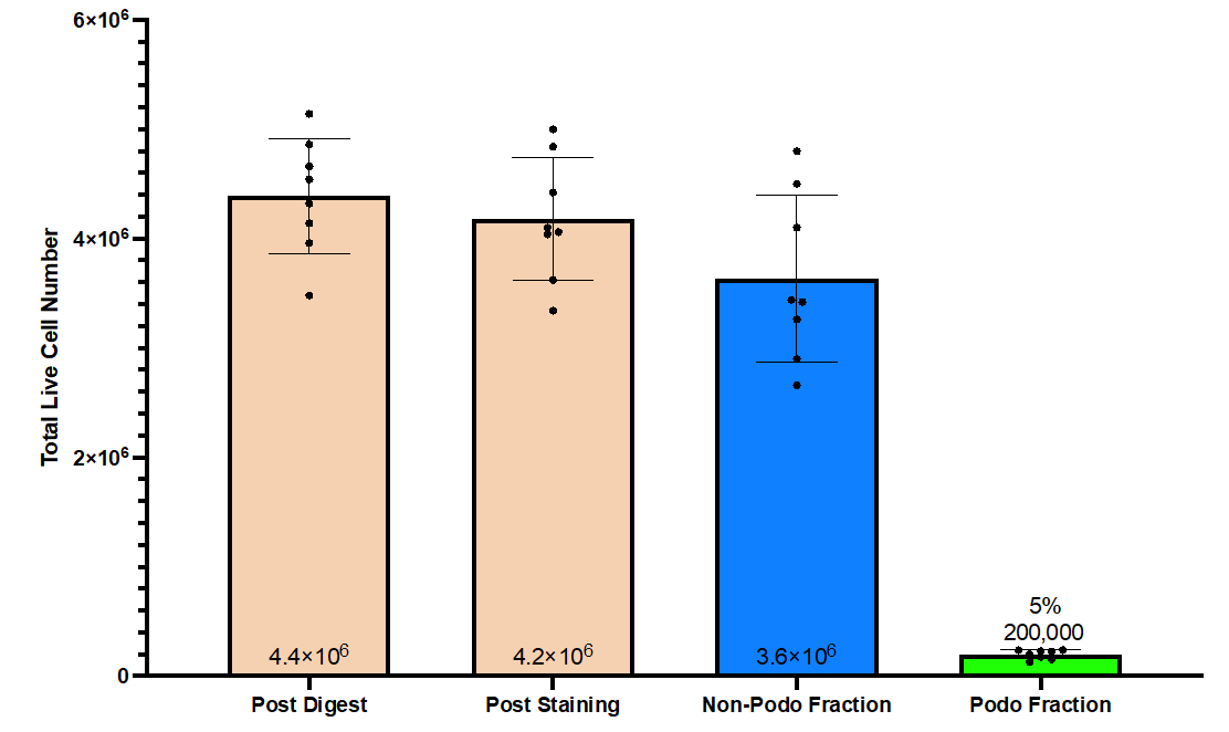
Figure 5. Live cell number was counted by trypan blue exclusion using a hemocytometer following kidney dissociation, antibody labeling, and MACS isolation of podocytes. Averaging 8 separate isolations, the mean number of cells was 4,387,500 ± 187,042 (mean ± standard error of the mean) post digest and 4,177,500 ± 199,336 post staining. Following isolation, two mouse kidneys yielded 3,463,077± 151,337 cells in the non-podocyte fraction and 195,819 ± 14,795 cells in the podocyte fraction. Note that a portion of the kidney cells were lost at each step.
Validation of protocol
This protocol or parts of it have been used and validated in the following research articles:
McKinzie et al. [25]. Podocytes from hypertensive and obese mice acquire an inflammatory, senescent, and aged phenotype. Am J Physiol Renal Physiol.
Kaverina et al. [26]. Inhibiting NLRP3 signaling in aging podocytes improves their life- and health-span. Aging (Albany NY).
Pippin et al. [27]. Upregulated PD-1 signaling antagonizes glomerular health in aged kidneys and disease. J Clin Invest.
Wang et al. [28]. Global Transcriptomic Changes in Aged Mouse Podocytes. Kidney Int (Figures S1 and S3B).
Podocyte isolation should be validated using the following criteria:
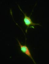
1. A small aliquot of each cell fraction should be placed on a microscope slide under a coverslip with fluorescent mounting media (ProLongTM Gold Antifade Mountant). The cell pellet should be imaged under fluorescence microscopy (we used the EVOS FL Cell Imaging System). Alexa Fluor 594®-conjugated donkey anti-rabbit IgG–positive podocytes will have a red fluorescent signal, indicating the presence of nephrin antibody on podocytes, while the non-podocyte fraction will have a negative signal (see example in Figure 6), thus validating the separation of podocytes from other kidney cells (non-podocytes).
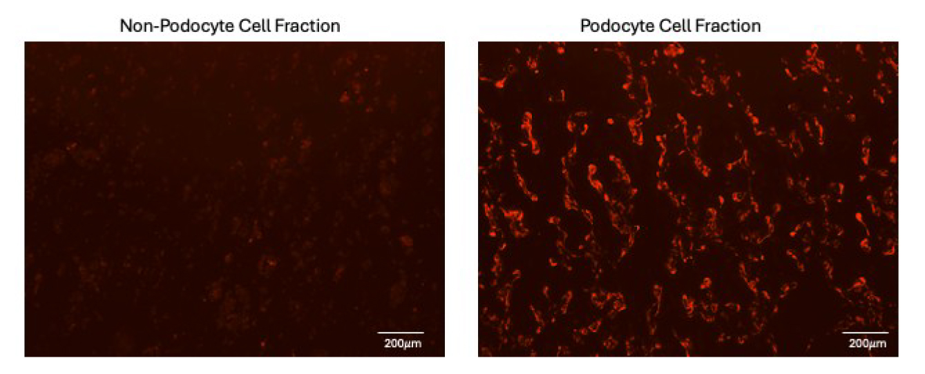
Figure 6. Fluorescent images of immunostaining for nephrin/Nphs1 and detection with Alexa Fluor 594®-conjugated donkey anti-rabbit IgG following MACS isolation. There is a positive signal for nephrin in the podocyte cell fraction (right panel) but an absence of signal in the non-podocyte cell fraction (left panel), indicating successful separation of podocytes and non-podocytes.
2. Additionally, qRT-PCR should be performed for a panel of podocyte genes comparing both podocyte and non-podocyte fractions. This will confirm the expression of podocyte genes in the podocyte fractions but their absence/low-level expression in the non-podocyte fraction (see example in Figure 7).
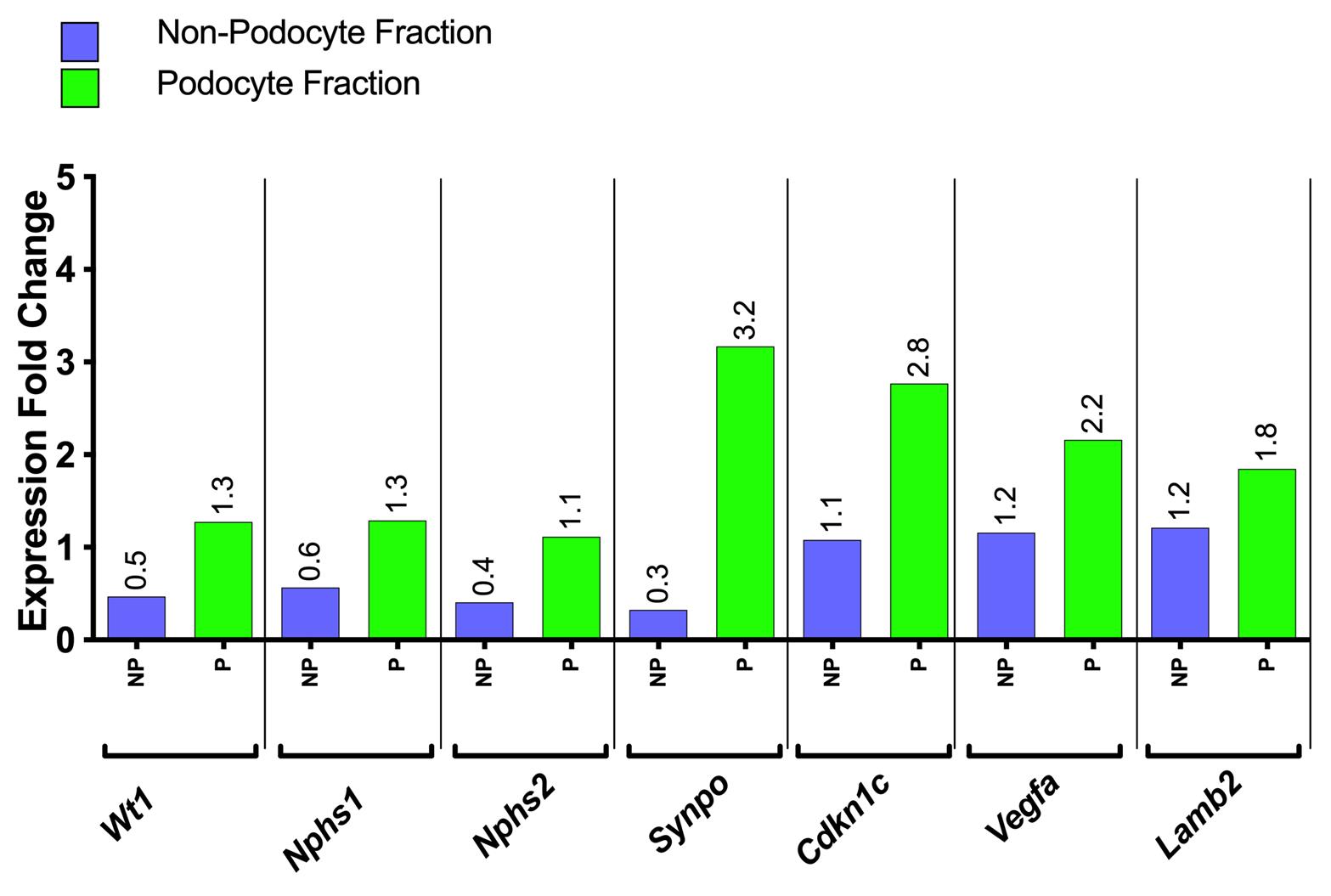
Figure 7. Representative example of qRT-PCR performed for a panel of canonical podocyte genes on the mRNA from both non-podocyte (NP) and podocyte (P) fractions in triplicate. Expression levels for Wilms tumor protein/Wt1, Nephrin/Nphs1, Podocin/Nphs2, Synaptopodin/Synpo, p57, Cdkn1c, vascular endothelial growth factor A/Vegfa, and Laminin beta-2/Lamb2 are shown. Gapdh was used as a housekeeping gene. Expression levels for all genes were significantly higher in podocyte cell fractions compared to non-podocyte cell fractions, indicating successful podocyte isolation. Note that this should be performed on podocyte isolations from each mouse (n = 1, in this example). However, this does not have to be performed for all the genes listed here to conserve RNA. If podocyte genes are not enriched in the podocyte fraction, the sample should not be used for further analysis, as shown in the example in Figure 8.
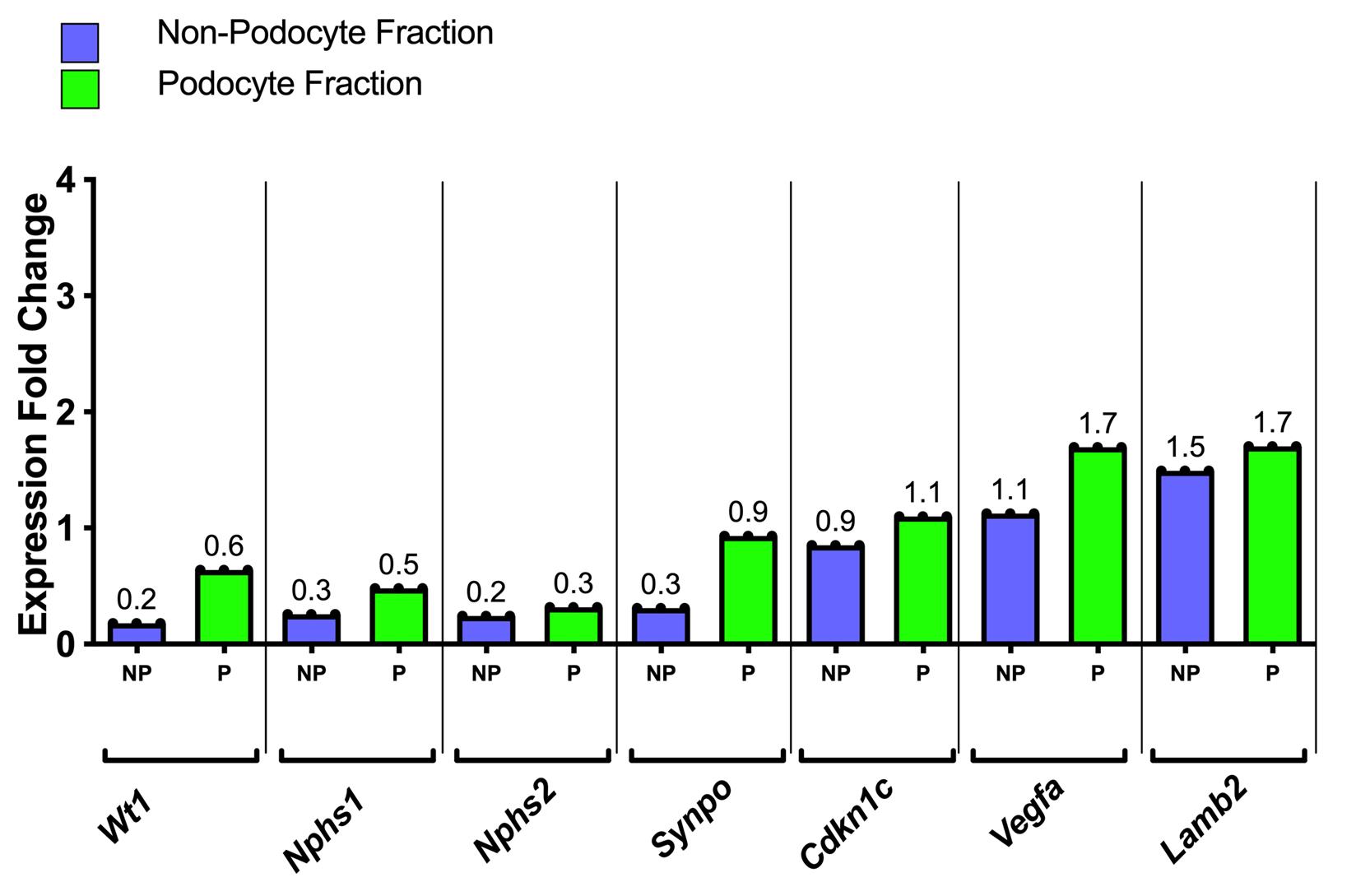
Figure 8. Representative example of qRT-PCR performed for a panel of canonical podocyte genes on the mRNA from both non-podocyte (NP) and podocyte (P) fractions in triplicate, where podocyte genes are not enriched in the podocyte fraction
General notes and troubleshooting
General notes
1. DNase is necessary during the kidney dissociation step. Digestion causes the lysis of some cells and release of DNA, which is sticky and causes clumping. Do not omit.
2. Be certain to properly rinse the Petri dishes following mincing of the kidney with the razor blade to avoid losing any kidney tissue.
3. Cutting the end of the pipette tip reduces shearing of cells during the protocol.
4. Be consistent with the timing of the warm digestion; do not go longer than 30 min.
Troubleshooting
1. While there is the slight possibility that the podocyte fraction may contain other cell types like endothelial cells, proximal tubular cells, and immune cells, this should be minimal if the protocol is followed carefully. The validation steps should reveal a lack of podocytes in the podocyte fraction. Additionally, qRT-PCR can be performed for canonical genes for contaminating cell types if they are suspected to be present in the podocyte fraction.
2. One could perform FACS sorting of cells following the labeling procedure. However, FACS takes significantly more time and is harder on the cells than MACS column separation. Therefore, podocyte yield and RNA integrity may be compromised.
Acknowledgments
Specific contributions of each author: Conceptualization, J.W.P.; Investigation, J.W.P., Writing—Original Draft, J.W.P., Writing— Review & Editing J.W.P., S.J.S., C.J.L., D.G.E., O.W.; Funding acquisition, S.J.S., O.W.; Supervision, J.W.P., S.J.S.
Funding sources that supported the work: S.J.S. and O.W. were supported by National Institute of Diabetes and Digestive and Kidney Diseases Grants 5R01DK056799-10, 5R01DK056799-12, 1R01DK097598-01A1, UC2DK126006-2, and 1R01DK090358-12 and by Department of Defense Grant DODPR180585/PR180585P1.
Citation of the original research paper in which the protocol was described and validated: Wang et al. [28].
Acknowledgment of any previous work from which the protocol was developed, modified, or derived from: Walz et al., von Schönfeldt et al., Cheng et al., and Deng et al. [29–32].
Competing interests
The authors declare no conflicts of interest.
Ethical considerations
Animal protocols (2968-04) were approved by the University of Washington Institutional Animal Care and Use Committee.
References
- Bayat, A. (2002). Science, medicine, and the future: Bioinformatics. 324(7344): 1018–1022. https://doi.org/10.1136/bmj.324.7344.1018.
- Eddy, S., Mariani, L. H. and Kretzler, M. (2020). Integrated multi-omics approaches to improve classification of chronic kidney disease. Nat Rev Nephrol. 16(11): 657–668. https://doi.org/10.1038/s41581-020-0286-5
- Rhee, E. P. (2018). How Omics Data Can Be Used in Nephrology. Am J Kidney Dis. 72(1): 129–135. https://doi.org/10.1053/j.ajkd.2017.12.008
- Mariani, L. H., Pendergraft, W. F. and Kretzler, M. (2016). Defining Glomerular Disease in Mechanistic Terms: Implementing an Integrative Biology Approach in Nephrology. Clin J Am Soc Nephrol. 11(11): 2054–2060. https://doi.org/10.2215/cjn.13651215
- Lake, B. B., Menon, R., Winfree, S., Hu, Q., Melo Ferreira, R., Kalhor, K., Barwinska, D., Otto, E. A., Ferkowicz, M., Diep, D., et al. (2023). An atlas of healthy and injured cell states and niches in the human kidney. Nature. 619(7970): 585–594. https://doi.org/10.1038/s41586-023-05769-3
- Li, H., Dixon, E. E., Wu, H. and Humphreys, B. D. (2022). Comprehensive single-cell transcriptional profiling defines shared and unique epithelial injury responses during kidney fibrosis. Cell Metab. 34(12): 1977–1998.e9. https://doi.org/10.1016/j.cmet.2022.09.026
- Liu, H., Doke, T., Guo, D., Sheng, X., Ma, Z., Park, J., Vy, H. M. T., Nadkarni, G. N., Abedini, A., Miao, Z., et al. (2022). Epigenomic and transcriptomic analyses define core cell types, genes and targetable mechanisms for kidney disease. Nat Genet. 54(7): 950–962. https://doi.org/10.1038/s41588-022-01097-w
- Kirita, Y., Wu, H., Uchimura, K., Wilson, P. C. and Humphreys, B. D. (2020). Cell profiling of mouse acute kidney injury reveals conserved cellular responses to injury. Proc Natl Acad Sci USA. 117(27): 15874–15883. https://doi.org/10.1073/pnas.2005477117
- Ransick, A., Lindström, N. O., Liu, J., Zhu, Q., Guo, J. J., Alvarado, G. F., Kim, A. D., Black, H. G., Kim, J., McMahon, A. P., et al. (2019). Single-Cell Profiling Reveals Sex, Lineage, and Regional Diversity in the Mouse Kidney. Dev Cell. 51(3): 399–413.e7. https://doi.org/10.1016/j.devcel.2019.10.005
- Park, J., Shrestha, R., Qiu, C., Kondo, A., Huang, S., Werth, M., Li, M., Barasch, J. and Suszták, K. (2018). Single-cell transcriptomics of the mouse kidney reveals potential cellular targets of kidney disease. Science. 360(6390): 758–763. https://doi.org/10.1126/science.aar2131
- Balzer, M. S., Rohacs, T. and Susztak, K. (2022). How Many Cell Types Are in the Kidney and What Do They Do? Annu Rev Physiol. 84: 507–531. https://doi.org/10.1146/annurev-physiol-052521-121841.
- Park, J., Liu, C., Kim, J. and Susztak, K. (2019). Understanding the kidney one cell at a time. Kidney Int. 96(4): 862–870. https://doi.org/10.1016/j.kint.2019.03.035
- Schumacher, A., Rookmaaker, M. B., Joles, J. A., Kramann, R., Nguyen, T. Q., van Griensven, M. and LaPointe, V. L. S. (2021). Defining the variety of cell types in developing and adult human kidneys by single-cell RNA sequencing. npj Regener Med. 6(1): 45. https://doi.org/10.1038/s41536-021-00156-w
- Haque, A., Engel, J., Teichmann, S. A. and Lönnberg, T. (2017). A practical guide to single-cell RNA-sequencing for biomedical research and clinical applications. Genome Med. 9(1): 75. https://doi.org/10.1186/s13073-017-0467-4
- Balzer, M. S., Ma, Z., Zhou, J., Abedini, A. and Susztak, K. (2021). How to Get Started with Single Cell RNA Sequencing Data Analysis. J Am Soc Nephrol. 32(6): 1279–1292. https://doi.org/10.1681/asn.2020121742
- Park, S., Yang, S. H., Jeong, C. W., Moon, K. C., Kim, D. K., Joo, K. W., Kim, Y. S., Lee, J. W. and Lee, H. (2020). RNA-Seq profiling of microdissected glomeruli identifies potential biomarkers for human IgA nephropathy. Am J Physiol Renal Physiol. 319(5): F809–F821. https://doi.org/10.1152/ajprenal.00037.2020
- Chung, J. J., Goldstein, L., Chen, Y. J., Lee, J., Webster, J. D., Roose-Girma, M., Paudyal, S. C., Modrusan, Z., Dey, A., Shaw, A. S., et al. (2020). Single-Cell Transcriptome Profiling of the Kidney Glomerulus Identifies Key Cell Types and Reactions to Injury. J Am Soc Nephrol. 31(10): 2341–2354. https://doi.org/10.1681/asn.2020020220
- Muto, Y. and Humphreys, B. D. (2021). Recent advances in lineage tracing for the kidney. Kidney Int. 100(6): 1179–1184. https://doi.org/10.1016/j.kint.2021.05.040
- Humphreys, B. D. and DiRocco, D. P. (2014). Lineage-tracing methods and the kidney. Kidney Int. 86(3): 481–488. https://doi.org/10.1038/ki.2013.368
- Dubner, A. M., Lu, S., Jolly, A. J., Noble, T., Hinthorn, T., Nemenoff, R. A., Moulton, K. S., Majesky, M. W. and Weiser-Evans, M. C. (2023). Confounding Effects of Tamoxifen: Cautionary and Practical Considerations for the Use of Tamoxifen-Inducible Mouse Models in Atherosclerosis Research—Brief Report. Arterioscler, Thromb, Vasc Biol. 43(11): 2223–2230. https://doi.org/10.1161/atvbaha.123.319922
- The Jackson laboratory. (2013). 12 things you dont know about Cre-lox. Accessed on https://www.jax.org/news-and-insights/jax-blog/2013/september/a-dozen-facts-you-didnt-know-about-cre-lox
- Janbandhu, V., Moik, D. and Fässler, R. (2013). Cre recombinase induces DNA damage and tetraploidy in the absence of LoxP sites. Cell Cycle. 13(3): 462–470. https://doi.org/10.4161/cc.27271
- Loonstra, A., Vooijs, M., Beverloo, H. B., Allak, B. A., van Drunen, E., Kanaar, R., Berns, A. and Jonkers, J. (2001). Growth inhibition and DNA damage induced by Cre recombinase in mammalian cells. Proc Natl Acad Sci USA. 98(16): 9209–9214. https://doi.org/10.1073/pnas.161269798
- Pätäri‐Sampo, A., Ihalmo, P. and Holthöfer, H. (2006). Molecular basis of the glomerular filtration: Nephrin and the emerging protein complex at the podocyte slit diaphragm. Ann Med. 38(7): 483–492. https://doi.org/10.1080/07853890600978149
- McKinzie, S. R., Kaverina, N., Schweickart, R. A., Chaney, C. P., Eng, D. G., Pereira, B. M. V., Kestenbaum, B., Pippin, J. W., Carroll, T., Wessely, O., et al. (2024). Podocytes from hypertensive and obese mice acquire an inflammatory, senescent, and aged phenotype. Am J Physiol Renal Physiol. 326(4): F644–F660. https://doi.org/10.1152/ajprenal.00417.2023
- Kaverina, N., Schweickart, R. A., Chan, G. C., Maggiore, J. C., Eng, D. G., Zeng, Y., McKinzie, S. R., Perry, H. S., Ali, A., O’Connor, C., et al. (2023). Inhibiting NLRP3 signaling in aging podocytes improves their life- and health-span. Aging (Albany NY). 15(14): 6658–6689. https://doi.org/10.18632/aging.204897
- Pippin, J. W., Kaverina, N., Wang, Y., Eng, D. G., Zeng, Y., Tran, U., Loretz, C. J., Chang, A., Akilesh, S., Poudel, C., et al. (2022). Upregulated PD-1 signaling antagonizes glomerular health in aged kidneys and disease. J Clin Invest. 132(16): e1172/jci156250. https://doi.org/10.1172/jci156250
- Wang, Y., Eng, D. G., Kaverina, N. V., Loretz, C. J., Koirala, A., Akilesh, S., Pippin, J. W. and Shankland, S. J. (2020). Global transcriptomic changes occur in aged mouse podocytes. Kidney Int. 98(5): 1160–1173. https://doi.org/10.1016/j.kint.2020.05.052
- Wang, Y., Eng, D. G., Kaverina, N. V., Loretz, C. J., Koirala, A., Akilesh, S., Pippin, J. W. and Shankland, S. J. (2020). Global transcriptomic changes occur in aged mouse podocytes. Kidney Int. 98(5): 1160–1173. https://doi.org/10.1016/j.kint.2020.05.052
- Walz, T., Malm, C., Nishikawa, B. and Wasteson, A. (1995). Transforming growth factor-alpha (TGF-alpha) in human bone marrow: demonstration of TGF-alpha in erythroblasts and eosinophilic precursor cells and of epidermal growth factor receptors in blastlike cells of myelomonocytic origin. Blood. 85(9): 2385–2392. https://doi.org/10.1182/blood.v85.9.2385.bloodjournal8592385
- Schönfeldt, V. v., Krishnamurthy, H., Foppiani, L. and Schlatt, S. (1999). Magnetic Cell Sorting Is a Fast and Effective Method of Enriching Viable Spermatogonia from Djungarian Hamster, Mouse, and Marmoset Monkey Testes1. Biol Reprod. 61(3): 582–589. https://doi.org/10.1095/biolreprod61.3.582
- Cheng, J., Baumhueter, S., Cacalano, G., Carver-Moore, K., Thibodeaux, H., Thomas, R., Broxmeyer, H., Cooper, S., Hague, N., Moore, M., et al. (1996). Hematopoietic defects in mice lacking the sialomucin CD34. Blood. 87(2): 479–490. https://doi.org/10.1182/blood.v87.2.479.bloodjournal872479
- Wilcox, G., Beaven, M. A. (1976). A sensitive and specific tritium release assay for dopamine-beta-hydroxylase (DbetaH) in serum. Anal Biochem. 75(2):484–97. https://doi.org/10.1016/0003-2697(76)90104-4
Article Information
Publication history
Received: Mar 27, 2025
Accepted: May 18, 2025
Available online: Jun 19, 2025
Published: Jul 5, 2025
Copyright
© 2025 The Author(s); This is an open access article under the CC BY-NC license (https://creativecommons.org/licenses/by-nc/4.0/).
How to cite
Pippin, J. W., Loretz, C. J., Eng, D. G., Wessely, O. and Shankland, S. J. (2025). Isolation of Podocyte Cell Fractions From Mouse Kidney Using Magnetic Activated Cell Sorting (MACS). Bio-protocol 15(13): e5364. DOI: 10.21769/BioProtoc.5364.
Category
Cell Biology > Cell isolation and culture > Cell isolation
Systems Biology > Transcriptomics
Do you have any questions about this protocol?
Post your question to gather feedback from the community. We will also invite the authors of this article to respond.