- Protocols

- Articles and Issues
- For Authors
- About
- Become a Reviewer

Fast and Sustainable Thermo-osmotic DNA Extraction Protocol for Trans-spectrum Contingency and Field Use
Published: Vol 13, Iss 17, Sep 5, 2023 DOI: 10.21769/BioProtoc.4796 Views: 1761
Reviewed by: Shyam SolankiOlga SinAnonymous reviewer(s)
Original research article
The authors used this protocol in:

Feb 2023
Advertisement


Protocol Collections
Comprehensive collections of detailed, peer-reviewed protocols focusing on specific topics


Related protocols
Abstract
In the field of molecular genetics, DNA extraction protocols and kits are sample-specific and proprietary, preventing lateral distribution among similar facilities from different sectors to alleviate supply shortages during a crisis. Expanding upon previous fast extraction protocols such as alkaline- and detergent-based ones, the use of boiling-hot water to rupture cells, virions, and nuclei, as proposed during the COVID-19 pandemic, might alleviate shortages and costs. Different soft, relatively abundant (highly enriched), and uncomplicated (genomically homogenous and with few inhibitors) biosamples are collected in 1.5 mL tubes, mixed with boiling-hot water, and stirred vigorously, so as to have membranes lysed and proteins deactivated; mechanical disruption may be used as well if necessary. Incubation in boiling water bath for 20–30 min follows. Depending on sample type and quantity, which affects the total extraction volume, 2–5 μL are pipetted off for direct PCR and the same volume for two decimal serial dilutions. The latter are intended to optimize the crude extract to a workable DNA/inhibitor concentration balance for direct PCR. Uncomplicated, highly enriched samples such as mycelial growth in fruits and human swab samples can be processed, contrary to complicated samples such as blood and physically unyielding samples such as plant tissue. The extract can be used for immediate PCR in both benchtop and portable thermocyclers, thus allowing nucleic acid amplification tests (NAAT) being performed in resource-limited settings with low cost and waste footprint or during prolonged crises, where supply chain failures may occur.
Key features
• DNA extraction from different sample types using only boiling water and occasional mechanical assistance.
• Crude extract serially diluted twice, 10- and 100-fold, to bypass purification and quantification steps.
• Direct PCR for 2–10 μL of crude lysate and dilutions (conditional to sample type and quantity) to enhance probability of workable DNA-inhibitors’ concentrations.
• Lowers the cost and curtails the overall footprint of testing to increase sustainability in field operations and in standard lab environments under supply chain derailment.
Keywords: Thermo-osmotic DNA extractionBackground
A fast and massive DNA extraction is quintessential for nucleic acid amplification tests (NAAT), especially if performed under duress; the latter implies either field conditions in resource-limited settings (RLS), in routine or expedient setups, or in abnormal conditions with samples surging and/or supply pipelines malfunctioning due to corporate concerns or supply chain overextension/collapse. Thus, although the technology for DNA extraction systems achieves significant yields from complicated and/or miniscule samples, their use is expensive even if purchased in bulk to achieve economies of scale. Alkaline approaches were suggested for contingency (Goudoudaki et al., 2021) and field conditions (Priye et al., 2016), adaptable to the intended use (Goudoudaki et al., 2021). Still, specific chemicals were used, which may not be available in proper quantities once a health or ecological crisis prompts massive spatial dispersion and increased processing output of incoming samples, which may overload standard public health or agronomic facilities and infrastructures. Thus, ease of procurement and use by personnel moderately trained in this specific discipline but experienced in other sectors of biosciences may increase the processing rate of initial screening and offload work from dedicated infrastructures, restricting their involvement to tackle only unresolved or suspect samples. Last but not least, this almost in-situ processing approach may limit the need for dispatching samples to distant facilities, which is expensive, time consuming, creates biosafety and biosecurity risks, and may degrade the samples, thus de-valuing the actual diagnostic procedure.
By using only distilled water and heat, both easily available, there is no chemical waste and no need for special reagents, and thus for diverse stockpiles. The latter may be inexpensive and long-lead or long-expiration date, but their storage, even in normal conditions, takes space and there is a need for correct management of the stock. Also, the skills necessary are basic, found in everyone with the most elementary training in Biosciences. Thus, the overall footprint of the analysis, both before- and after-use, is kept minimal. This protocol is actually an expansion of a previous one, developed during the COVID-19 pandemic (Fomsgaard and Rosenstierne, 2020) as a result of kits supply restrictions (Benda et al., 2021), to include different extraction substrates and target genomes. It can be used in the field, given that consumables, heat, and electricity sources for portable thermocyclers (Kambouris et al., 2020) can be provided. Compared to the alkaline protocol (Goudoudaki et al., 2021), the thermo-osmotic protocol described herein requires even less reagents and produces no chemical waste, nor does it require stocking reagents that may expire and require specific storage. The protocol is named after the thermal deterioration of the membrane bonds allowing osmosis to perform the cell lysis and has been used already in one original publication (Goudoudaki et al., 2023).
Additionally, the present protocol may be used for improving the turnaround time and volume throughput in existing, indoor facilities. This comes at the expense of other performance metrics including sensitivity, which are markedly inferior compared to the ones achieved by dedicated DNA extraction protocols, even those of relatively low cost (Velegraki et al., 1999a and 1999b). Furthermore, facilities commandeered during crisis management may be among the beneficiaries. The method may be used for public health and environmental and agronomic emergencies (Kambouris et al., 2018), thus qualifying for compatibility with the One-Health framework and any other that may refer to genetic/genomic biomarkers that may be typed by basic PCR protocols, with or without follow ups such as, but not restricted to, restriction digestion (Velegraki et al., 1999a and 1999b; Arabatzis et al., 2004) and single-locus sequencing (Irinyi et al., 2015). The method, being plain and simple, shows moderate performance and should be used for highly enriched (containing high numbers of target genomes) and uncomplicated (without similar loads of pseudo-targets and inhibitors) samples.
Materials and reagents
The provisions below do not include expendables and instrumentation necessary for PCR and downstream procedures, such as gel electrophoresis.
Biological materials
Swab/scrap samples from human oral mucosa (tongue or inner cheek/gum)
Fungal mycelia grown on fruits and vegetables
Reagents
Commercially available distilled water for house use (i.e., steam ironing) from any convenience store and manufacturer, upon availability
Absolute ethanol, 99.8% denatured with IPA, MEK, and Bitrex pure (Panreac/Applichem, catalog number: 147194) or any other of comparable concentration (high purity is not essential)
Solutions
70% ethanol (see Recipes)
Recipes
70% ethanol
Reagent Final concentration Volume Ethanol (absolute) 70% 700 mL H2O 30% 300 mL Total n/a 1,000 mL
Laboratory supplies
Falcon-shaped plastic tube, 50 mL (KIMA Vacutest, catalog number: KIMA-17102-50)
Beaker, 1,000 mL (Hamed, catalog number: 303173)
Polypropylene floating rack, 8 positions (Flinn Scientific, catalog number: FB1671)
Pasteur pipettes, glass (Hamed, catalog number: 303107)
Rubber bulb for Pasteur pipettes (Isolab Laborgerate, catalog number: 084-03-001)
Pipette P20, 0–20 μL (Gilson PIPETMAN®, catalog number: F123600)
Pipette P200, 20–200 μL (Gilson PIPETMAN®, catalog number: F123601)
Pipette P1000, 200–1,000 mL (Gilson PIPETMAN®, catalog number: F123602)
Pipette tips, 0–200 μL (KIMA Vacutest, catalog number: KIMA-18260)
Pipette tips, 1,000 μL (KIMA Vacutest, catalog number: KIMA-18172)
Plastic microtubes, 1.5 mL (PierceTM, catalog number: 69715)
Plastic microtubes for PCR, 0.2 mL (Thermo ScientificTM, catalog number: AB0620)
Scalpel blades (no. 22 stainless disposable; FEATHER, lot 03067430) or box cutter (e.g., Q-Connect, catalog number: 233471)
Rack (50-position for 18 mm diameter microtubes) (Fruugo)
Hydrophilic cotton (e.g., pharmacy package)
Plastic spoons (e.g., any convenience store, 10 pc package), spoons (e.g., stainless steel 18/0 convenience store), glass slides (76 mm × 26 mm, Knittel Glass, catalog number: 303157), or wood tongue depressor (disposable, Heine, catalog number: 508424)
Plastic pellet pestles, blue polypropylene autoclavable (Sigma-Aldrich, catalog number: Z359947-100EA)
Cotton (any pharmacy/convenience store) or paper tissue/napkins (any convenience store)
Equipment
Camping gas stove (e.g., KEMPER, 12.3 cm × 12.3 cm × 21.7 cm, model: 06450208) or electric kitchen stove burner (e.g., Severin, model: 3519368)
Lighter (e.g., KEMPER, model: 10422)
Procedure
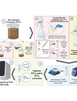
Sample collection (see General note 1) and preparation
If swab/tissue collector item is not sterile, rub it with 70% alcohol on a piece of cotton or paper tissue/napkin and light it with the lighter.
Allow the flame to die out and let it cool down in the air.
Sample gathering; either:
Collect a swab from the upper surface of the tongue or the inner surface of the cheek by mildly scrapping with any of the proposed or other similar objects (see General note 2);
Cut off a piece (3–5 mm in diameter) of the mycelium grown on the plant tissue. Alternatively, scrap it from animal tissue or scrap bacterial or yeast growth from any such surface by using disposable sterile scalpel bladed or flame-sterilized scalpel, razor, or box cutter (see General note 3).
Store sample in a 1.5 mL microtube (see Troubleshooting 1) until step B1. If needed, use some guidance/assistance to put it in (see General note 4).
Fill a beaker to half capacity with tap water; this will serve as a water bath.
Fill a 50 mL Falcon tube to half capacity with commercially available distilled water and place it into the beaker.
Heat the beaker on the stove (camping gas or electrical) at full intensity until the water bath starts boiling; then, reduce to half intensity to keep the water boiling during the entire procedure.
Cell lysis
Add boiling hot water to the extraction tube
If mechanical assistance in lysis is required (due to the nature, quantity, or physical qualities of the sample), add 250 μL of boiling-hot distilled water or half the volume you intend to use (precision is of no particular importance) to the extraction microtube containing the sample from step A4 (see General note 5).
If no mechanical assistance is required, add the sum of the extraction volume of boiling water and go straight to step B4. Caution: Steam may come out of the Falcon tube upon unscrewing and the tube may be hot. Take precautions during touching and handling and keep it safely placed on the respective rack or any similar support (e.g., a small beaker or glass).
Crush with a spiral motion of the pestle or other similar instrument for 2–3 min.
Use an equal volume of boiling-hot distilled water (e.g., 250 μL, as suggested in step B1a) to wash cellular debris of the pestle within the microtube.
Close the lid of the microtube and vortex or shake vigorously by hand for 1 min (see General note 6)
Incubation
Place the microtubes in the boiling water bath for 20–30 min or more, according to the extraction volume and rigor of extraction (see General note 7; see Troubleshooting 2). Use the incubation time to prepare the molecular testing reactions, if applicable (step E1).
Upon completion, carefully open microtubes and pipette 10 μL from the center of the extraction volume to a PCR microtube.
Caution: Steam pressure will have built up in the microtube; thus, handle with care and away from the face. Preferably, place in a rack, either custom made or improvised, before opening and further handling.
Dilutions
Transfer 5 μL of the 10 μL to the first dilution tube and mix with 45 μL of distilled water by softly pipetting in and out 5–10 times, so as to have a 1/10 dilution. Use a 20 μL pipette for the whole procedure.
Remove 5 μL of the 1/10 dilution to a second PCR tube (of 200 μL volume or as used per standard practice).
Remove another 5 μL of the 1/10 dilution to the second dilution tube and mix with 45 μL of distilled water by softly pipetting in and out 5–10 times, so as to have a 1/100 dilution.
Remove 5 μL of the 1/100 dilution to a third PCR tube (see General note 8).
PCR amplification
Prepare the mastermix, according to the desired protocol, while incubating the extract in the water bath and keep on ice until use (see General note 8). This step should be implemented during step C1.
Dispense the required volumes of mastermix to the three PCR tubes with the serial dilutions for each sample and proceed to amplification in thermocycler as per standard procedure/protocol. This step should be implemented after step D4.
Data analysis
Each extraction was performed in triplicates independently, with no more than a week between two successive efforts. Any of the three dilutions producing amplicon was deemed successful. Here, we report that the protocol can be used in cases where at least two of the three independent extraction efforts produced amplicon in at least one out of three serial dilutions (the undiluted crude extract being understood as the first in the series of the three dilutions).
Validation of protocol
There is no way to verify the performance of the present protocol directly: it is an extraction protocol and since its product is not purified, it cannot be tested properly—neither by spectrophotometry, as the cell debris would absorb or scatter the emitted UV, nor by gel electrophoresis, as said debris would obstruct the motion of the DNA, mar the electric field, and also absorb the fluorescent dyes, creating noise and smear. The only applicable validation approach is indirect, by the success or failure of some downstream nucleic acid amplification test (NAAT). To secure validation, negative controls with distilled water-only added in equal volume to the template were used, to ensure that amplicon production was not due to contamination of the NAAT reagents, expendables, or equipment.
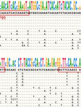
Furthermore, to ensure that the NAAT (here, PCR) does work in terms of protocol, cycler, and reagents, positive controls were used. These were semi-simultaneous extractions of mycelial tissue, by other established methods such as alkaline extraction and/or CTAB (Velegraki et al., 1999a; Goudoudaki et al., 2021), and also of stock DNA extracts kept in the lab from previous projects, extracted with conventional protocols and occasionally quantified by mini-spectrophotometer (Psarias et al., 2020). The full procedure for validation and controls may be found in the source publication (Goudoudaki et al., 2023). In short, from a number of tried applications, positive PCR results were returned for two cases of samples: (a) swab samples tested either by consensus sequence PCR targeted at the local bacteriome (product band of size compatible with the expected target range) or for one specific endogenous (human) locus (product band of the expected size); (b) consensus sequence PCR on extracts of fungal mycelia grown on the surface of fruits held in refrigeration (product band of size compatible with the expected target range).
General notes and troubleshooting
General notes
The method is valid with relatively soft and uncomplicated samples in which the target genome is abundant (highly enriched). Mouth swabs produced specific human genome amplicons and consensus sequences amplification product for bacteria but not for fungi, which are much less populous in the oral mucosa.
Glass slides for microscopy, plastic or metallic spoon—especially disposable ones for deserts—or the handle of all-metallic cutlery or tongue depressors may be used.
During sampling, samples as pure as possible of target cells must be collected (pure is understood as “containing as few admixtures and as homogenous a cellular population as possible”). The quantity of the sample must be limited, as there is no purification step to dispose of cell debris and consequent background noise and possible inhibitions.
If needed, help/guide the sample within the microtube with a similarly sterilized microbiology needle or a similar piece (i.e., a second glass slide) or a plastic tip.
If mechanical assistance is required, take caution that the volume of sample + boiled water is approximately 500 μL. Additional boiling water will be added to wash off the pestle from attached debris, but it is better that the final volume does not exceed 1 mL.
If the extraction microtube is too full (> 1 mL), punch holes using a needle, thin nail, or pin in the microtube lid before incubation so the pressure will not blow the lid open, throwing the content out of the tube.
Use purpose-built floating rack or punch/cut holes to felizol (EPC–Expanded PolyStyrene) sheets of less than 50 mm thickness recovered from packaging boxes.
Functional concentration during the NAAT may be adjusted to other values: either by i) increasing the template volume in the reaction tube while subtracting an equal volume of sterile distilled H2O from the mastermix or ii) if no mastermix is prepared, by changing the volumes in the reaction microtube. Occasionally, the opposite may also be applicable (diminishing the template volume while increasing that of the sterile distilled H2O). Similar manipulations are applicable at the serial dilutions step.
Troubleshooting
If the sample size within the microtube is excessively large (operative’s call, depending on the difficulty to properly handle the extraction mix, but see General note 6 for an example), it may be divided in two. If it is solid, this can be done by cutting with a sterilized surgical disposable blade; if it is liquid, the excess may be pipetted off to another tube, creating a technical replicate.
Longer incubation time is advised (a) when treating larger samples in terms of volume/mass and (b) if the cell lysis phase is less rigorous, as in the absence of mechanical assistance. Refer to step C1.
Acknowledgments
This project was in part funded by the European Commission, Horizon 2020 (H2020-668353; Ubiquitous Pharmacogenomics).
This protocol is based on the research paper (Goudoudaki et al. 2023).
Competing interests
The authors declare no conflicts of interest or competing interests.
Ethical considerations
The study was approved by the Institutional Research Ethics Committee of the University of Patras. Swabs were collected from the operatives performing the development of the protocol and the subsequent analysis as explicitly stated in the paper (Goudoudaki et al., 2023).
References
- Arabatzis, M., Kollia, K., Menounos, P., Logotheti, M. and Velegraki, A. (2004). Delineation of Clavispora lusitaniae clinical isolates by polymerase chain reaction-single strand conformation polymorphism analysis of the ITS1 region: a retrospective study comparing five typing methods. Med. Mycol. 42(1): 27–34.
- Benda, A., Zerajic, L., Ankita, A., Cleary, E., Park, Y. and Pandey, S. (2021). COVID-19 Testing and Diagnostics: A Review of Commercialized Technologies for Cost, Convenience and Quality of Tests. Sensors 21(19): 6581.
- Fomsgaard, A. S. and Rosenstierne, M. W. (2020). An alternative workflow for molecular detection of SARS-CoV-2 – escape from the NA extraction kit-shortage, Copenhagen, Denmark, March 2020. Eurosurveillance 25(14): e2000398.
- Goudoudaki, S., Kambouris, M. E., Siamoglou, S., Gioula, G., Kantzanou, M., Manoussopoulou, M., Patrinos, G. P. and Manoussopoulos, Y. (2023). Can Water-Only DNA Extraction Reduce the Logistical Footprint of Biosurveillance and Planetary Health Diagnostics? Toward a New Method. OMICS: J. Integr. Biol. 27(3): 116–126.
- Goudoudaki, S., Milioni, A., Kritikou, S., Velegraki, A., Patrinos, G. P., Gioula, G., Manoussopoulos, Y. and Kambouris, M. E. (2021). Fast, Scalable, and Practical: An Alkaline DNA Extraction Pipeline for Emergency Microbiomics Biosurveillance. OMICS: J. Integr. Biol. 25(8): 484–494.
- Irinyi, L., Serena, C., Garcia-Hermoso, D., Arabatzis, M., Desnos-Ollivier, M., Vu, D., Cardinali, G., Arthur, I., Normand, A. C., Giraldo, A., et al. (2015). International Society of Human and Animal Mycology (ISHAM)-ITS reference DNA barcoding database—the quality controlled standard tool for routine identification of human and animal pathogenic fungi. Med. Mycol. 53(4): 313–337.
- Kambouris, M. E., Manoussopoulos, Y., Kritikou, S., Milioni, A., Mantzoukas, S. and Velegraki, A. (2018). Toward Decentralized Agrigenomic Surveillance? A Polymerase Chain Reaction–Restriction Fragment Length Polymorphism Approach for Adaptable and Rapid Detection of User-Defined Fungal Pathogens in Potato Crops. OMICS: J. Integr. Biol. 22(4): 264–273.
- Kambouris, M. E., Siamoglou, S., Kordou, Z., Milioni, A., Vassilakis, S., Goudoudaki, S., Kritikou, S., Manoussopoulos, Y., Velegraki, A., Patrinos, G. P., et al. (2020). Point-of-need molecular processing of biosamples using portable instrumentation to reduce turnaround time. Biosaf. Health 2(3): 177–182.
- Priye, A., Wong, S., Bi, Y., Carpio, M., Chang, J., Coen, M., Cope, D., Harris, J., Johnson, J., Keller, A., et al. (2016). Lab-on-a-Drone: Toward Pinpoint Deployment of Smartphone-Enabled Nucleic Acid-Based Diagnostics for Mobile Health Care. Anal. Chem. 88(9): 4651–4660.
- Psarias, G., Iliopoulou, E., Liopetas, I., Tsironi, A., Spanos, D., Tsikrika, A., Kalafatis, K., Tarousi, D., Varitis, G., Koromina, M., et al. (2020). Development of Rapid Pharmacogenomic Testing Assay in a Mobile Molecular Biology Laboratory (2MoBiL). OMICS: J. Integr. Biol. 24(11): 660–666.
- Velegraki, A., Kambouris, M., Kostourou, A., Chalevelakis, G. and Legakis, N. J. (1999a). Rapid extraction of fungal DNA from clinical samples for PCR amplification. Med. Mycol. 37(1): 69–73.
- Velegraki, A., Kambouris, M., Skiniotis, G., Savala, M., Mitroussia-Ziouva, A. and Legakis, N. (1999b). Identification of medically significant fungal genera by polymerase chain reaction followed by restriction enzyme analysis. FEMS Immunol. Med. Microbiol. 23(4): 303–312
Article Information
Copyright
© 2023 The Author(s); This is an open access article under the CC BY-NC license (https://creativecommons.org/licenses/by-nc/4.0/).
How to cite
Goudoudaki, S., Kambouris, M. E., Manoussopoulou, M., Patrinos, G. P., Velegraki, A. and Manoussopoulos, Y. (2023). Fast and Sustainable Thermo-osmotic DNA Extraction Protocol for Trans-spectrum Contingency and Field Use. Bio-protocol 13(17): e4796. DOI: 10.21769/BioProtoc.4796.
Category
Molecular Biology > DNA > DNA extraction
Microbiology > Pathogen detection > PCR
Do you have any questions about this protocol?
Post your question to gather feedback from the community. We will also invite the authors of this article to respond.