- Protocols

- Articles and Issues
- For Authors
- About
- Become a Reviewer

Rhizoctonia solani Infection Assay of Young Sugar Beet and Arabidopsis plantlets
Published: Vol 12, Iss 2, Jan 20, 2022 DOI: 10.21769/BioProtoc.4300 Views: 3972
Reviewed by: Juan Facundo Rodriguez AyalaAndrea Paola ZuluagaWeiyan JiaShuhei Ota
Original research article
The authors used this protocol in:

Oct 2020
Advertisement


Protocol Collections
Comprehensive collections of detailed, peer-reviewed protocols focusing on specific topics


Abstract
Rhizoctonia solani is a soil-borne fungus, which rarely produces any spores in culture. Hence, all inoculation procedures are based on mycelia, often as a coat on cereal kernels, placed in close vicinity to the plant to be infected. In this protocol, an inoculation method is described where the fungus is first allowed to infest a perlite-maize flour substrate for 10 days, followed by thorough soil mixing to generate uniform fungal distribution. Pre-grown seedlings are then replanted in the infested soil. Plant materials can be harvested, five (sugar beet) and ten days (Arabidopsis) post infection, followed by a rapid cleaning step ahead of any nucleic acid preparation. Commercial DNA or RNA extraction kits can be used or, if higher DNA yield is required, a CTAB extraction method. Our purpose was to develop a reliable and reproducible protocol to determine the infection levels in planta upon infection with R. solani. This protocol is less laborious compared to previous ones, improves the consistency of plant infection, reproducibility between experiments, and suits both a root crop and Arabidopsis.
Graphic abstract:

Overview of the R. solani infection procedure.
Background
Sugar beet (Beta vulgaris) is a biennial plant species grown for its high sugar content in the taproot. In temperate regions where this crop is mainly grown, it takes about six months from sowing to harvesting (Draycott, 2006). This long timespan exposes the root to several soil-borne pathogens, such as Rhizoctonia solani. Rhizoctonia solani can cause damping-off disease on seedlings or root and crown rot disease on older roots, and this has developed into a major and increasing problem in sugar beet growing areas (Kluth and Varrelmann, 2010). The isolates of this fungal basidiomycete are divided into hyphal anastomosis groups (AGs). AG2-2IIIB is the subgroup that mainly incites disease on sugar beet, but such strains can also infect maize, resulting in problems where these two crops are used in the same crop rotation scheme (Führer Ithurrart et al., 2004).
Preparing materials for more advanced molecular analyses have become problematic due to the absence of asexual spores (conidia) and the sexual stage being very rare (Parmeter, 1970), which limits more precise inoculation procedures using specified spore concentrations. Hence, inoculation of sugar beets in the greenhouse is traditionally performed by replacing soil around the plants in the pots with infected millet seeds or infected ground barley kernels, followed by disease scoring (Scholten et al., 2001; Bolton et al., 2010). Under field conditions, infected ground barley kernels are spread in furrows or broadcast before sugar beet seeds are drilled. Alternatively, the barley kernels are placed in the crown of the plant, at the eight-leaf growth stage (Strausbaugh et al., 2013). These procedures can cause variation in terms of amount of fungal DNA being established on the host plant surface, thereby affecting the observed plant responses. There are methods available for measuring fungal DNA concentration in soil (Budge et al., 2009; Abbas et al., 2014). One of the problems, is that DNA concentration is rarely correlated with disease symptoms. Other factors that influence disease development is soil composition, soil humidity, and temperature (Bolton et al., 2010). Here, we describe in detail how to generate diseased plants under controlled growth conditions, followed by extraction of reproducible amounts of RNA or DNA. This protocol has been used to identify defense genes against R. solani in sugar beet and Arabidopsis (Holmquist et al., 2021). Our plant infection method was instrumental to identify the R. solani virulence factor RsLysM (Dölfors et al., 2019), the protease inhibition effector RsRlpA (Charova et al., 2020), and the mitochondria and chloroplast targeting effector RsCRP1 (Tzelepis et al., 2021). RNA and DNA obtained through this protocol can be used to monitor plant transcript responses to R. solani and measure fungal colonization in planta, including fungal activities during plant pathogen interaction.
Materials and Reagents
Materials
Plant pots, small pots (6 × 6 × 5 cm) and big pots (13 × 13 × 13 cm) (SW Horto, catalog number: 700-245)
Plant trays (34 × 22 × 4 cm) (Nelson Garden, catalog number: 5770)
Soil (S-soil, Hasselfors Garden, Örebro, pH 5.5-5.6) composed of sighted light peat, black peat, perlite, sand, and lime
Miracloth Calbiochem® (Merck, catalog number: 475855-1)
Glass beakers (250 mL VWR, catalog number: 213-0014)
Laboratory bottles (500 mL SARSTEDT, catalog number: 3607507)
Plastic petri dishes (Ø × H: 92 × 16 mm, SARSTEDT, catalog number: 82.1473.001)
Glass Petri dishes (Ø × H: 92 × 16 mm, Fisher Scientific, catalog number: 1201333)
Eppendorf micro-tubes (SARSTEDT, catalog numbers: 72.690.001 [1.5 mL]; 72.695.500 [2.0 mL])
Spoon (sterile)
Aluminum foil
Scalpel and blades (Fisher Scientific, catalog numbers: 12348019 and 12398009)
Mortar and pestle (80 mm × 92 mm, VWR, catalog number: 470148-960)
Parafilm (10.2 cm × 38.1 m, VWR, catalog number: 52858-000)
Filtropur sterilization filter S0.2 (SARSTEDT, catalog number: 83.1826.001)
Plants
Beta vulgaris seeds (DLF Beet Seed)
Arabidopsis thaliana, Col-0 (Arabidopsis Information Resource, TAIR)
Pathogen
The Rhizoctonia solani AG2-2IIIB BBA 69670 isolate (Wibberg et al., 2016) was used throughout the work.
Molecular biology working kit
DNeasy plant mini kit (Qiagen, catalog number: 69104)
RNeasy plant mini kit (Qiagen, catalog number: 74903)
Other reagents
Inoculum medium (see Recipes)
Potato dextrose agar, PDA (Applichem, catalog number: A5838)
Distilled water
Perlite, 0-6.5 mm (SW Horto AB, Hammenhög, Sweden)
Maize flour (Risenta, Sollentuna, Sweden)
Liquid nitrogen
Chloroform, EMSURE® ACS, ISO, Reag. Ph. (VWR, catalog number: 1.02445.1000, CAS number: 67-66-3)
Isopropanol (2-Propanol, EMSURE ACS, ISO, Reag. Ph. Eur. for analysis, VWR, catalog number: 1.09634.5000)
Ethanol (70%)
Sodium chloride (NaCl) (Saveen Werner AB (Duchefa), catalog number: 31434)
Sodium hydroxide (NaOH) (Sigma-Aldrich, catalog number: S8045)
Ethylenediaminetetraacetic acid (EDTA) (VWR, catalog number: BDH9232, CAS number: 60-0-04)
Tris base TRIS-RO ROCHE (Sigma-Aldrich, CAS number: 77-86-1)
Hydrochloric acid (37%) (Sigma-Aldrich, catalog number: H1758)
Distilled water
Hexadecyltrimethylammonium bromide (CTAB) (Sigma-Aldrich, catalog number: H6269)
Tris-EDTA buffer solution (Sigma-Aldrich, catalog number: T9285)
Optional: RNase A (17,500 U) (Qiagen catalog number: 19101)
Potato dextrose agar (PDA) (1 L) (see Recipes)
3% CTAB extraction buffer (50 mL) (see Recipes)
1 M Tris-HCl (pH8) (1 L) (see Recipes)
5 M NaCl (100 mL) (see Recipes)
0.5 M EDTA (pH8) (1 L) (see Recipes)
1× TE buffer (100 mL) (see Recipes)
Equipment
Growth chamber (Percival AR82L2/Split) or greenhouse
Analytic balance (Mettler Toledo, model: AE100)
Autoclave
Laminar flow hood
Heating cabinet
Water bath (Sigma-Aldrich, model: Julabo TW12, catalog number: Z615498)
Heating plate
Liquid nitrogen container
Safety glasses, gloves, and lab coat
Procedure
Preparation of infested soil
Dissolve 39 g PDA powder in 1 L distilled water and autoclave for 15 min at 125°C (135 kPa). Once the media cools down (at approximately 50°C), pour approximately 20 mL per Petri plate and let it solidify. Grow R. solani on PDA plates for 10 days at room temperature. Use sterile conditions. Store cultures at 4°C for up to one month.
Calculate how many grams inoculum will be needed for the experiment (depending on pot size, number of biological replicates, and experimental set up). For instance, 1 g inoculum per 20 g soil for Arabidopsis and 1 g inoculum per 10 g soil for sugar beet.
Mix perlite, maize flour, and distilled water (1:1:5) in a beaker (see inoculum medium recipe).
Place 40 g of perlite mixture in clean glass Petri dishes, seal with aluminum foil, and autoclave for 15 min at 125°C (135 kPa).
Work under sterile conditions and cut 1 × 1 cm pieces from PDA plates with R. solani. Place one piece upside down in the middle of each glass Petri dish filled with Perlite media (step 2) using a sterile scalpel. Seal the glass Petri dishes with Parafilm and incubate for 10 days at room temperature in the dark, to generate fungal infested substrate (FIS).
Add pre-weighed FIS to soil with a spoon and mix thoroughly. The mix should contain 1 part FIS for every 10 parts of soil for sugar beet inoculation and 1:20 (FIS:soil) for Arabidopsis inoculation.
Note: It is critical to mix very thoroughly. Weigh and mix all soil/inoculum needed all at once to avoid soil batch variation.
Use the soil-FIS mix in the same day it was prepared.
Preparation of diseased sugar beet or Arabidopsis plants
Wrap sugar beet seeds in Miracloth and dip the “bag” in cold water in a glass beaker for 30 min.
Transfer the seeds to a 57°C water bath for 5 min.
Dip the seeds in cold water again and let them dry overnight at room temperature.
Transfer sugar beet seeds to soil and cultivate for three weeks under greenhouse conditions (16 h light/8 h dark cycle with 22°C/18°C). Add nutrients (2 mL of Blomstra, Cederroth, Upplands Väsby/L water) once a week,
Alternatively, incubate Arabidopsis Col-0 seeds in sterile water overnight or up to four days at 4°C.
Plant Arabidopsis seeds in soil and cultivate for four weeks in short day conditions (16 h dark at 18°C/8 h light at 22°C cycle, and 60% relative humidity). Keep the soil moist but not wet.
Carefully lift the plants from the soil with tweezers. Gently wash away the soil from the roots with distilled water.
Note: If the soil has become compact, loosen the soil around the plant by pressing lightly on the sides of the pot to avoid damaging the root.
Transfer clean plantlets to infested soil. Monitor plants for symptom development (Figure 1; Figure 2). Symptoms on sugar beet seedlings may occur on the hypocotyl after two days. At day eight, susceptible plants start damping-off. Symptoms are diffuse on Arabidopsis seedlings, until the rosette leaves start showing signs of chlorosis and/or necrosis around day five.

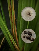
Figure 1. Sugar beet plantlets, 26 days post germination. A. Tolerant genotype, 5 days post R. solani infection. B. Tolerant genotype, grown in non-infested soil. C. Susceptible genotype, 5 days post R. solani infection. D. Susceptible genotype, grown in non-infested soil.
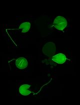
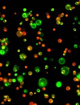
Figure 2. Arabidopsis thaliana Col-0, 38-days post germination. Plants in R. solani infested soil at five different inoculum ratios and control plants grown in H2O-treated soil. Plant phenotypes at 10 dpi. The red rectangle marks the optimal infestation level.
Harvest of plant material and isolation of total RNA and DNA
Harvest roots and hypocotyls of sugar beet seedlings at 5 dpi and Arabidopsis plantlets, including roots, at 10 dpi, using a clean scalpel (harvest timing and number of plants per biological replicate depend on your project). Wash away soil and other contaminants using sterile distilled water. Let dry on paper towels. Wrap in aluminum foil and freeze immediately in liquid nitrogen.
Note: It is important that roots are completely clean. Work fast. Avoid piling up materials ahead of freezing.
Grind the samples in liquid nitrogen with a mortar and pestle (autoclaved or heat sterilized). Keep the plant material frozen until all samples have been ground to a fine powder.
Note: Do not let the plant material thaw before DNA or RNA extraction.
Proceed with RNA and/or DNA isolation from 100 mg sample, using commercially available kits, such as RNeasy- and DNeasy-plant mini kit (Qiagen) following the manufacturer’s manual. If higher DNA yield is preferred, the modified CTAB DNA extraction method can be used.
CTAB DNA extraction (Möller et al., 1992)
Add 500 µL of 3% CTAB extraction buffer to 100 mg powdered plant material in a 2.0 mL microcentrifuge tube and vortex vigorously.
Optional: Add 4 μL of RNAse A (100 mg/mL) and mix by inverting.
Incubate the samples in a 65°C water bath for 30 min. Mix by inverting the tubes once every 5-10 min.
Centrifuge the samples for 10 min at 13,000 × g and transfer the supernatant to a new 1.5 mL tube.
Add 600 µL of chloroform and vortex.
Centrifuge at 13,000 × g for 10 min. Transfer the upper phase of the supernatant to a clean 1.5 mL Eppendorf tube.
Add 1 volume of chloroform and vortex.
Centrifuge for 10 min at 13,000 × g and transfer the upper phase to a clean 1.5 mL tube.
Add 1 volume isopropanol (2-propanol) and mix by pipetting up and down. Precipitate the mix at -20°C for 30 min.
Centrifuge for 20 min at 13,000 × g. Discard the supernatant by pipette (avoid disturbing the DNA pellet) and let the pellet dry briefly.
Add approximately 150 µL of chilled 70% ethanol and centrifuge at 13,000 × g for 5 min to wash the DNA pellet. Discard the ethanol and let the pellet dry.
Dissolve the pellet in 100 µL of 1× TE buffer.
Data analysis
Infection levels are best evaluated by comparing fungal DNA in the different plant materials at 5- or 10-days post infections.
Wash the roots vigorously in sterile water, to eliminate excess soil and external fungal growth.
Repeat root washing until totally clean. This can be determined under a stereo microscope.
Use at least five sugar beet or Arabidopsis roots and hypocotyls in each replicate, for a minimum of four replicates.
Prepare total DNA according to Möller et al. (1992). See section C and D.
The amount of fungal DNA (RsG3PDH) can be determined with qPCR and normalized to the amount of plant DNA (Actin2), using an iQ5 qPCR System (Bio-Rad, Hercules, CA). Each 20 μL reaction contained 2 μL of gDNA template (50 ng), 150 nM of each primer and 10 μL of SYBR Green PCR Master Mix (Fermentas, St. Leon-Rot, Germany). The amplification program consisted of: 95°C for 10 min, 40 cycles of 95°C for 30 s, 60°C for 1 min, and 72°C for 30 s. Melt curve analysis was conducted to confirm a single amplification product.
Statistical analysis may be performed by applying Student’s t-test using at least four replicates.
Recipes
Potato dextrose agar (PDA) (1 L)
Mix 39 g/L PDA powder in 1 L distilled water.
Adjust to pH 5.6.
Autoclave for 15 min at 125°C (135 kPa).
Inoculation medium (1 kg)
Mix 143 g perlite, 143 g maize flour, and 714 mL distilled water.
Wrap glass Petri dishes in aluminum foil.
Autoclave for 15 min at 125°C (135 kPa).
3% CTAB extraction buffer (50 mL)
Dissolve 1.5 g CTAB in 16.5 mL of distilled water on a heating plate.
Add 7.5 mL of 1 M Tris-HCl (pH 7.4), 26 mL of 5 M NaCl, and 0.2 mL of 0.5 M EDTA (pH 8).
Filter sterilize using a 0.2 µm filtropour filter.
1 M Tris-HCl (pH8) (1 L)
Add 121.1 g Tris base to 800 mL of distilled water.
Adjust to pH 8 with concentrated HCl (approximately 42 mL).
Adjust to 1 L with distilled water.
Autoclave for 15 min at 125°C (135 kPa).
5 M NaCl (100 mL)
Add 29.22 g NaCl and adjust to 100 mL of distilled water.
Autoclave for 15 min at 125°C (135 kPa).
0.5 M EDTA (pH 8) (1 L)
Add 186.1 g EDTA to 800 mL of distilled water.
Adjust to pH 8 with NaOH (approximately 20 g).
Adjust to 1 L with distilled water.
Autoclave for 15 min at 125°C (135 kPa).
1× TE buffer (100 mL)
10 mM Tris-HCl
0.1 mM EDTA
Add distilled water up to the desired final volume.
Autoclave for 15 min at 125°C (135 kPa).
Acknowledgments
The work was funded by the Swedish Research Council (VR) and the Swedish University of Agricultural Sciences.
Competing interests
The authors declare no conflict of interest.
References
- Abbas, S.J., Ahmad, B. and Karlovsky P. (2014). Real-time (qPCR) assay for Rhizoctonia solani anastomosis group AG2-2 IIIB. Pak J Bot 46: 353-356.
- Bolton, M. D., Panella, L., Campbell, L. and Khan, M. F. (2010). Temperature, moisture, and fungicide effects in managing Rhizoctonia root and crown rot of sugar beet. Phytopathology 100(7): 689-697.
- Budge, G. E., Shaw, M. W., Colyer, A., Pietravalle, S. and Boonham, N. (2010). Molecular tools to investigate Rhizoctonia solani distribution in soil. Plant Pathology 58(6): 1071-1080.
- Charova, S. N., Dölfors, F., Holmquist, L., Moschou, P. N., Dixelius, C. and Tzelepis, G. (2020). The RsRlpA Effector Is a Protease Inhibitor Promoting Rhizoctonia solani Virulence through Suppression of the Hypersensitive Response. Int J Mol Sci 21(21): 8070.
- Draycott, A. P. (2006). Sugar beet. Blackwell Publishing. Oxford, UK.
- Dölfors, F., Holmquist, L., Dixelius, C. and Tzelepis, G. (2019). A LysM effector protein from the basidiomycete Rhizoctonia solani contributes to virulence through suppression of chitin-triggered immunity. Mol Genet Genomics 294(5): 1211-1218.
- Führer Ithurrart, M. E., Büttner G. and Petersen, J. (2004). Rhizoctonia root rot in sugar beet (Beta vulgaris ssp. altissima) - Epidemiological aspects in relation to maize (Zea mays) as a host plant. J Plant Dis Prot 111: 302-312.
- Holmquist, L., Dölfors, F., Fogelqvist, J., Cohn, J., Kraft, T. and Dixelius, C. (2021). Major latex protein-like encoding genes contribute to Rhizoctonia solani defense responses in sugar beet. Mol Genet Genomics 296(1): 155-164.
- Kluth, C. and Varrelmann, M. (2010). Maize genotype susceptibility to Rhizoctonia solani and its effect on sugar beet crop rotations. Crop Protection 29(3): 230-238.
- Möller, E. E., Bahnweg, G., Sandermann, H, and Geiger, H. H. (1992). A simple and efficient protocol for isolation of high molecular weight DNA from filamentous fungi, fruit bodies and infected plant material. Nucleic Acids Res 20: 6115-6116.
- Parmeter, J. R. (1970). Rhizoctonia solani: biology and pathology. University of California Press. Berkeley, Los Angeles and London.
- Scholten, O. E., Panella, L. W., Bock, T. and Lange, W. (2001). A greenhouse test for screening sugar beet (Beta vulgaris) for resistance to Rhizoctonia solani. Eur J Plant Pathol 107(2): 161-166.
- Strausbaugh, C. A., Eujayl, I. A. and Panella, L. W. (2013). Interaction of Sugar Beet Host Resistance and Rhizoctonia solani AG-2-2 IIIB Strains. Plant Dis 97(9): 1175-1180.
- Tzelepis, G., Dölfors, F., Holmquist, L. and Dixelius, C. (2021). Plant mitochondria and chloroplasts are targeted by the Rhizoctonia solani RsCRP1 effector. Biochem Biophys Res Commun 544: 86-90.
- Wibberg, D., Andersson, L., Rupp, O., Goesmann, A., Puhler, A., Varrelmann, M., Dixelius, C. and Schluter, A. (2016). Draft genome sequence of the sugar beet pathogen Rhizoctonia solani AG2-2IIIB strain BBA69670. J Biotechnol 222: 11-12.
Article Information
Copyright
© 2022 The Authors; exclusive licensee Bio-protocol LLC.
How to cite
Dölfors, F., Holmquist, L., Tzelepis, G. and Dixelius, C. (2022). Rhizoctonia solani Infection Assay of Young Sugar Beet and Arabidopsis plantlets . Bio-protocol 12(2): e4300. DOI: 10.21769/BioProtoc.4300.
Category
Plant Science > Plant physiology > Biotic stress
Microbiology > Microbe-host interactions > Fungus
Biological Sciences > Biological techniques > Microbiology techniques
Do you have any questions about this protocol?
Post your question to gather feedback from the community. We will also invite the authors of this article to respond.