- Protocols

- Articles and Issues
- For Authors
- About
- Become a Reviewer

Proteoliposomes for Studying Lipid-protein Interactions in Membranes in vitro
Published: Vol 11, Iss 20, Oct 20, 2021 DOI: 10.21769/BioProtoc.4197 Views: 3397
Reviewed by: Khyati Hitesh ShahSheng-Yang Wu
Original research article
The authors used this protocol in:
Feb 2020
Advertisement


Protocol Collections
Comprehensive collections of detailed, peer-reviewed protocols focusing on specific topics


Related protocols
Abstract
Lipids in biomembranes can control the structure and, therefore, the functionality of membrane-embedded protein complexes. Unraveling how the lipid composition determines the mode of operation of membrane proteins provides mechanistic insights into their functionality. We applied a proteoliposome technique for studying how proteins function in biomembranes. The incorporation of isolated membrane proteins in preformed liposomes made from a well-defined lipid composition (proteoliposomes) is a powerful tool for studying lipid-protein interactions. Over several decades, the proteoliposome technique was employed for many different membrane proteins. Recently, it was recognized that different lipid compositions control the light-harvesting functionality of the major photosynthetic light-harvesting complex II (LHCII) isolated from plant thylakoid membranes in vitro. This technique allows systematic examination of the role of so-called non-bilayer lipids on light-harvesting characteristics of LHCII. This protocol describes the isolation of LHCII from leaves and details a four-step procedure to incorporate the detergent-solubilized membrane protein in large unilamellar vesicles (LUV). The protocol was optimized to ensure a very high lipid/protein ratio, designed to specifically examine lipid-protein interactions by minimizing LHCII aggregation. The procedure provides structurally and functionally highly intact LHCII in a detergent-free lipid bilayer with a defined composition.
Keywords: PhotosynthesisBackground
Biological membranes fulfill a battery of essential functions in living cells. These functions are defined and regulated by specialized membrane proteins that are embedded in the hydrophobic matrix of a lipid-bilayer. This lipid matrix is not just an inert solvation space for membrane proteins but can adopt an active role in controlling their structure and function by specific lipid-protein interactions (Mourtisen and Bloom, 1993; Cantor, 1997; Mondal et al., 2014). In particular, a special lipid class called non-bilayer lipids can have a strong impact on membrane protein structure and function (van den Brinck-van der Laan et al., 2004). Non-bilayer lipids are abundant in biomembranes in general and in photosynthetic thylakoid membranes in particular (Boudiere et al., 2014). A concept that explains lipid-protein interactions in biomembranes and the role of non-bilayer lipids is the lateral membrane pressure hypothesis (Cantor, 1997; van den Brinck-van der Laan et al., 2004; Anishkin et al., 2014). This hypothesis predicts that the characteristic hydrostatic pressure profile along the z-axis of the lipid bilayer is dependent on the physical and chemical nature of the lipid mixture and that this pressure controls and modifies the structure of membrane-integral protein complexes (Cantor, 1997; van den Brinck-van der Laan et al., 2004; Anishkin et al., 2014). Studying lipid-protein interactions is challenging partially because native biomembranes are complex, composed of multi-component systems with different proteins and many different lipid types (diversity of headgroups and fatty acids). However, proteoliposomes are a system that allows examination of lipid-protein interactions under compositionally well-defined conditions. Proteoliposomes are composed of large unilamellar vesicles (LUVs) made of a lipid bilayer in which isolated membrane proteins have been incorporated (Rigaud et al., 1995; Rigaud and Levy, 2003). They have been used for studying energy-transducing membrane proteins (Rigaud et al., 1995), including photosynthetic membrane proteins (McDonnel and Staehelin, 1980; Moya et al., 2001; Yang et al., 2006; Natali et al., 2016; Crisafi and Pandit, 2017; Tietz et al., 2020). Different methods exist to integrate isolated detergent-solubilized membrane protein complexes into the lipid bilayer matrix of LUVs (Rigaud et al., 1995; Rigaud and Levy, 2003). Here, a detergent-based reconstitution protocol (Rigaud et al., 1995; Rigaud and Levy, 2003) was optimized to study lipid-protein interactions of photosynthetic light-harvesting complex II (LHCII) isolated from spinach leaves (Tietz et al., 2020). LHCII is a ~25 kDa-sized trimeric protein complex embedded in the thylakoid membranes of plants and green algae that binds 42 chlorophylls and four carotenoids (Barros and Kühlbrandt, 2009). It is the most abundant membrane protein complex on earth and optimized for highly efficient harvesting of sunlight. The LHCII-proteoliposomes preparation consists of four steps (Rigaud et al., 1995; Rigaud and Levy, 2003; Tietz et al., 2020): (i) preparation of LUVs with defined lipid composition and diameter, (ii) destabilization of the LUVs by doping them with a maximal amount of detergent without formation of mixed lipid-detergent micelles, (iii) incorporation of detergent-solubilized LHCII in the destabilized LUVs, and (iv) detergent removal by polystyrene beads (Biobeads) and purification by gel filtration. Compared to other protocols, the procedure introduced here generates LHCII-proteoliposomes with a very low protein density (lipid/protein ratio ~60,000), specifically allowing the study of lipid-protein interactions. At the same time, the structural and functional integrity of the LHCII is highly preserved during the proteoliposome preparation, as indicated by CD- and low-temperature fluorescence spectroscopy (Tietz et al., 2020). The protocol is versatile since it is expected to work for other membrane protein complexes and lipid mixtures as well.
Materials and Reagents
For LHCII protein isolation
2 × 250 ml glass beakers (cooled on ice)
8 × glass centrifuge tubes with thick walls to hold the low temperature (cooled on ice)
3 × 150 ml glass beakers (with magnetic stir bar)
4 × 50 ml measuring cylinders
2 × 100 ml measuring cylinders
6 × SS34 rotor tubes (cooled on ice)
6 × SW28 rotor tubes (cooled on ice)
2 × gauzes (mesh-size 20 μm) (10 × 10 cm) with funnel for 250 ml beaker
Soft paint brush
2 × ice buckets with lids (to keep samples dark)
Magnetic stirrer with stir bars
Standard glass test tubes
20 ml glass pipettes
1.5 ml Eppendorf® cups
80% acetone
Solid KCl salt
Stock solution (see Recipes)
Solution A (see Recipes)
Solution B (see Recipes)
Solution NET (see Recipes)
Solution SE (see Recipes)
Solution S (see Recipes)
0.1 M and 1 M HCl (see Recipes)
Solution KCl (see Recipes)
For proteoliposomes preparation
Isolated thylakoid membrane lipids dissolved in chloroform. Store in small glass vial with Teflon® sealed lid at -20°C to minimize evaporation. Lipids can be purchased from Avanti® Polar Lipids (Alabaster, Alabama, USA) or can be isolated from leaves. For isolation of thylakoid lipids, see Hara and Radin (1978), with further purification described in Kotapati and Bates (2020). Lipid concentrations: monogalactosyldiacylglycerol (MGDG), 70 mM; digalactosyldiacylglycerol (DGDG), 30 mM; sulfoquinovosylgalactosyldiacylglycerol (SQDG), 20 mM; and phosphatidyldiacylglycerol (PG) 20 mM
Ice bucket with lid
5 ml glass grinding tube with cap
200 nm and 400 nm 13 mm (diameter) nucleopore membrane filters (Whatman®)
PETE mesh spacer 13 mm (Sterlitech®)
3-ml glass standard spectrometer glass cuvette (side length 1 cm) with magnetic micro stir bar
Magnetic stirrer with micro stir bar (for spectrometer glass cuvette)
Sephadex G-25 M PD10 gel filtration column (GE Healthcare®) with fraction collector filled with standard glass test tubes
Chloroform
Nitrogen gas tank with regulator, hose, and attached Pasteur pipette
SM2 BioBeads (Bio-Rad®)
Dewar with liquid nitrogen
Glass tubes for liquid nitrogen measurements
Catalase bovine liver (10,000-40,000 units per mg, Sigma-Aldrich)
Glucose oxidase from Aspergillus (550 units per ml, Sigma-Aldrich)
Glucose (200 mM, Sigma-Aldrich)
10 mM HEPES (see Recipes)
73.6 mM alpha-DM solubilized (see Recipes)
Equipment
LHCII Isolation
1 L Waring commercial blender (Thermo Fisher Scientific)
pH meter (Mettler Toledo, SevenEasy)
Gradient mixer with stirrer (home-made)
Eppendorf Table centrifuge 5810
Sorvall RC 5CPlus centrifuge with SS34 rotor (pre-chilled to 4°C)
Beckman L8-70M ultra-centrifuge with swing bucket SW28 rotor (pre-chilled to 4°C)
Proteoliposomes
250 ml glass beaker with thermometer
High-pressure extruder (Lipex® Extruder), requires N2 gas
Nitrogen tank with regulator attached to extruder
UV-Vis spectrometer (U-3900 spectrophotometer (Hitachi®))
Fluorescence spectrometer (Fluromax-4 spectrofluorometer (Horiba®)) with liquid nitrogen Dewar assembly.
Rotor evaporator (Büchli® Rotavapor R) with heated water bath with glass grinding to connect 5 ml glass tube and ventilation for N2 gas
Fraction collector (Gilson® FC 205)
Oven (set to 60°C) (Fisher Scientific®, catalog number: 3510S)
Procedure
Isolation of light-harvesting complex II (LHCII)
Note: If not indicated, all steps should be done with pre-cooled material, if possible in a cold room and in dim light.
Chloroplast Isolation
Harvest approximately 200 g of spinach (or other plant species) leaf material from dark-adapted plants (end of night or several hours in darkness) and remove midveins and stem.
Homogenize half of this material (~100 g) in 220 ml of solution A (Recipe A2) with Waring blender, filter through 20 μm mesh-size gauze, and collect in 250 ml beaker.
Distribute suspension equally on four pre-cooled (on ice) glass centrifuge tubes and spin for 5 min at 3,200 × g with table centrifuge.
After discarding the supernatant, carefully resuspend the four pellets in 16 ml of solution B (Recipe A3) per centrifuge tube with a paint brush, pool the four suspensions, and keep on ice.
Repeat Steps A1a to A1d with the second half (~100 g) of the leaf material, using fresh beaker and centrifuge tubes. Pool both batches and distribute equally on four centrifuge glass tubes.
Spin for 7 min at 3,200 × g with pre-cooled (4°C) table centrifuge.
Osmotic shock treatment
After discarding the supernatant, resuspend the four pellets with a soft paint brush in 30 ml of solution NET (Recipe A4) per glass tube.
Pool the four suspensions in a 150 ml beaker and stir slowly under dim light for 45 min at 4°C (cold room).
Distribute the suspension equally to four glass tubes and spin for 7 min at 3,200 × g with table centrifuge.
Homogenize the four pellets again with a paint brush in 30 ml of solution NET per glass tube and repeat Steps A2b and A2c.
Destacking of thylakoid membranes
Homogenize the pellets in 30 ml of solution SE (Recipe A5) per glass tube and pool in a 150 ml beaker.
Stir suspension and adjust pH to 6, first with 1 M HCl and later with 0.1 M HCl at room temperature.
Keep stirring for another 15 min at room temperature.
Distribute suspension equally to four SS34 (or similar) rotor centrifuge tubes.
Spin for 15 min at 24,000 × g with Sorvall centrifuge.
Detergent treatment
After discarding the supernatant, resuspend the pellets in a total of 30 ml of solution S (Recipe A6) and adjust the volume with solution S to exactly 50 ml.
Measure the total chlorophyll content in milligrams spectroscopically, according to Porra et al. (1989) by mixing 50 μl of the suspension in 2 ml of 80% acetone (vortex) in an Eppendorf cup.
Distribute the suspension equally on two fresh SS34 centrifuge tubes and spin for 20 min at 40,000 × g with Sorvall centrifuge.
At room temperature, resuspend pellets in pure H2O with the volume determined by the chlorophyll content: ml H2O = 2 × mg chlorophyll (measured). The final chlorophyll concentration should be 0.5 mg/ml. Transfer to 150 ml beaker.
Stir the suspension slowly and slowly add (dropwise) Triton X-100 to a final concentration of 0.5% (v/v): ml added Triton X-100 = ml H2O × 0.005.
Incubate under slow stirring for 30 min at room temperature in the dark.
Sucrose gradient ultracentrifugation
Take six prepared tubes with the 30 ml sucrose density gradient (0.1 to 1 M).
Overlay the suspension equally on the six tubes (approximately 6 ml per tube) (see Note 1).
Prepare the swing bucket SW28 rotor.
Spin for 16 h (overnight) at 100,000 × g.
The next day carefully harvest the LHCII band that is the middle band (dark green) in the gradient with a glass pipette.
Determine the volume of the combined suspension with a 50 ml measuring cylinder.
Determine the chlorophyll concentration according to Porra et al. (1989) , as described above.
KCl-treatment
Transfer suspension to 150 ml beaker and stir slowly.
Slowly add KCl crystals until a final concentration of 300 mM is reached.
Stir at 4°C (cold room) for 30 min.
Distribute the suspension equally to two SS34 centrifuge tubes and spin for 40,000 × g for 15 min.
Resuspend pellets in a total of 50 ml solution KCl.
Distribute the suspension again equally to two SS34 centrifuge tubes and spin for 40,000 × g for 15 min.
Repeat Steps A6e and A6f.
Resuspend the pellets in a few milliliters (1-2 ml) of pure water per pellet and combine them. Determine the total volume and the chlorophyll concentration according to Porra et al. (1989) by using 20 μl of suspension in 2 ml of 80% acetone.
Spin for 40,000 × g for 15 min.
Resuspend the pellet in 0.35% Triton X-100 solution so that the final chlorophyll concentration is 2 mM.
Centrifuge with low speed to remove bubbles. Aliquot the isolated LHCII (50 μl per aliquot) to Eppendorf Cups. Flash-freeze samples in liquid nitrogen and store the isolated LHCII at -80°C (see Note 2).
Preparation of LHCII-proteoliposomes
Preparation of large unilamellar vesicles (LUVs)
Assemble Lipex® Extruder according to the manufacture’s manual with 400 nm filter and preheat to 60°C in oven.
Fill 250 ml beaker ¾ with hot water.
Heat water bath for rotary evaporator to 50°C.
Mix desired lipid solution (dissolved in chloroform) in a 5 ml glass grinding tube with a total amount of 2.4 μmol lipids. The typical mixture is MGDG (25 mol%), DGDG (48 mol%), PG (15 mol%), and SQDG (12 mol%).
Adjust water temperature in 250 ml beaker to ~40°C by adding cold water and using a thermometer.
Place glass tube with lipid mixture halfway in 250 ml beaker. Evaporate chloroform carefully (no splashing) under nitrogen gas until it looks dry.
Connect 5 ml glass grinding tube with dried lipid film to rotary evaporate, start rotating, activate vacuum, and lower the tube ~¼ into the 50°C water bath. Evaporate remaining solvent with a rotor evaporator for 15 min. Turn off the evaporator and rotor and release the vacuum by replacing it with N2 gas. Take the 5 ml glass tube from the rotary evaporator and cap it.
Add 1.2 ml of HEPES buffer to a 1.5 ml Eppendorf cup and bubble gently for 1 min with N2 gas to reduce oxygen concentration.
Add 700 ml of 10 mM HEPES buffer (bubbled with N2 gas before) to the dry lipid film and vortex thoroughly until the lipid film is completely dissolved. The solution should look milky.
Connect the preheated Lipex® Extruder (60°C) to the nitrogen gas tank. Fill in lipid emulsion and wait for 30 s (emulsion heats up to extruder temperature). Extrude the lipid emulsion 10 times. After the final extrusion, add the remaining 300 μl of N2-bubbled HEPES buffer to flush out remaining liposomes from extruder. Replace the 400 nm filter with a 200 nm filter. Extrude another 10 times as before. Collect the LUVs in an Eppendorf cup and store the LUVs at 4°C.
Reconstitution of LHCII in LUVs (LHCII-proteoliposomes)
Keep samples with LHCII protein in the dark (dark cover).
Allow frozen LHCII aliquot to thaw slowly on ice in the dark (ice bucket with lid). The thawing can take 30-60 min.
Solubilize and dilute LHCII preparation (in this order): In an Eppendorf cup, mix HEPES buffer, 500 μM α-DM (stock 73.6 mM), and LHCII (final chlorophyll concentration 25 μM) to a total volume of 220 μl. Keep the diluted LHCII on ice in the dark until use in Step B2e.
Mixed detergent-liposomes: In a 3-ml spectrometer glass cuvette with micro stir bar, add total LUV sample and fill up to a total volume of 1.6 ml with HEPES buffer. Stir solution slowly and add 20 μl 73.6 mM α-DM to an end concentration of 920 μM α-DM (see Note 3). Let it stir for 5 min.
Slowly add 200 μl of diluted LHCII solution to the stirring detergent-liposome suspension (chlorophyll end concentration 2.8 μM) and incubate for 30 min in the dark.
Add 25 mg of wet Biobeads and continue stirring for 60 min in the dark at room temperature.
Add an additional 100 mg of wet Biobeads and continue stirring for another 25 min in the dark at room temperature.
Transfer the BioBead-proteoliposome suspension to a fresh Eppendorf cup (minimize transfer of BioBeads) and spin for 20 min at 18,000 × g at 4°C. Harvest the supernatant carefully and transfer to a fresh Eppendorf cup.
Finally, purify the LHCII-proteoliposomes further by gel-filtration with a Sephadex G-25 M PD10 gel filtration column equilibrated with 10 mM HEPES buffer. Collect samples with a fraction collector filled with glass test tubes in 30 s intervals. Collect and combine the faint green samples that are the final LHCII-proteoliposome preparation, transfer to Eppendorf cup, and store on ice in the dark.
Determine lipid and protein content
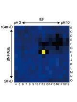
Lipid content can be measured by two-dimensional thin-layer chromatography of a lipid extract, as described in Kirchhoff et al. (2018) .
The protein content can be determined from a spectroscopic chlorophyll quantification of an 80% acetone extract, according to Porra et al. (1989) . For the 80% acetone extract, use a 100 μl sample and dilute in 400 μl of 100% acetone. Calculate total chlorophyll concentration in nmol/ml according to Porra et al. (1989) . Use an appropriate spectrometer cuvette that can handle 500 μl samples. For calculation of the LHCII-trimer concentration from total chlorophyll, assume a total chlorophyll to LHCII-trimer ratio of 42 to 1 (Barros and Kühlbrandt, 2009).
Characterization by low temperature (77 K) fluorescence spectroscopy (see Tietz et al., 2020)
Prepare the fluorescence spectrometer for emission scan between 640 and 800 nm, with a 475 nm excitation wavelength and optical bandwidth of 4 nm.
Fill Dewar carefully with liquid nitrogen (wear protection goggles).
Mix in Eppendorf cup by careful pipetting 346 μl LHCII-proteoliposomes, 2 μl catalase, 2 μl glucose oxidase, and 10 μl glucose; wait 5 min to establish anaerobic conditions (see Note 4).
Transfer the sample into the 77 K sample tube and freeze carefully by dipping into Dewar filled with liquid N2 (wear protection goggles). Place the frozen sample in the 77 K sample tube very slowly in 77 K Dewar (sample tubes break if moved in too fast). Adjust test tube position in the fluorimeter beam if required.
Record and average four emission scans.
Notes
The suspension that is left over can be shock frozen in liquid nitrogen and stored at -80°C for another ultracentrifugation.
LHCII is a very stable protein and can be stored for many months at -80°C. Thawed aliquots can be flash-frozen several times.
Due to some variability in LHCII incorporation into LUVs, it might be required to adjust the detergent concentration. This can be judged based on low-temperature fluorescence spectra. If significant LHCII aggregation is seen (shoulder at 700 nm), the detergent concentration should be increased (for example, by 10%). See Tietz et al. (2020) for details. If free LHCII is visible (emission at ~650 nm with excitation at 475 nm), the detergent concentration should be lowered (e.g., by 10%).
Anaerobic conditions minimize damage by reactive oxygen production that drastically lead to distortion of fluorescence spectra during fluorimetry.
Recipes
LHCII isolation
Stock solution
1 mM MgCl2 + 1 mM MnCl2 + 0.5 mM KH2PO4 + 10 mM KCl + 2 mM EDTA + 330 mM sorbitol in 2 L ultrapure water.
Solution A
25 mM MES + 40 mM KCl in 1 L stock solution. Adjust pH to 6.1 (KOH)
Solution B
25 mM HEPES in 200 ml stock solution. Adjust to pH 6.7 (KOH)
Solution NET
10 mM KCl + 1 mM Tricin + 5 mM EDTA in 500 ml ultrapure water. Adjust to pH 7.8 (KOH)
Solution SE
100 mM sorbitol + 5 mM EDTA in 250 ml ultrapure water. Adjust to pH ca 7 (KOH)
Solution S
100 mM sorbitol in 200 ml ultrapure water. No pH adjustment
0.1 M and 1 M HCl
Sucrose gradient (with gradient mixer) 0.1 M to 1 M sucrose; 6 tubes:
100 ml 0.1 M sucrose + 0.05% Triton X-100 (add freshly)
100 ml 1 M sucrose + 0.05% Triton X-100 (add freshly)
Solution KCl
100 mM KCl in 200 ml ultrapure water. No pH adjustment
LHCII proteoliposome preparation
20 ml 10 mM HEPES in ultrapure water. Adjust to pH 6.7 (KOH)
73.6 mM alpha-DM solubilized in HEPES buffer
Acknowledgments
The work was supported by grants from the US Department of Energy (DE-SC0017160), the National Science Foundation (MCB-1953570), and USDA-NIFA (#1005351 and #0119). This protocol was derived from Tietz et al. (2020; doi: 10.1074/jbc.RA119.011707).
Competing interests
The corresponding author declares no competing interests.
References
- Anishkin, A., Loukin, S. H., Teng, J. and Kung, C. (2014). Feeling the hidden mechanical forces in lipid bilayer is an original sense. Proc Natl Acad Sci USA 111: 7898-7905.
- Barros, T. and Kühlbrandt, W. (2009). Crystallisation, structure and function of plant light-harvesting complex II. Biochim Biophys Acta 1787: 753-772.
- Boudiere, L., Michaud, M., Petroutsos, D., Rebeille, F., Falconet, D., Bastien, O., Roy, S., Finazzi, G., Rolland, N., Jouhet, J., Block, M. A. and Marechal, E. (2014). Glycerolipids in photosynthesis: Composition, synthesis and trafficking. Biochim Biophys Acta 1837: 470-480.
- Cantor, R. S. (1997). Lateral pressure profiles in cell membranes: A mechanism for modulation of protein function. J Phys Chem B 101: 1723-1725.
- Crisafi, E. and Pandit, A. (2017). Disteangling protein and lipid interactions that control a molecular switch in photosynthetic light harvesting. Biochm Biophys Acta 1859: 40-47.
- Hara, A. and Radin, N. S. (1978).Lipid extraction of tissues with a low-toxicity solvent. Analytical Biochem 90: 420-426.
- Kirchhoff, H. and Yarbrough, R. (2018). Evaluation of lipids and membranes for the study of photosynthetic superorganization. In: Methods in Molecular Biology. Covshoff, S. (Ed.). 1770, Springer Press, 305-316.
- Kotapati, H. K. and Bates, P. D. (2020).Normal phase HPLC method for combined separation of both polar and neutral lipid classes with application to lipid metabolic flux. J Chromatogr B Analy Technol Biomed Life Sci 1145: 122099.
- McDonnel, A. and Staehelin, L. A. (1980). Adhesion between liposomes mediated by the chlorophyll a/b light-harvesting complex isolated from chloroplast membranes. J Cell Biol 84: 40-56.
- Mondal, S., Khelashvili, G. and Weinstein, H. (2014). Not just an oil slick: How the energetics of protein-membrane interactions impacts the function and organization of tranmembrane proteins. Biophys J 106: 2305-2316.
- Mourtisen, O. G. and Bloom, M. (1993). Models of lipid-protein interactions in membranes. Annu Rev Biophys Biomol Struct 22: 145-171.
- Moya, I., Silvestri, M., Vallon, O., Cinque, G. and Bassi, R. (2001). Time-resolved fluorescence analysis of the photosystem II antenna proteins in detergent micelles and liposomes. Biochemistry 40: 12552-12661.
- Natali, A., Gruber, J.M., Dietzel, L., Stuart, M.C.A., van Grondelle, R. and Croce, R. (2016). Light-harvesting complexes (LHCs) cluster spontaneously in membrane environment leading to shortening of their excited state lifetimes. J Bio Chem 291: 16730-16739.
- Porra, R. J. (1989). Determination of accurate extinction coefficients and simultaneous equations for assaying chlorophylls a and b extracted with four different solvents: verification of the concentration of chlorophyll standards by atomic absorption spectroscopy Biochim. Biophys Acta 975: 384-394
- Rigaud, J. L. and Levy, D. (2003). Reconstitution of membrane proteins into liposomes. Methods Enzymol 372: 65-86.
- Rigaud, J.L., Pitard, B., Levy, D. (1995). Reconstitution of membrane proteins into liposomes: application to energy-transducing membrane proteins. Biochim Biophys Acta 1231: 223-246.
- Tietz, S., Leuenberger, M., Höhner, R., Olson, A.H., Fleming, G.R., Kirchhoff, H. (2020). A proteoliposome-based system reveals how lipids control photosynthetic light harvesting. J Biol Chem 295: 1857-1866.
- van den Brinck-van der Laan, E., Killian, J. A. and de Kruijff, B. (2004). Nonbilayer lipids affect peripheral and integral membrane proteins via changes in the lateral pressure profile. Biochim Biophys Acta 1666: 275-288.
- Yang, C., Boggasch, S., Haase, W. and Paulsen, H. (2006). Thermal stability of trimeric light-harvesting chlorophyll a/b (LHCIIb) in liposomes of thylakoid lipids. Biochim Biophys Acta 1757: 1642-1648.
Article Information
Copyright
© 2021 The Authors; exclusive licensee Bio-protocol LLC.
How to cite
Readers should cite both the Bio-protocol article and the original research article where this protocol was used:
- Kirchhoff, H. (2021). Proteoliposomes for Studying Lipid-protein Interactions in Membranes in vitro. Bio-protocol 11(20): e4197. DOI: 10.21769/BioProtoc.4197.
- Tietz, S., Leuenberger, M., Höhner, R., Olson, A.H., Fleming, G.R., Kirchhoff, H. (2020). A proteoliposome-based system reveals how lipids control photosynthetic light harvesting. J Biol Chem 295: 1857-1866.
Category
Plant Science > Plant biochemistry > Protein > Isolation and purification
Plant Science > Plant physiology > Metabolism
Biochemistry > Other compound
Do you have any questions about this protocol?
Post your question to gather feedback from the community. We will also invite the authors of this article to respond.