- Protocols

- Articles and Issues
- For Authors
- About
- Become a Reviewer

Dissection and Staining of Mouse Brain Ventricular Wall for the Analysis of Ependymal Cell Cilia Organization
Published: Vol 6, Iss 6, Mar 20, 2016 DOI: 10.21769/BioProtoc.1757 Views: 15689
Reviewed by: Pamela MaherOneil G. BhalalaAnonymous reviewer(s)
Original research article
The authors used this protocol in:
Jul 2014
Advertisement

Protocol Collections
Comprehensive collections of detailed, peer-reviewed protocols focusing on specific topics


Abstract
In the developing and mature central nervous system (CNS) the ventricular lumen is lined by the neuroepithelium and ependymal, respectively. These ventricular epithelia perform important functions related to the development, morphogenesis and physiology of the brain. In the mature CNS, ependyma constitutes a barrier between brain parenchyma and cerebro- spinal fluid (CSF). The most prominent feature of the apical surface of ependymal cells is the presence of multiple motile cilia that extend towards the ventricular lumen. The beating of cilia ensures the circulation of the CSF and its impairment leads to hydrocephalus. For an effective CSF flow, ciliary beating must be coordinated at the level of individual cells and at the tissue level. This coordination is achieved through the precise organization of cilia positioning within the plane of the ependyma. Two major features have been described regarding the planar organization of cilia in ependymal cells (Mirzadeh et al., 2010) and both have a cellular and tissular aspect (Boutin et al., 2014). The first one, rotational polarity, refers to the orientation of ciliary beating. At the cellular level, all cilia beat in the same direction (Figure 1B, black arrows). At the tissue level, each ependymal cell coordinates the direction of their beating with that of neighboring cells (Figure 1C, grey arrows). The second feature, translational polarity, is unique to ependymal cells and refers to the clustering of cilia in a tuft. At the cellular level, this tuft is displaced relative to the center of the ependymal cell (Figure 1B, red arrow). At the tissue level, the positioning of the ciliary tuft is coordinated between adjacent cells (Figure 1C). Alteration of any of these polarities at either level impairs CSF flow circulation (Mirzadeh et al., 2010; Boutin et al., 2014; Guirao et al., 2010; Hirota et al., 2010; Ohata et al., 2014). Cilia axonemes arise from basal bodies (BB) which are cylindrical structures anchored perpendicular to the sub-apical surface of the cells (Figure 1D). BBs are polarized by the presence of appendices such as basal foot or striated rootlets. The basal foot protrudes in a direction correlated with the direction of cilia beating, while the striated rootlet protrudes in the opposite direction of cilia beating (Marshall, 2008). The ‘en face view’ observation of BBs’ organization allows the visualization of ependymal polarities (Mirzadeh et al., 2010; Boutin et al., 2014). Here, we describe an immunofluorescence (IF) protocol for observation of ciliated cells in mouse brain ventricular lateral wall whole mounts (LWWM). This protocol can be used for classical confocal microscopy analysis. In addition, it is well suited for super-resolution STimulated Emission Depletion (STED) microscopy if observation of structures that have features which are smaller than the optical diffraction limit is needed. Finally, we describe a combination of antibodies that allow the concomitant observation, in a single sample, of ependymal polarities at the level of individual cilia, individual cells and at the tissue level.
Materials and Reagents
- Microscope slide
- Coverslips
- Mice at desired stage [between postnatal day 1 (P1) and 21 (P21)]
- 1x PBS
- Triton X-100
- 4% Paraformaldehyde (PFA) (prepared by dilution of powder in 1x PBS)
- Bovine Serum Albumin (BSA) (Sigma-Aldrich, catalog number: A-8022 )
- Hoechst 33258 solution (Sigma-Aldrich, catalog number: 94403 )
- Mowiol (Merck Millipore Corporation, Calbiochem®, catalog number: 475904 )
- Prolong Gold antifade reagent (Thermo Fisher Scientific, Molecular ProbeTM, catalog number: P36934 )
- ZO1 (Thermo Fisher Scientific, InvitrogenTM, catalog number: 61-7300 )
- γ-tubulin [GTU-88] (Abcam, catalog number: ab11316 )
- FGFR1OP (FOP) (Abnova Corporation, catalog number: H00011116-M01 )
- β-catenin (BD, catalog number: 610153 )
- FoxJ1 (eBioscience, catalog number: 14-9965 )
- Acetylated-α-tubulin (Sigma-Aldrich, catalog number: T6793 )
- Secondary antibodies
- Primary and secondary antibodies (see Recipes)
Equipment
- Dissection tools including forceps (Zillow, Dumont, model: 55 ) and Ultra Fine Micro Knives (Fine Science Tools, catalog number: 10316-14 )
- Confocal microscope (OLYMPUS, model: Fluoview FV1000 )
- STED microscope (Leica Microsystems, model: SP8 3x STED ) equipped with a 100x oil objective (HC-PL-APO; NA 1.40; STED white) and a 592 nm depletion laser
Software
- Biotool software (Boutin et al., 2014)
Procedure
The general procedure described here is suitable for staining ependymal cells from lateral wall whole mounts of P1 to P21 mice. However, the last sub-dissection step of the tissue has to be performed before the staining for mice older than P5 and after the staining for mice younger than P5 to ensure an optimal result.
- After cervical dislocation of the mice, the brain is removed from the skull.
- Dissection of the brain at RT in 1x PBS to reveal the ventricular lateral wall (Figure 2) [a protocol including a video is provided in Mirzadeh et al. (2010)].
- Fixation of the whole mount by immersion in 500 μl fresh solution of 4% PFA-0.1% Triton X-100 12 min at room temperature (RT).
- Wash 3 times 10 min in 1x PBS-0.1% Triton X-100 (1 ml) at RT with agitation.
- Sub-dissection of the lateral wall by separating it from the underlying striatum [see Mirzadeh et al. (2010) for a precise description of this step]. Performing this sub-dissection at this step of the protocol improves the staining for tissues older than P5.
- Incubation in blocking solution 1x PBS-3% BSA (1 ml) 1 h RT with agitation.
- Incubation with primary antibody diluted in 1x PBS-3% BSA (250-500 μl) over-night RT with agitation.
- Wash 3 times 1x PBS -0.1% Triton X-100 (1 ml) 10 min RT with agitation.
- Incubate with secondary antibody (1:800) and Hoechst (1:1,000) diluted in 1x PBS (For confocal observation of samples) OR with secondary antibody (1:800) diluted in 1x PBS only (for STED observation of samples) (800 μl) 1 h RT with agitation.
- Wash 3 times 1x PBS-0.1% Triton X-100 (1 ml) 10 min RT with agitation.
- For tissues younger than P5: sub-dissection of the LWWM by separating the lateral wall and the underlying striatum in 1x PBS [see Mirzadeh et al. (2010)].
- Place the whole mount on the slide with ependymal face up.
- Add 7-8 drops of Mowiol (for confocal observation of samples) or of Prolong mounting medium (for STED observation of samples) directly on the whole mount.
- Place a coverslip on the sample.
- Keep slides at room temperature, protected from the light, for at least 24 h before imaging.
Representative data

Figure 1. Planar organization of ependymal cells in the lateral wall. A. Schematic representation of the mouse forebrain depicting the localization of the lateral wall of the lateral ventricle. Dashed square highlights the region of interest considered for analysis of ependymal planar polarity. B-C. Schemes represent the planar organization of ependymal cells in the region of interest. B. At the cellular level, ependymal cilia are clustered in a tuft (dotted red line) which localize off center of the apical surface (red arrow). Ciliary beatings (small black arrows) are coordinated. The sum of individual ciliary beatings generates the tuft beating direction (Thick grey arrow). In individual cells, the direction of tuft positioning correlates with the direction of tuft beating (C). At the tissue level, beating direction (grey arrows) and positioning of the ciliary tuft (red arrows) are coordinated between neighboring ependymal cells. D-E. Schematic representation of the lateral (D) and ‘en face’ (E) view of individual ependymal cell organization. D. Multicilia axonemes arise from basal bodies (BBs) anchored perpendicularly to the apical surface of the cells (green). BBs display polarized appendices such as the basal foot (red dot) that indicates the effective beating direction of the cilium (small black arrow). E. Observation of cilia BBs from a top view reveals all ependymal polarity features.
Figure 2. Dissection steps for LWWM preparation. LWWM preparations allows the “en face” observation of ependymal cells. A. Brain after removal from the skull. B. Cut the brain between the two hemispheres (dashed red line) using a microscalpel. C. Remove the cerebellum (dashed red line). D-F. Use a microscapel to reveal the hippocampus (dashed red line). G-H. Insert the microscalpel in the ventricle (arrow) to remove the hippocampus and medial ventricular wall. I. Remove cortical wall following the dashed red line. J. Whole mount ready to be fixed, the lateral wall is outlined by the dashed red line. 
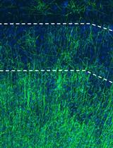
Figure 3. Examples of staining on P1, P4, P5 and P21 LWWM Confocal (A-E) and STED (F) images. LWWM stained for A. ZO1 (green) and Acetyl-α- tubulin (red) at P1. B. FoxJ1 (green) and DAPI (blue) at P4. C. β-catenin (green) and Acetyl-α- tubulin (red) at P21. D. β-catenin (green) and ɣ-tubulin (red) at P21. E-F. FGFR1 Oncogene Partner (FOP) at P5. Scale bar: 10 μm in A-B, 15 μm in C-D and 2.5 μm in E-F.
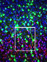
Figure 4. Analysis of planar organization of ependymal cells in the lateral wall (A, B, C). Triple immunostaining for ZO1 (white), FGFR1 Oncogene Partner (FOP, green) and γ-tubulin (red) on P21 LWWM allows the observation of translational and rotational polarities at the cellular and tissular level in a single sample. A. Large field picture displaying the tissue organization. B. Zoom on the cell marked with * in (A). C. Zoom on the BBs patch from cell shown in (B). Analysis of ZO1/FOP/γ-tubulin triple staining using Biotool1 software [described in Boutin et al. (2014)] allows the definition of polarity parameters at BB, cell and tissue level. C’. Definition of individual cilia polarity: each black vector represents the rotational polarity axis of individual BB defined as the direction from FOP (green) to γ-tubulin (red) positive dots. The biotool software calculates the mean BB orientation of the patch to define the cellular rotational polarity axis that corresponds to the beating direction of individual cell (grey arrow). B’-B”. Definition of polarity axis at the cellular level: Outline of cell (green) and BB patch (red). The grey vector in (B’) represents the rotational polarity axis of the cell defined as shown in C’. The red arrow in (B’’) represents the translational polarity axis of the cell defined as direction from the cell center (green cross) to the BB patch center (red cross). A’-A”. Analysis of polarity at the tissue level: The translational and rotational polarity axes are defined for each cell and compared to mean direction of the field (black and red thick arrows). Scale bar: 15 μm in A, 2.5 μm in B and 1 μm in C.
Notes
- Freshly made 4% PFA aliquots can be stored at -20 °C up to one year and defrosted just before fixation.
- Short fixation time is mandatory for good staining. 12 min fixation is sufficient to fix large and small tissues while preserving the structure and giving the best results. We recommend not to exceed 20 min fixation for ependymal observation.
Recipes
- Primary antibodies with the corresponding secondary antibodies
Table 1. List of primary and secondary antibodies
Antibody Isotype Reference Dilution Secondary antibody ZO1 Rabbit Invitrogen 61-7300 1:600 Anti-Rabbit A647 γ-tubulin [GTU-88] Mouse IgG1 Abcam ab11316 1:400 Anti-Mouse IgG1 A568 FGFR1OP (FOP) Mouse IgG2b Abnova H00011116-M01 1:2,000 Anti-Mouse IgG2b A488 β-catenin Mouse IgG1 BD Transduction Laboratories 610153 1:1,000 Anti-Mouse IgG1 A488 FoxJ1 Mouse IgG1 eBioscience 14-9965 1:2,000 Anti-Mouse IgG1 A488 Acetylated-α- tubulin Mouse IgG2b Sigma T6793 1:1,000 Anti-Mouse IgG2b A568
Acknowledgments
The method described in this article has been optimized for detection of ependymal cells organization and is based on a protocol described previously (Mirzadeh et al., 2010). The original version of the protocol has been described in (Boutin et al., 2014). The authors wish to thank the Spassky lab (ENS, Paris) for introduction to the LWWM dissection method. CB is a recipient of a postdoctoral fellowship from ‘La Ligue Nationale Contre le Cancer’. HC acknowledges funding from the Agence Nationale de la Recherche (ANR-13-BSV4-0013-01-ATMIR). This work was supported by the French National Research Agency through the "Investments for the Future" program (France-BioImaging, ANR-10-INSB-04).
References
- Boutin, C., Labedan, P., Dimidschstein, J., Richard, F., Cremer, H., Andre, P., Yang, Y., Montcouquiol, M., Goffinet, A. M. and Tissir, F. (2014). A dual role for planar cell polarity genes in ciliated cells. Proc Natl Acad Sci U S A 111(30): E3129-3138.
- Guirao, B., Meunier, A., Mortaud, S., Aguilar, A., Corsi, J. M., Strehl, L., Hirota, Y., Desoeuvre, A., Boutin, C., Han, Y. G., Mirzadeh, Z., Cremer, H., Montcouquiol, M., Sawamoto, K. and Spassky, N. (2010). Coupling between hydrodynamic forces and planar cell polarity orients mammalian motile cilia. Nature Cell Biology 12(4): 341-U386.
- Hirota, Y., Meunier, A., Huang, S., Shimozawa, T., Yamada, O., Kida, Y. S., Inoue, M., Ito, T., Kato, H., Sakaguchi, M., Sunabori, T., Nakaya, M. A., Nonaka, S., Ogura, T., Higuchi, H., Okano, H., Spassky, N. and Sawamoto, K. (2010). Planar polarity of multiciliated ependymal cells involves the anterior migration of basal bodies regulated by non-muscle myosin II. Development 137(18): 3037-3046.
- Marshall, W. F. (2008). Basal bodies platforms for building cilia. Curr Top Dev Biol 85: 1-22.
- Mirzadeh, Z., Doetsch, F., Sawamoto, K., Wichterle, H. and Alvarez-Buylla, A. (2010). The subventricular zone en-face: wholemount staining and ependymal flow. J Vis Exp(39).
- Mirzadeh, Z., Han, Y. G., Soriano-Navarro, M., Garcia-Verdugo, J. M. and Alvarez-Buylla, A. (2010). Cilia organize ependymal planar polarity. J Neurosci 30(7): 2600-2610.
- Ohata, S., Nakatani, J., Herranz-Perez, V., Cheng, J., Belinson, H., Inubushi, T., Snider, W. D., Garcia-Verdugo, J. M., Wynshaw-Boris, A. and Alvarez-Buylla, A. (2014). Loss of Dishevelleds disrupts planar polarity in ependymal motile cilia and results in hydrocephalus. Neuron 83(3): 558-571.
Article Information
Copyright
© 2016 The Authors; exclusive licensee Bio-protocol LLC.
How to cite
Labedan, P., Matthews, C., Kodjabachian, L., Cremer, H., Tissir, F. and Boutin, C. (2016). Dissection and Staining of Mouse Brain Ventricular Wall for the Analysis of Ependymal Cell Cilia Organization. Bio-protocol 6(6): e1757. DOI: 10.21769/BioProtoc.1757.
Category
Neuroscience > Development > Explant culture
Neuroscience > Development > Immunofluorescence
Do you have any questions about this protocol?
Post your question to gather feedback from the community. We will also invite the authors of this article to respond.