- Submit a Protocol
- Receive Our Alerts
- Log in
- /
- Sign up
- My Bio Page
- Edit My Profile
- Change Password
- Log Out
- EN
- EN - English
- CN - 中文
- Protocols
- Articles and Issues
- For Authors
- About
- Become a Reviewer
- EN - English
- CN - 中文
- Home
- Protocols
- Articles and Issues
- For Authors
- About
- Become a Reviewer
Thioglycollate-elicited Peritoneal Macrophages Preparation and Arginase Activity Measurement in IL-4 Stimulated Macrophages
Published: Vol 5, Iss 17, Sep 5, 2015 DOI: 10.21769/BioProtoc.1585 Views: 22923
Reviewed by: Ivan ZanoniSabine Le SauxAnonymous reviewer(s)

Protocol Collections
Comprehensive collections of detailed, peer-reviewed protocols focusing on specific topics
Related protocols

In vitro Assessment of Efferocytic Capacity of Human Macrophages Using Flow Cytometry
Ana C.G. Salina [...] Larissa D. Cunha
Dec 20, 2023 5145 Views

Quantification of Macrophage Cellular Ferrous Iron (Fe2+) Content using a Highly Specific Fluorescent Probe in a Plate-Reader
Philipp Grubwieser [...] Christa Pfeifhofer-Obermair
Feb 5, 2024 2212 Views

Novel Experimental Approach to Investigate Immune Control of Vascular Function: Co-culture of Murine Aortas With T Lymphocytes or Macrophages
Taylor C. Kress [...] Eric J. Belin de Chantemèle
Sep 5, 2025 3543 Views
Abstract
Macrophages are an essential cell population of innate immunity that plays important roles in inflammatory processes. Two main different phenotypes have been described with opposing activities: The classically activated macrophages (M1) and the alternatively activated macrophages (M2). Alternative activation of mouse macrophages can be induced by type 2 cytokines such as IL-4 and it is characterized by the regulation of the L-arginine metabolism. M2 macrophages convert arginine to ornithine and urea through the action of Arginase-1. Here we described a method for the isolation of peritoneal macrophages from thioglycollate-elicited mice and alternative activation by stimulation with IL-4. Intraperitoneal injection of thioglycollate elicits large numbers of macrophages into peritoneal cavity.
Keywords: MacrophagesMaterials and Reagents
- Mice C57BL/6 male (age 8-12 weeks)
- Difco Fluid Thioglycollate medium (BD, catalog number: 225650 )
- 70% and 100% ethanol
- PBS (Lonza, catalog number: BE 17-515Q )
- RPMI 1640 (Lonza, catalog number: BE 12-115F )
- FBS (endotoxin<10 EU/ml) (Hyclone, catalog number: SV30160.03 )
- Penicillin-Streptomycin (Lonza, catalog number: BE 17-602E )
- Murine IL-4 (Peprotech, catalog number: 214-14 )
- Tris (Panreac Applichem, catalog number: 131940.1211 )
- NaCl (Merck KGaA, catalog number: 1.06404.1000 )
- EDTA (Sigma-Aldrich, catalog number: ED255 )
- Triton X-100 (Sigma-Aldrich, catalog number: 8787 )
- Protease inhibitor mixture (Sigma-Aldrich, catalog number: P8340 )
- Urea (Sigma-Aldrich, catalog number: 4883 )
- MnCl2 (Sigma-Aldrich, catalog number: 244589 )
- L-arginine monohydrochloride (Sigma-Aldrich, catalog number: A6969 )
- H2SO4 (Sigma-Aldrich, catalog number: 320501 )
- H3PO4 (Sigma-Aldrich, catalog number: 345245 )
- Isonitrosopropiophenone (Sigma-Aldrich, catalog number: I3502 )
- 12-well plates
- 1.5 ml Eppendorf tubes
- 50 ml sterile Falcon tubes
- Cell strainer (Becton Dickinson, catalog number: 352340 )
- 10 ml syringe
- 21G Needles
- 25G needles
- Lysis buffer (see Recipes)
- Heat-inactivated FBS (see Recipes)
- Stopping solution (see Recipes)
Equipment
- Bench-top refrigerated centrifuge
- Scissors
- Incubator (37 °C, 5% CO2 and 95 % humidity)
- Tissue culture hood (biosafety cabinet)
- Optical microscopy
- Neubauer cell counting chamber
- Espectrophotometer plate reader
- Thermoblock (Eppendorf Thermomixer Compact) or water bath
Procedure
- Injection of thyoglicollate into the peritoneum
- Preparation of 3% thioglycollate medium.
- Suspend 30 grams of thioglycollate medium in 1,000 ml of pyrogen-free water.
- Aliquot to 100 ml sterile bottles.
- Autoclave (15 psi/121 °C/15 min).
- After cooling, store in a dark place at room temperature for 2 months before using.
Note: Thioglycollate solution needs to age for several weeks until it turns to brown in color. This process is important to increase the extravasation of peritoneal cells.
- Suspend 30 grams of thioglycollate medium in 1,000 ml of pyrogen-free water.
- Inject 2.5 ml of 3% thioglycollate medium i.p. per mouse using a 10 ml syringe with a 25 G needle. Wait for 4 days and harvest peritoneal cells (see step B).
- Preparation of 3% thioglycollate medium.
- Isolation and culture of thioglycollate elicited peritoneal macrophages
- Sacrifice mouse with CO2 or isofluorane (the use of cervical dislocation is not indicated in this protocol in order to avoid potential internal bleeding).
- Clean abdomen with 70% ethanol.
- Remove skin to expose the peritoneal wall. Practice a small incision in the skin and pull firmly.
- Inject 10 ml of RPMI 1640 into the peritoneal cavity with a 25 G needle.
- Massage abdomen for approximately 30 sec.
- Recover peritoneal fluid as much as possible using a 10 ml syringe with a 21 G needle. (Usually approximately 8-10 ml fluid can be recovered from one mouse.)
- Remove needle from syringe and put fluid into a 50 ml conical centrifuge tube on ice.
- Centrifuge peritoneal cells 5 min at 300 x g (1,500 rpm, 4 °C). Discard the supernatant and collect the pellet.
- Resuspend cell pellet in 10 ml of RPMI 1640 medium and count cells. (Usually approximately 30-50 million cells can be recovered from one mouse.)
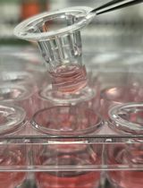
- Culture peritoneal cells (1 x 10⁶/cm2) in a 12-well plate in 1 ml of RPMI containing 10% heat-inactivated FBS at 37 °C with 5% CO2 for 3 h.
- Remove non-adherent cells by extensive washing with RPMI containing 10% heat-inactivated FBS at 37 °C.
- Proceed to overnight cell starvation in the presence of 1 ml of RPMI 2% FBS per well.
- Sacrifice mouse with CO2 or isofluorane (the use of cervical dislocation is not indicated in this protocol in order to avoid potential internal bleeding).
- M2 activation of peritoneal macrophages
- After cell starvation, maintain the cells in RPMI 2% FBS and stimulate macrophages by adding IL-4 (20 ng/ml) to the 12-well plate and incubate at 37 °C for 24 h.
- Arginase activity measurement
- For cell dissociation, wash cells once with PBS and add an appropriate volume of lysis buffer at RT (see Recipes).
Note: Usually 200 μl in each 12-well plate.
- Remove cells with a cell scraper, collect them into a 1.5 ml Eppendorf and pipette up and down 5 times with a 200 µl tip for complete suspension.
- Lyse the cells for 15 min at 4 °C.
- Then spin down cell lysates at full speed (10 min, 14,000 rpm, 16,800 x g) at 4 °C and isolate the supernatant. Store at 4 °C before use.
- Meanwhile, prepare a stock of 8 M urea in 50 mM Tris-HCl (pH 7.5).
- Dilute stock of urea in 50 mM Tris-HCl (pH 7.5) to yield a standard range from 25 to 1,500 μg/ml. (e.g. 25, 50 100, 250, 500, 1,000 and 1,500 μg/ml)
- Transfer 50 μl of cell lysates and standards to a 2 ml Eppendorf tube and add 50 μl of 10 mM MnCl2 diluted in 50 mM Tris-HCl (pH 7.5).
- Incubate tubes in a thermoblock or water bath for 10 min at 55 °C to trigger arginase-1 activity.
- Then add 50 μl of 0.5 M L-arginine to the tubes and incubate in a thermoblock or water bath at 37 °C for 60 min. This step induces arginine hydrolysis.
- Stop the reaction by adding 400 μl of stopping solution (H2SO4/H3PO4/H2O = 1/3/7, v/v/v).
- Next, add 50 μl of 9% isonitrosopropiophenone in 100% ethanol to each sample and standard, and incubate the tubes in a thermoblock at 100 °C for 60 min.
- Place the tubes in the dark at RT for 30 min.
- Transfer 100 μl/well of samples and standards in triplicate to a 96-well plate and read optical density at 540 nm with a 690 nm correction.
- Calculate sample concentrations from the standard curve and converted to Arginase Units using the following formula: [Urea Produced (μg/ml)/Total Protein (μg/ml)].
- For cell dissociation, wash cells once with PBS and add an appropriate volume of lysis buffer at RT (see Recipes).
Recipes
- Lysis buffer
20 mM Tris (pH 7.5)
150 mM NaCl
2 mM EDTA
0.1% Triton X-100
Protease inhibitor mixture 1 μg/ml
- Heat-inactivated FBS
Inactivate FBS at 55 °C for 30 min
Stored in aliquots at -20 °C
- Stopping solution (for 11 ml)
H2SO4 1 ml
H3PO4 3 ml
H2O 7 ml
Acknowledgments
This study was supported by grant PI11.0036 from the FIS and MPY 1410/09 from ISCIII to SH, and by grant TPY-M-1068/13 to AL. AL was supported by MINECO-ISCIII, through the Miguel Servet Programme (CP12/03087). L. J-G. was supported by FIS (FI12/00340). This protocol is adapted from Jimenez-Garcia et al. (2015).
Note: The study was performed under an ISCIII approved protocol.
References
- Jiménez-García, L., Herránz, S., Luque, A. and Hortelano, S. (2015). Critical role of p38 MAPK in IL-4-induced alternative activation of peritoneal macrophages. Eur J Immunol 45(1): 273-286.
Article Information
Copyright
© 2015 The Authors; exclusive licensee Bio-protocol LLC.
How to cite
Jiménez-García, L., Herránz, S., Luque, A. and Hortelano, S. (2015). Thioglycollate-elicited Peritoneal Macrophages Preparation and Arginase Activity Measurement in IL-4 Stimulated Macrophages. Bio-protocol 5(17): e1585. DOI: 10.21769/BioProtoc.1585.
Category
Immunology > Immune cell function > Macrophage
Do you have any questions about this protocol?
Post your question to gather feedback from the community. We will also invite the authors of this article to respond.
Share
Bluesky
X
Copy link


