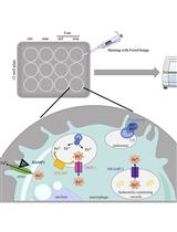
Chitin is a key component of insects, fungi, and house-dust mites. Chitin has been shown to induce M2-type immune responses in vivo. Intranasal or intraperitoneal (i.p.) administration of chitin particles results in infiltration of eosinophils to the local sites and activation of macrophages with a M2 phenotype. Chitin-challenged mice model can be used to induce M2 macrophages polarization and thus to analyze the M2 phenotype from isolated peritoneal cells.
Weight 800 ng of chitin per mouse and wash 3 times with PBS (5 min, 13,000 rpm, 16,800 x g).
Suspend chitin in 1 ml of PBS and sonicate in bath for 1 h [at the maximum setting of the sonicator (Ultrasons, Selecta)]. Note: Alternatively chitin can be sonicated at 25% output power three times for 5 min with a Branson sonicator.
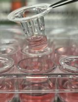
Filter the suspension with 70 μm sterile cell strainers.
Inject 1 ml of chitin suspension i.p. per mouse using a 10 ml syringe with a 25 G needle. Wait for 2 days and harvest peritoneal cells.
Isolation of chitin elicited peritoneal exudate cells (PECs)
Sacrifice mouse with CO2 or isofluorane (the use of cervical dislocation is not indicated in this protocol in order to avoid potential internal bleeding).
Clean abdomen with 70% ethanol.
Remove skin to expose the peritoneal wall. Practice a small incision in the skin and pull firmly.
Inject 10 ml of RPMI 1640 into the peritoneal cavity with a 25 G needle.
Massage abdomen for approximately 30 sec.
Recover peritoneal fluid as much as possible using a 10 ml syringe with a 21G needle. (Usually approximately 8-10 ml fluid can be recovered from one mouse.)
Remove needle from syringe and put fluid into a 15 ml conical centrifuge tube on ice.
Centrifuge peritoneal cells 5 min at 300 x g (1,500 rpm, 4 °C). Discard the supernatant and collect the pellet.
Resuspend cell pellet in 10 ml of RPMI 1640 medium and count cells. (The cell number expected to be recovered is about 5-8 millions.)
Analyze peritoneal cells to confirm M2 activation by performing either real-time PCR or flow cytometry techniques to detect typical M2 markers (e.g. Arginase-1, Ym-1, Fizz-1, mannose receptor).
Acknowledgments
This study was supported by grant PI11.0036 from the FIS and MPY 1410/09 from ISCIII to SH, and by grant TPY-M-1068/13 to AL. AL was supported by MINECO-ISCIII, through the Miguel Servet Programme (CP12/03087). L. J-G. was supported by FIS (FI12/00340). This protocol is adapted from Jimenez-Garcia el at. (2015).
Jiménez-García, L., Herránz, S., Luque, A. and Hortelano, S. (2015). Chitin-challenged Mice Model to Study M2 Macrophages Polarization. Bio-protocol 5(17): e1584. DOI: 10.21769/BioProtoc.1584.
Post your question to gather feedback from the community. We will also invite the authors of this article to respond.
0/150
Tips for asking effective questions
+ Description
Write a detailed description. Include all information that will help others answer your question including experimental processes, conditions, and relevant images.