- Protocols

- Articles and Issues
- For Authors
- About
- Become a Reviewer

Past Issue in 2014
Volume: 4, Issue: 11
Biochemistry
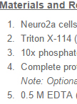
Small-scale Triton X-114 Extraction of Hydrophobic Proteins

Protocol for Preparation of Nuclear Protein from Mouse Lungs
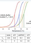
Determination of Pseudokinase-ligand Interaction by a Fluorescence-based Thermal Shift Assay

Cell Biology
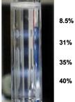
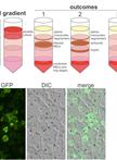
Small-scale Subcellular Fractionation with Sucrose Step Gradient
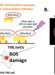
Novel Method for Site-specific Induction of Oxidative DNA Damage to Study Recruitment of Repair Proteins to Heterochromatin and Euchromatin


Immunology
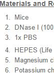
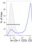
Protocol for Macrophage Depletion from Mice
In vitro Inflammasome Assay

Identification of Helminth-induced Type 2 CD4+ T Cells and ILC2s
Microbiology
Preparation of Parasite Protein Extracts and Western Blot Analysis
Intracellular Glycogen Assays

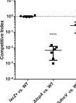
Infant Rabbit Colonization Competition Assays

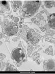
Immuno-EM Analysis of PF13_0191-GFP Expressing Parasites
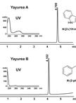
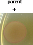
Purification and Structural Analysis of QS-inhibiting Compounds from Staphylococcus delphini
Stem Cell
Competitive Bone-marrow Transplantations