Past Issue in 2014

Volume: 4, Issue: 10

left

Dec

20

Dec

5

Nov

20

Nov

5

Oct

20

Oct

5

Sep

20

Sep

5

Aug

20

Aug

5

Jul

20

Jul

5

Jun

20

Jun

5

May

20

May

5

Apr

20

Apr

5

Mar

20

Mar

5

Feb

20

Feb

5

Jan

20

Jan

5

right

Cancer Biology

Karyotype Analysis

Karyotype Analysis

CC Chun-Hung Chou
Muh-Hwa Yang Muh-Hwa Yang
28857 Views
May 20, 2014
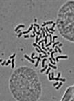
A chromosome is the structure that organizes DNA and protein in cells. It is a single piece of coiled DNA containing coding and non-coding sequences. Human cells have 23 pairs of chromosomes including 22 pairs of autosomes and one pair of sex chromosome, giving a total of 46 per cell. In tumor cells, chromosomal instability has been considered to be one of the hallmarks of tumor formation. Here we use the karyotype analysis to separate the chromosomes and observe the chromosomes in tumor cells with a microscope.

Cell Biology

Isolation and 3-dimensional Culture of Primary Murine Intestinal Epithelial Cells

Isolation and 3-dimensional Culture of Primary Murine Intestinal Epithelial Cells

Agnieszka  Pastuła Agnieszka Pastuła
MQ Michael Quante
25487 Views
May 20, 2014
The intestine, together with skin and blood, belongs to the organs with the highest cell turnover, which makes it a perfect model to study cellular processes, such as proliferation and differentiation. Epithelial cell turnover in intestine is possible due to the presence of intestinal stem cells, which are located at the bottom of the crypt. Here, we recapitulate a detailed protocol for the isolation and culture procedures of primary epithelial intestinal cells in a three - dimensional (3D) in vitro system, described for the first time by Hans Clevers group (Sato et al., 2009). This specific 3D culture preserves intestinal stem cells, which give rise to differentiated progeny for example goblet cells. The culture has many applications and represents a useful model to study stem cell biology, epithelial cell regeneration, and transplantation studies. Moreover, the presented 3D culture can be used to investigate the barrier function of intestinal epithelial cells, as well as heterotypic cell interactions between epithelial cells and stromal cells.

Developmental Biology

Barrier Function Assay

Barrier Function Assay

TD Tia DiTommaso
Floris  Foijer Floris Foijer
10761 Views
May 20, 2014
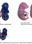
This protocol serves to determine the integrity of the barrier function of (murine) epidermis. Defects in the barrier function lead to dehydration and infection in neonatal mice/humans. One possible way to assess epidermal barrier integrity is by a dye penetration assay as described hereunder. This assay should be done on unfixed, untreated tissues (e.g. formaldehyde- or glutaraldehyde-fixed) or on whole mouse embryos (E18.5). This protocol was adapted from Hardman (1998).

Microbiology

ELISA on Virus-Infected Cells

ELISA on Virus-Infected Cells

DG Daniel L. Glauser
PS Philip G. Stevenson
11581 Views
May 20, 2014
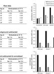
The gammaherpesvirus murid herpesvirus 4 (MuHV-4) enters cells by endocytosis from the cell surface and fusion of the viral envelope with the membrane of late endosomes. The viral envelope glycoproteins undergo antigenic changes both upon virion endocytosis and upon fusion of the viral envelope with the endosomal membrane. These changes in virion antigenicity during virus entry were first described by immunofluorescence of infected cells. Although immunofluorescence provides valuable information on the subcellular distribution of the viral glycoproteins, the quantification of immunofluorescence signals in a large number of cells is not only dependent on relatively expensive microscopy equipment, but is also relatively time-consuming. In order to quantify the antigenicity of MuHV-4 virions entering NMuMG epithelial cells in a reliable, as well as time- and cost-effective way, we have developed an ELISA with infected cells as the solid phase. In this assay, cells are grown on 96-well tissue culture plates, exposed to virions at 4 °C, followed by incubation at 37 °C allowing virion endocytosis. Cells are fixed either directly after virion binding at 4 °C or after incubation at 37 °C. After subsequent permeabilization, the cells are incubated with monoclonal antibodies specific for the viral envelope glycoproteins, followed by detection with an alkaline phosphatase-coupled secondary antibody. Upon incubation of cells with p-nitrophenyl phosphate substrate, the absorbance is measured on a conventional ELISA microplate reader. The different ways of data interpretation are discussed.
IFN-α/β Detection Assay Using Sensor Cell Lines

IFN-α/β Detection Assay Using Sensor Cell Lines

Valeria  Lulla Valeria Lulla
Andres  Merits Andres Merits
Aleksei  Lulla Aleksei Lulla
11488 Views
May 20, 2014
Type I interferons (IFN-α/β) play an important role in host resistance to viral infections. Signaling through the JAK-STAT pathway, IFN-α/β stimulates response elements (ISRE) in the promoters of ISG to regulate their expression (reviewed in Reference 2). This method was adapted from InvivoGen to specifically detect and quantify IFN-α/β secreted in response to virus infection. HEK-Blue™ IFN-α/β cells were generated by stably introducing the human STAT2 and IRF9 genes into HEK293 cells to obtain a fully active type I IFN signaling pathway. The activation of this pathway is made detectable by the addition of a reporter gene expressing a secreted embryonic alkaline phosphatase (SEAP) under the control of the ISG54 promoter. ISG54 is a well-known ISG activated through an ISRE-dependent mechanism by type I IFNs.
Purification and TEM of Afp and Its Variants

Purification and TEM of Afp and Its Variants

DR Daria Rybakova
AM Alok K. Mitra
MH Mark R. H. Hurst
9987 Views
May 20, 2014
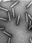
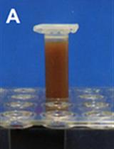
The Serratia entomophila antifeeding prophage (Afp), forms a phage-tail-like particle (tailocin) that causes cessation of feeding activity of the New Zealand grass grub, Costelytra zealandica. Here, we describe the more detailed purification protocol for Afp particles and its variants which is based on the procedure described in our original publication (Rybakova et al., 2013). The purification procedure includes inducing Escherichia coli (E. coli) cells harbouring afp genes under arabinose-inducible promoter for 24 h. The cells are harvested and sonicated on ice followed by DNAse treatment and centrifugation. The supernatant is then filter sterilised and applied to the size exclusion chromotography (SEC) column. The fractions containing Afp or its variants are pooled and ultracentrifuged. The supernatant is removed and the transparent pellet is resuspended in the residual buffer. The procedure results in Afp particles or variants thereof that are approximately 70% pure. The Afp particles are negatively stained and visualized using Transmission electron microscopy (TEM).
Construction of Deletion-knockout Mutant Fowlpox Virus (FWPV)

Construction of Deletion-knockout Mutant Fowlpox Virus (FWPV)

Stephen M.  Laidlaw Stephen M. Laidlaw
Michael A.  Skinner Michael A. Skinner
8415 Views
May 20, 2014
The construction of deletion-knockout poxviruses is a useful approach to determining the function of specific virus genes. This protocol is an adaptation of the transient dominant knockout selection protocol published by Falkner and Moss (1990) for use with vaccinia virus. The protocol makes use of the dominant selectable marker Escherichia coli guanine phosphoribosyltransferase (gpt) gene (Mulligan and Berg, 1981), under the control of an early/late poxvirus promoter. The deletion viruses that are produced no longer contain a selectable marker, which may be preferable for the production of vaccines.
Purification of HCV-remodeled and Control ER Membranes

Purification of HCV-remodeled and Control ER Membranes

David  Paul David Paul
RB Ralf Bartenschlager
8067 Views
May 20, 2014
As for all positive strand RNA viruses, hepatitis C virus (HCV) RNA replication is tightly associated with rearranged host cell membranes, termed viral replication factories. However, up to now little is known about both viral and cellular constituents of viral replication factories. Here, we describe a protocol to specifically isolate HCV-remodeled host cell membranes and endoplasmic reticulum (ER) membranes of naïve cells, by using a functional NS4B HA-tagged subgenomic replicon and a C-terminally HA-tagged calnexin-overexpressing cell line, respectively. Post-nuclear whole cell membrane fractions are first enriched by density gradient centrifugation, followed by HA-specific affinity tag purification. Upon elution under native conditions, purified samples can be subject to a variety of biochemical and functional assays.

Molecular Biology

Identification of Proteins Interacting with Genomic Regions of Interest in vivo Using Engineered DNA-binding Molecule-mediated Chromatin Immunoprecipitation (enChIP)

Identification of Proteins Interacting with Genomic Regions of Interest in vivo Using Engineered DNA-binding Molecule-mediated Chromatin Immunoprecipitation (enChIP)

Toshitsugu  Fujita Toshitsugu Fujita
Hodaka  Fujii Hodaka Fujii
14641 Views
May 20, 2014
Elucidation of molecular mechanisms of genome functions requires identification of molecules interacting with genomic regions of interest in vivo. To this end, it is useful to isolate the target regions retaining molecular interactions. We established locus-specific chromatin immunoprecipitation (ChIP) technologies consisting of insertional ChIP (iChIP) and engineered DNA-binding molecule-mediated ChIP (enChIP) for isolation of target genomic regions (Hoshino and Fujii, 2009; Fujita and Fujii, 2011; Fujita and Fujii, 2012; Fujita and Fujii, 2013a; Fujita and Fujii, 2013b; Fujita et al., 2013). Identification and characterization of molecules interacting with the isolated genomic regions facilitates understanding of molecular mechanisms of functions of the target genome regions. Here, we describe enChIP, in which engineered DNA-binding molecules, such as zinc-finger proteins, transcription activator-like (TAL) proteins, and a catalytically inactive Cas9 (dCas9) plus small guide RNA (gRNA), are utilized for affinity purification of target genomic regions. The scheme of enChIP is as follows:1. A zinc-finger protein, TAL or dCas9 plus gRNA is generated to recognize DNA sequence in a genomic region of interest. 2. The engineered DNA-binding molecule is fused with a tag(s) and the nuclear localization signal (NLS), and expressed in the cell to be analyzed.3. The resultant cell is crosslinked, if necessary, and lysed, and DNA is fragmented.4. The complexes including the engineered DNA-binding molecule are subjected to affinity purification such as mmunoprecipitation. The isolated complexes retain molecules interacting with the genomic region of interest.5. Reverse crosslinking and subsequent purification of DNA, RNA, or proteins allow identification and characterization of these molecules.In this protocol, we describe enChIP with a TAL protein to isolate a genomic region of interest and analyze the interacting proteins by mass spectrometry (Fujita et al., 2013).

Plant Science

Immunofluorescence Labeling of Pollen Tubes

Immunofluorescence Labeling of Pollen Tubes

HW Hao Wang
LJ Liwen Jiang
11188 Views
May 20, 2014
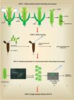
Pollen tube is regarded as an excellent single-cell model system in plant cell studies. This protocol describes the use of a rapid and reliable immunofluorescence labeling method for studying in situ localization of proteins in pollen tubes. The whole experiment contains two major steps: pollen tube in vitro germination, and pollen tube fixation and immunolabeling. It takes about 2 days from pollen tube germination to immunofluorescence detection.
Arabidopsis thaliana Embryo Sac Mitochondrial Membrane Potential Stain

Arabidopsis thaliana Embryo Sac Mitochondrial Membrane Potential Stain

María  Victoria  Martin María Victoria Martin
Diego  Fernando  Fiol Diego Fernando Fiol
EZ Eduardo Julián Zabaleta
GP Gabriela Carolina Pagnussat
10923 Views
May 20, 2014
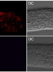
The aim of this experiment is to study mitochondrial functional status in Arabidopsis embryo sacs using the membrane potential indicator JC-1. Changes in the membrane potential are presumed to be due to the opening of the mitochondrial permeability transition pore (MPTP), allowing passage of ions and small molecules. The resulting equilibrium of ions leads in turn to the decoupling of the respiratory chain and the release of cytochrome c into the cytosol, a distinctive feature of the early stages of programmed cell death.JC-1 is a lipophilic dye that can selectively enter into mitochondria and reversibly change color from green to red as the membrane potential increases. In healthy cells with high mitochondrial potential, JC-1 spontaneously forms complexes with intense red fluorescence. On the other hand, in mitochondria with low mitochondrial potential, JC-1 remains in the monomeric form, which exhibits only green fluorescence (Martin et al., 2013; Hauser et al., 2006). This protocol could be used in isolated mitochondria, and in a variety of cell types and different tissues of plants and other organism.