- Protocols

- Articles and Issues
- For Authors
- About
- Become a Reviewer

Past Issue in 2014
Volume: 4, Issue: 6
Biochemistry
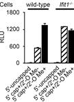
Cellular Translational Reporter Assay

Synthesis of the Adenosine A2A Receptor Fluorescent Agonist MRS5424

Cancer Biology
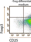
In vitro Regulatory T cells Differentiation From Naïve T Cells


Immunology
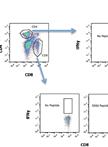
Measurement of CD8 and CD4 T Cell Responses in Mouse Lungs
Microbiology
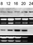
RNA Isolation and Northern Blot Analysis

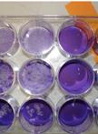
Catalase Activity Assay in Candida glabrata

Helicase Assays
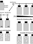
Virus Infection and Titration of SARS-CoV in Mouse Lung
Western Blotting for Staphylococcus aureus AgrA
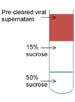
Human Astrovirus Propagation, Purification and Quantification
Determination of Mutation Frequency During Viral DNA Replication


Measuring Genetic Robustness in Vesicular Stomatitis Virus

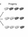
Fitness Determinations in Vesicular Stomatitis Virus
Neuroscience
Isolation and Culture of Neurospheres for the Study of Pathogenesis of Prion Disease
Neural Stem Cell Differentiation and Prion Infection
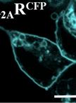
Adenosine A2A Receptor Ligand Binding Experiments by Using Real-time Single-cell FRET

Transpharyngeal Exposure of GnRH Neurons
