Past Issue in 2014

Volume: 4, Issue: 4

left

Dec

20

Dec

5

Nov

20

Nov

5

Oct

20

Oct

5

Sep

20

Sep

5

Aug

20

Aug

5

Jul

20

Jul

5

Jun

20

Jun

5

May

20

May

5

Apr

20

Apr

5

Mar

20

Mar

5

Feb

20

Feb

5

Jan

20

Jan

5

right

Biochemistry

Binding to Secreted Bone Matrix in vitro

Binding to Secreted Bone Matrix in vitro

AT Aurélie Jeanne Tormo
CB Christian Beauséjour
JG Jean-François Gauchat
9461 Views
Feb 20, 2014
This method examines the bone matrix binding capacity of proteins. Using osteogenic differentiation medium, multipotent stromal cells (MSC) are induced to differentiate into osteocytes in vitro and to secrete bone matrix. The latter is confirmed using Alizarin red S staining, which detects the presence of calcific deposits (hydroxyapatite). These calcific deposits are used to test the bone binding properties of proteins. The binding to the calcific deposits is assessed by Western blot analysis.
A Surface Plasmon Resonance Method to Study HCV NS5B Inhibitors

A Surface Plasmon Resonance Method to Study HCV NS5B Inhibitors

Melanie  Wong Melanie Wong
Giuseppe A.  Papalia Giuseppe A. Papalia
8853 Views
Feb 20, 2014
Surface Plasmon Resonance (SPR) technology is a well-established platform used to evaluate the kinetic parameters of protein-small molecule interactions. Below, we describe the use of the ProteOn XPR36 biosensor from Bio-Rad (Hercules, CA) to evaluate the binding of small molecule inhibitors to recombinant NS5B protein. The high pI (> 9) of this construct allows for chemical immobilization using HEPES-buffered saline at pH 7.5. This is in contrast to traditional biosensor protocols that use both low pH and ionic strength. The use of a more physiological buffer to immobilize this enzyme leads to improved surface activity.

Cancer Biology

Isolation of Mammary Epithelial Cells and Fibroblasts From Mouse Tumor

Isolation of Mammary Epithelial Cells and Fibroblasts From Mouse Tumor

Shiva Kazerounian Shiva Kazerounian
20203 Views
Feb 20, 2014
Developing cancer therapeutics requires the ability to investigate their effects using in vitro models of a specific type of tumor. This protocol provides a method for the isolation and adoption to growth in culture of cells from primary tumors. This is particularly valuable for studying mouse models where original tumor cells can be evaluated, for example for gene modifications, and subsequently injected back to the same background mice to create more tumors for in vivo efficacy studies.
In vivo Extravasation Assay

In vivo Extravasation Assay

Elisa  Penna Elisa Penna
Daniela Taverna Daniela Taverna
14825 Views
Feb 20, 2014
Tumor metastases develop when disseminated intravascular cancer cells acquire the ability to arrest by adhering to the capillary walls of distant organs, actively extravasate into their parenchyma, proliferate and establish secondary colonies. The extravasation assay described here is an in vivo technique aimed to analyze the ability of tumor cells to achieve early colonization of the lungs following tail vein injection in mice. Importantly, tumor cells need to be easily visible, therefore either they are fluorescent (e.g. expressing RFP or GFP) or they have to be pre-labelled with a fluorescent tracker prior to injection. Lungs are analyzed at different time points, experimentally determined by the researcher, depending on cell features and malignancy. Generally, an early time point is required to check equal lodging in the pulmonary vasculature for the various cells injected. At one or more later time points (from 6 to 48 h) extravasated cells dispersed in the lung parenchyma are quantitated. With our protocol extravasation is directly evaluated in the whole lungs ex vivo considering cell fluorescence. However, immunofluorescence stainings for endothelial markers and microscopic analyses of lung sections are recommended to evaluate positioning and status of tumor cells (i.e. inside, outside the vessels or associated to them; single cells or clusters). Since extravasation is not only influenced by tumor cell motility but also by their survival ability, the results obtained with this technique should be complemented with proliferation and apoptosis analyses.

Cell Biology

Lysosomal Amino Acid Efflux Assay

Lysosomal Amino Acid Efflux Assay

Chunlei  Cang Chunlei Cang
DR Dejian Ren
9806 Views
Feb 20, 2014
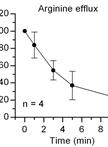
As the cellular “recycling” organelle, lysosomes break down proteins into amino acids, which are then transported into cytosol for reuse by various amino acid transporters. The transport rate of an amino acid is presumably regulated by cellular conditions such as organelle pH, membrane potential and metabolic states. Because of their intracellular localization and the relative inaccessibility, lysosomal amino acid transporters have been studied largely via indirect measurements. Using lysosome purification and 14C-labeled amino acids, this protocol provides a method to measure the efficiency of specific amino acid transporters on lysosomes.

Immunology

Isolation and Culture of Bone Marrow-derived Mast Cells

Isolation and Culture of Bone Marrow-derived Mast Cells

KV Krisztina V. Vukman
MM Martin Metz
MM Marcus Maurer
SO Sandra M. O’Neill
22160 Views
Feb 20, 2014
The generation of mast cells for in vitro studies comes from a variety of sources including mast cell lines (MC/9) (McCurdy et al., 2001), bone marrow-derived mast cells (BMMCs) (Supajatura et al., 2001), skin-derived mast cells (FSMCs) (Matsushima et al., 2004), peritoneal-derived mast cells (PMCs) (Hochdorfer et al., 2011) and peritoneal cell-derived cultured mast cells (PCMCs) (Vukman et al., 2012). BMMCs are generally used for in vitro studies because of the high yield of mast cells generated and also because they can be generated from knockout and transgenic mice making this a good source to examine specific factors important for mast cell function. Due to the large yield of cells generated they are the cells of choice for reconstitution studies in mast cell knockout mice (Sur et al., 2007). Furthermore, they are more responsive to both allergic and non-allergic stimuli when compared to mast cell lines. The major disadvantage of BMMCs is that they are not fully matured when compared to mast cells generated or obtained from other sources. For example, compared to PCMCs [see the protocol “Isolation and Culture of Peritoneal Cell-derived Mast Cells” (Vukman et al., 2014)], BMMCs express a restricted range of TLRs and cytokines when stimulated with TLR ligands (Mrabet-Dahbi et al., 2009). The different sources of mast cells can display phenotypical and functional differences and therefore it is important that when designing an experiment the correct cellular source is obtained. Here, we describe a protocol for the isolation and culture of murine mast cells from mouse bone marrow.
Isolation and Culture of Peritoneal Cell-derived Mast Cells

Isolation and Culture of Peritoneal Cell-derived Mast Cells

KV Krisztina V. Vukman
MM Martin Metz
MM Marcus Maurer
SO Sandra M. O’Neill
16205 Views
Feb 20, 2014
The generation of mast cells for in vitro studies comes from a variety of sources including mast cell lines (MC/9) (McCurdy et al., 2001), bone marrow-derived mast cells (BMMCs) (Supajatura et al., 2001), skin-derived mast cells (FSMCs) (Matsushima et al., 2004), peritoneal-derived mast cells (PMCs) (Hochdorfer et al., 2011) and peritoneal cell-derived cultured mast cells (PCMCs) (Vukman et al., 2012). PCMCs are generally used for in vitro studies because they are a more mature source of mast cells when compared to mast cells generated or obtained from other sources. They can differ, for example, in their pro-inflammatory responses to bacterial antigens and toll like receptors (TLRs) ligands (Mrabet-Dahbi et al., 2009). In comparison to BMMCs [see the protocol “Isolation and Culture of Bone Marrow-derived Mast Cells” (Vukman et al., 2014)] or mast cell lines they express a wider range of TLRs, and secrete significantly more cytokines when stimulated with TLR ligands (Mrabet-Dahbi et al., 2009). Therefore, when examining pro-inflammatory responses, mast cells generated from cells obtained from the peritoneal cavity give stronger responses. PCMCs can also be generated from knockout and transgenic mice making them a good source to examine specific factors important for mast cell function. However, due to the low yield of cells generated using this method (1 million per mouse) their use is restricted and therefore in most studies more than one source of mast cells may be required. The different sources of mast cells can display phenotypical and functional differences and therefore it is important that when designing an experiment, the correct cellular source is obtained. Here, we describe a protocol for the isolation and culture of murine mast cells from peritoneal cavity cells.
Chikungunya ELISA Protocol

Chikungunya ELISA Protocol

CB Caitlin Mattos Briggs
AP Amanda Piper
Raquel  Hernandez Raquel Hernandez
10556 Views
Feb 20, 2014
This protocol will result in the accurate qualitative measurement of anti-Chikungunya virus antibody (Ab) from infected mouse tissue or serum. This assay was developed by Dr. Caitlin Briggs, Arbovax, Inc. Chikungunya is a BL3 agent and should be handled in a biosafety level 3 laboratory under BL3 conditions. This protocol was used in the publication “Chikungunya virus host range E2 transmembrane deletion mutants induce protective immunity against challenge in C57BL/6J mice” (Piper et al., 2013).
DNA PCR Assays for Igh Rearrangement

DNA PCR Assays for Igh Rearrangement

Yusuke  Satoh Yusuke Satoh
TS Takao Sudo
Takafumi  Yokota Takafumi Yokota
10052 Views
Feb 20, 2014
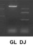
This protocol is used for the detection of immunoglobulin heavy (H) chain rearrangements. This PCR-based assay enables detection of DH-JH recombination in cultured hematopoietic cells (Schlissel et al., 1991; Satoh et al., 2013) [e.g. ES-derived cells (Satoh et al., 2013)].
Isolation of Multipotent Stromal Cells from Mouse Bone Marrow

Isolation of Multipotent Stromal Cells from Mouse Bone Marrow

AT Aurélie Jeanne Tormo
MR Moutih Rafei
JG Jean-François Gauchat
9543 Views
Feb 20, 2014
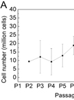
Generating mouse multipotent stromal cells (MSC) from bone-marrow cells is usefull for a wide range of applications. Effectively, these MSC can differentiate into adipocytes, osteocytes [See “Binding to Secreted Bone Matrix in vitro” (Tormo et al., 2014)] or chondrocytes upon culture in specific differentiation medium.

Microbiology

An ex vivo Model of HIV-1 Infection in Human Lymphoid Tissue and Cervico-vaginal Tissue

An ex vivo Model of HIV-1 Infection in Human Lymphoid Tissue and Cervico-vaginal Tissue

Andrea  Introini Andrea Introini
CV Christophe Vanpouille
JG Jean Charles Grivel
Leonid  Margolis Leonid Margolis
15001 Views
Feb 20, 2014
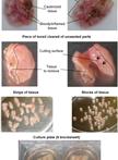
Human tissue explants are a valuable tool to study the interactions between host and infectious agents. They reliably mimic many important aspects of tissue cytoarchitecture and functions and allow us the investigation of the mechanisms of microbial pathogenesis under controlled laboratory conditions. One of the advantages of this system is that, unlike isolated cells, infection of tissue blocks with HIV-1 does not require exogenous stimulation with mitogens or activating factors. Here we describe a protocol to infect with HIV-1 human lymphoid tissue from tonsils and cervico-vaginal tissue and maintain them in culture in a non-polarized setting. These ex vivo infected tissues can be used as fruitful models to study HIV-1 pathogenesis and HIV-1 vaginal transmission, respectively, as well as an efficient platform for testing anti-HIV therapeutic and preventative strategies.

Stem Cell

Limiting Dilution Assays to Determine Frequencies of Lymphohematopoietic Progenitors

Limiting Dilution Assays to Determine Frequencies of Lymphohematopoietic Progenitors

Takafumi  Yokota Takafumi Yokota
Yusuke  Satoh Yusuke Satoh
9910 Views
Feb 20, 2014
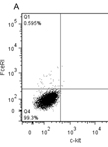
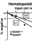
This protocol is useful to determine the frequencies of lymphohematopoietic progenitors in tested samples. To effectively support the growth and differentiation of primitive lymphohematopoietic progenitors, complex signals from stromal cells are important. Several stromal cell lines are known to support both lymphoid and myeloid cells simultaneously in mouse. In this protocol, we introduce two stromal co-culture systems for murine lymphohematopoietic progenitors and their application for limiting dilution assays.