Improve Research Reproducibility A Bio-protocol resource
- Protocols

- Articles and Issues
- For Authors
- About
- Become a Reviewer

Past Issue in 2014
Volume: 4, Issue: 3
Biochemistry
Phosphoinositides Coated Beads Binding Assay
The PIs coated beads assay or “PIP-Beads” developed by Echelon Biosciences (Salt Lake City, USA) is a quick assay to recognize which PIs are able to bind to a given protein domain, in a quantitative way. It is much faster and cheaper than liposomes and more reproducible than PIP-strip assays. The “PIP-Beads” assay is a biochemical assay that basically involves an incubation of a purified protein or protein domain with the appropriate PI-coated set of beads. After washing, drying and resuspending the samples, they can be easily analyzed by SDS-PAGE separation. Phosphoinositides (PIs) have been characterized as important determinants of cell membrane domains, such as the apical and basolateral domains in epithelial polarized cells (Martin-Belmonte and Mostov, 2007), controlling membrane trafficking (Szentpetery et al., 2010) or determining the presynaptic or postsynaptic terminal in neurons, among other functions (Di Paolo and De Camilli, 2006). These phosphoinositides enriched membranes bring the proteomic machinery together, confers to these membrane their different identities and functions. This protein-PIs interaction in many cases involves direct binding of specific protein membrane domains with certain PIs. Some of these domains are characterized such as PH domains from phospholipase-C- or synaptotagmin-like C2 domains (Galvez-Santisteban et al., 2012), while some of them are not. To determine which PI is binding to a given protein domain, it is important to have a quick and efficient assay. The liposome binding assays are very good to establish the kinetic properties of binding, but they are expensive and permit only to test a few PIs per experiment. On the other hand, PIP-strip (phosphatidil-inositol-phosphate) based analysis is easy and fast, however the PIs are presented in a flat surface and the reproducibility is sometimes limited.
Cell Biology
Microsome Isolation from Tissue

This protocol details the extraction of microsomes from frozen tissue in order to further examine the protein-protein interactions occurring within the endoplasmic reticulum. This protocol was adapted from Abisambra et al. (2013) with modifications made in order to optimize for subsequent use.
Immunology
In vitro Biomineralization Assay


Biomineralization in vertebrates has both physiological and pathological aspects. Physiological mineralization is essential for proper development and function of hard tissues, such as bone, teeth, and growth plate cartilage, but it does not occur in soft tissues. Pathological ectopic mineralization, in contrast, occurs in soft tissues, including blood vessels, kidney, articular cartilage, and cardiovascular tissue. Here, we describe the simple method for detecting and measuring the presence of mineralized nodules in cardiac ventricular fibroblasts by using von Kossa and alizarin red S staining, and a colorimetric method for calcium quantification, respectively.
Intracellular Staining for Phosphorylated STAT4 and STAT5 in Mouse Splenocytes
The Stat (Signal Transducer and Activator of Transcription) family of proteins are critical signal transducers involved in fundamental cellular processes, including cell growth and differentiation, development, apoptosis, immune responses and inflammation. In here, we describe a simple and reproducible flow cytometry protocol to measure Stat protein phosphorylation in splenocyte preparations from malaria infected mice.
Neuroscience
Optical Clearing Using SeeDB
We describe a water-based optical clearing agent, SeeDB (See Deep Brain), which clears fixed brain samples in a few days without quenching many types of fluorescent dyes, including fluorescent proteins and lipophilic neuronal tracers. SeeDB is a saturated solution of fructose (80.2% w/w) in water with 0.5% α-thioglycerol. In standard SeeDB optical clearing procedure, we treat paraformaldehyde-fixed embryo and brain samples with increasing concentrations of aqueous fructose solutions, and finally equilibrate them in SeeDB. The entire procedure takes approximately three days. Unlike previous methods, this method maintains a constant sample volume during the clearing procedure, an important factor to keep cellular morphology intact. After optical clearing, we can reach > 1,000 μm under confocal microscopy. When combined with two-photon microscopy, SeeDB allows us to image fixed mouse brains at millimeters-scale level. This method facilitates comprehensive and quantitative analyses for understanding neuronal circuitry, both in the adult and developing mouse brain. A SeeDB variant (SeeDB37) and optimized procedures (SeeDBp and SeeDB37ht protocols) are also supplied for specific requirements.
In situ Chemotaxis Assay in Caenorhabditis elegans (for the Study of Circadian Rhythms)

Olfaction is a well-studied sensory mechanism in Caenorhabditis elegans (C. elegans). The nematodes respond to a wide range of chemicals by either attraction, repulsion or a mixture thereof (Bargmann et al., 1993). We have used olfaction to characterize behavioural and molecular circadian rhythms in C. elegans. The circadian clock is a biological oscillator that provides an endogenous temporal structure that approximately matches the 24-hour periodicity in the environment (due to the rotational movement of the Earth). Circadian rhythms are present in most organisms from cyanobacteria to humans and they typically regulate sensory functions among many other processes. Olfaction is under circadian control in many animals (Granados-Fuentes et al., 2006; Granados-Fuentes et al., 2011; Tanoue et al., 2008; Krishnan et al., 1999). This protocol was designed to allow the assessment of olfaction for a population of worms within a short time interval, in the same plate where the worms grew (to avoid washing steps that may disturb the rhythms), and in the presence of food.
Plant Science
Determination of Water Use Efficiency for Arabidopsis thaliana
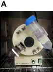
Water use efficiency (WUE) is a quantitative measurement of how much biomass or yield is produced per amount of water used. It is an important physiological factor for agriculture, especially in areas with a limited accessibility of water. It is also crucial in a better understanding of drought tolerance and drought resistance. The most common method to measure the WUE of individual plants is to weigh each pot or container to monitor the water loss and to harvest and measure biomass or yield at the end of the experiment. Since water can be lost from the soil surface through evaporation, there is a need to perform WUE measurements in a closed system. Here, we describe a simple method for WUE determination for Arabidopsis thaliana.
Assays for Determination of Acetylesterase Activity and Specificity Using pNP-acetyl and Acetylated Polysaccharides as Substrates
The acetylesterases are hydrolytic enzymes which in plants cleave acetyl groups from acetylated cell wall components, primarily polysaccharides. To estimate acetylesterase activity in plant apoplast, two assays can be used. First assay is a direct measurement of the acetylesterase activity in protein extract using synthetic substrate, pNP-acetyl. In this assay, amount of pNP released after hydrolysis of pNP-acetyl is determined by measuring the intensity of developed yellow color using spectrophotometer. The absorbance of reaction mixture is directly proportional to the activity of acetylesterases in the reaction mixture. Second assay is a determination of acetylesterase activity and its specificity towards natural polysaccharides and based on interaction between ferric perchlorate and acetyl residues resulting in ferric acetohydroxamic complex that can be quantified using spectrophotometer. In this assay, commercially available acetylated polysaccharides (xylan from Birchwood for acetylxylan esterase; pectin from citrus fruit for rhamnogalacturonan acetylesterase; or any other available polysaccharide of interest) incubated with apoplastic extract and amount of acetyl residues released from this polysaccharide is estimated using ferric perchlorate reagent (protocol was modified from McComb and McCready, 1957). The absorbance of produced colored complex is directly proportional to the amount of acetyls released from acetylated polysaccharide.