Past Issue in 2013

Volume: 3, Issue: 19

left

Dec

20

Dec

5

Nov

20

Nov

5

Oct

20

Oct

5

Sep

20

Sep

5

Aug

20

Aug

5

Jul

20

Jul

5

Jun

20

Jun

5

May

20

May

5

Apr

20

Apr

5

Mar

20

Mar

5

Feb

20

Feb

5

Jan

20

Jan

5

right

Biochemistry

Blood AST, ALT and UREA/BUN Level Analysis

Blood AST, ALT and UREA/BUN Level Analysis

YS Yuh-Pyng Sher
MH Mien-Chie Hung
39157 Views
Oct 5, 2013
AST (aspartate aminotransferase; GOT, glutamate oxalacetate transaminase) and ALT (alanine aminotransferase; GPT, glutamate pyruvate transaminase) are sensitive indicators to monitor the liver function under drugs treatment or with acute viral hepatitis. The elevated AST and ALT values in the blood sample indicate liver damage or injury. The determination of urea is the most widely used for the evaluation of kidney function. This protocol is for the quantitative determination of AST, ALT and UREA/BUN in serum and plasma on Roche automated clinical chemistry analyzers. The principle is shown below:For AST: α-ketoglutarate + L-aspartate L-glutamate + oxaloacetate (AST catalyzes this equilibrium reaction) oxaloacetate + NADH + H+ L-malate + NAD+ (malate dehydrogenase catalyzes this equilibrium reaction) The rate of the photometrically determined NADH decrease is directly proportional to the rate of formation of oxaloacetate and thus the AST activity. The above reactions were carried out at 37 °C and measured at a wavelength of 340 nm.For ALT: α-ketoglutarate + L-alanine L-glutamate + pyruvate (ALT catalyzes this equilibrium reaction) Pyruvate + NADH + H+ L-lactate + NAD+ (lactate dehydrogenase catalyzes this equilibrium reaction) The rate of the photometrically determined NADH decrease is directly proportional to the rate of formation of pyruvate and thus the ALT activity. The above reactions were carried out at 37 °C and measured at a wavelength of 340 nm.For UREA/BUN: Urea + H2O → 2 NH4+ + CO2 (urea is hydrolyzed by urease) α-ketoglutarate + NH4+ + NADH → L-glutamate + NAD+ + H2O (the presence of GLDH yields glutamate and NAD+) The decrease in absorbance due to consumption of NADH is measured kinetically. The above reactions were carried out at 37 °C and measured at a wavelength of 340 nm.
IEF-2DE Analysis and Protein Identification

IEF-2DE Analysis and Protein Identification

XW Xia Wu
SC Steven J. Clough
SH Steven C. Huber
Man-Ho Oh Man-Ho Oh
15631 Views
Oct 5, 2013
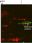
Isoelectric focusing followed by SDS-PAGE (IEF-2DE) separates proteins in a two-dimensional matrix of protein pI (Protein Isoelectric Point) and molecular weight (MW). The technique is particularly useful to distinguish protein isoforms (Radwan et al., 2012) and proteins that contain post-translational modifications such as phosphorylation (Oh et al., 2012) and lysine acetylation (Wu et al., 2011). Proteins that are separated by IEF-2DE can be identified by immunoblot analysis using sequence-specific antibodies or by mass spectrometry.
Hydroxyproline Assay Using NaBr/NaOCl

Hydroxyproline Assay Using NaBr/NaOCl

Derek T. A.  Lamport Derek T. A. Lamport
10074 Views
Oct 5, 2013
Hydroxyproline (Hyp) is a major constituent of a relatively few proteins that are major structural components of the extracellular matrix and primary cell wall of animals and plants respectively. Significant amounts of the cyclic amino acids proline and hydroxyproline decrease polypeptide flexibility; thus proline/hydroxyproline-rich proteins are ideal scaffold components. Collagens typify animal tissues but extensins, arabinogalactan proteins (AGPs) and their close relatives, collectively referred to as hydroxyproline-rich glycoproteins (HRGPs), typify plants (Lamport et al., 2011). While collagens are minimally glycosylated generally via a galactosyl hydroxylysine linkage, plant HRGP glycosylation involves short neutral oligosaccharides (in extensins) or much larger acidic polysaccharide substituents (in AGPs) O-linked via the hydroxyproline hydroxyl group. Hydroxyproline assay is thus an integral part of their characterization and dominates the biochemical properties of these glycoproteins. The colourimetric assay described here quantifies free hydroxyproline (e.g. released by acid hydrolysis) based on Kivirikko and Liesmma (1959) with hypobromite as an oxidant but modified by avoiding the use of hazardous liquid bromine. A number of oxidants have been used over the years, Vogel (1961, page 395) explains the preference for hypobromite as follows: “Hypochlorites tend to react slowly with reducing agents. Hypobromites although rather unstable when prepared directly from bromine and alkali, often react more rapidly; it is therefore advantageous to produce hypobromite in situ by adding an excess of bromide to the sample of hypochlorite:” OCl- + Br- → OBr- + Cl- “By this means the relative stability of hypochlorite is combined with the more effective oxidizing properties of hypobromite.”

Cancer Biology

LDH-A Enzyme Assay

LDH-A Enzyme Assay

DZ Di Zhao
YX Yue Xiong
QL Qun-Ying Lei
KG Kun-Liang Guan
14838 Views
Oct 5, 2013
LDH (Lactate dehydrogenase) enzyme catalyzes the reversible conversion of pyruvate to lactate using NAD+ as a cofactor. Although the physiological significance of lactate accumulation in tumor cells, a dead-end product in cellular metabolism, is currently a topic of debate, it has long been known that many tumor cells express a high level of LDH-A (Koukourakis et al., 2003; Koukourakis et al., 2006; Koukourakis et al., 2009). So detection of its enzyme activity in vitro is important for researching on LDH-A. Recently, it has been reported that Lys-5 acetylation could decrease LDH-A enzyme activity (Zhao et al., 2013).
Pancreatic Acinar Cell 3-Dimensional Culture

Pancreatic Acinar Cell 3-Dimensional Culture

Chunjing  Qu Chunjing Qu
SK Stephen F. Konieczny
14271 Views
Oct 5, 2013
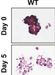
Normal pancreatic acinar cells are difficult to maintain on traditional plastic culture surfaces due to their physical properties of housing large quantities of digestive enzymes and the formation of intercellular tight junctions and gap junctions (Apte and Wilson 2005; Rukstalis et al., 2003). However, placing primary acinar cells within a 3-dimensional matrix (3D-culture) maintains the cells for sufficient time so that they can be monitored for physiological changes to different stimuli. We have used a modified collagen 3D-culture system that has been adapted from Means et al. (2005) to model the very early events associated with pancreatic cancer development. In this model, KrasG12D-expressing pancreatic acinar cells, or wildtype acinar cells treated with EGFR-dependent growth factors (i.e., TGFα), convert to ductal cysts that mimic the acinar-to-ductal metaplasia (ADM) stage that precedes formation of Pancreatic Intraepithelial Neoplasia (PanIN) and Pancreatic Ductal Adenocarcinoma (PDAC) (Means et al., 2005; Shi et al., 2013).

Cell Biology

Spindle Angle Measurements

Spindle Angle Measurements

JC Julien Cau
NM Nathalie Morin
GB Guillaume Bompard
11997 Views
Oct 5, 2013
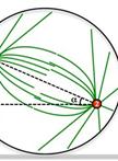
Spindle angles measures derive from the measures of spindle poles positions that were taken from fixed and immunostained adherent cells. To determine spindle angles (α), z-stack images of metaphasic cells immunostained with anti γ-tubulin (spindle poles) and anti β-tubulin antibodies (mitotic spindle) were acquired. A very simple ImageJ software macro was developed to measure the spindle angle using spindle pole coordinates (see Figure 1).

Microbiology

Virus Overlay Assay (Far-Western blotting)

Virus Overlay Assay (Far-Western blotting)

KJ Kun-Tong Jia
CG Chang-Jun Guo
XY Xiao-Bo Yang
JH Jian-Guo He
15600 Views
Oct 5, 2013
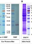
Virus overlay assay is a method to detect protein-protein interaction in vitro. We performed the virus overlay assay to identify the receptor proteins interacting with the infectious spleen and kidney necrosis virus (ISKNV).
Batch Culture Fermentation of Endophytic Fungi and Extraction of Their Metabolites

Batch Culture Fermentation of Endophytic Fungi and Extraction of Their Metabolites

Susheel  Kumar Susheel Kumar
Nutan  Kaushik Nutan Kaushik
15130 Views
Oct 5, 2013
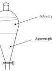
Antibiosis is one of the possible modes of action shown by endophytic fungi having antifungal activity. To test if antifungal activity in endophytic fungi is due to antibiosis, assay of the metabolites of endophytic fungi was needed. To obtain metabolites for bioassay batch culture fermentation and extraction of metabolites was done. Fungus was multiplied on wickerham media at incubation temperature of 25 ± 2 °C for 4 weeks and then extracted with solvents of different polarity. All the solvent extracts were dried under vacuum rotary evaporator to get dried crude fungal extract, which was subjected to further fractionation and bioassay.
Staphylococcus aureus Killing Assay of Caenorhabditis elegans

Staphylococcus aureus Killing Assay of Caenorhabditis elegans

AW Amanda C. Wollenberg
OV Orane Visvikis
AA Anna-Maria F. Alves
JI Javier E. Irazoqui
13132 Views
Oct 5, 2013
The Gram-positive bacterium Staphylococcus aureus is a human pathogen that displays virulence towards the nematode Caenorhabditis elegans. This property can be used to discover genes that are important for virulence in humans, because S. aureus possesses common virulence factors that are used in C. elegans and in humans to cause disease. S. aureus colonizes the C. elegans intestine, establishes an infection, and causes pathogenesis of the intestinal epithelium that ultimately kills the infected animal after 3 to 4 days (Sifri et al., 2003; Irazoqui et al., 2008; Irazoqui et al., 2010). The protocol described here is used to establish the rate of S. aureus-induced C. elegans death, which allows the comparison of wild type and mutant strains and thus ultimately aids in the identification of genes required either for S. aureus virulence or for C. elegans host defense. The assay can also be applied for antimicrobial drug discovery.
Cell Fractionation of Pseudomonas aeruginosa

Cell Fractionation of Pseudomonas aeruginosa

Esteban  Paredes-Osses Esteban Paredes-Osses
Kim R.  Hardie Kim R. Hardie
12960 Views
Oct 5, 2013
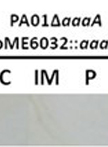
Pseudomonas aeruginosa is a Gram negative bacterium. Separating the cell envelope compartments enables proteins to be localized to confirm where in the cell they function. Cell fractionation can also provide a first step in a protein purification strategy (Williams et al., 1998). This protocol has been designed to obtain the different fractions of P. aeruginosa, namely the inner membrane, outer membrane, cytoplasmic and periplasmic compartments. Specific detection of the arginine specific autotransporter (AaaA) (Luckett et al., 2012) in the outer membrane of P. aeruginosa has been performed using this protocol.
Bioassay of Extracts of the Endophytic Fungi

Bioassay of Extracts of the Endophytic Fungi

Susheel  Kumar Susheel Kumar
Nutan  Kaushik Nutan Kaushik
9906 Views
Oct 5, 2013
Many of the microbes including fungi produce metabolites possessing antifungal activity and in some cases fungal metabolites are main cause of antifungal activity resulted by fungus. To infer that antifungal activity is due to fungal metabolites, there is a need to develop a repeatable procedure to assay these metabolites. Here we are presenting the poisoned food technique for bioassay of extract from endophytic fungi, where culture media is supplemented with extract to testpathogen viability as proxy for antifungal activity.

Plant Science

In vitro Protein Ubiquitination Assays

In vitro Protein Ubiquitination Assays

QZ Qingzhen Zhao
Qi  Xie Qi Xie
48552 Views
Oct 5, 2013
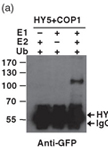
Ubiquitin can be added to substrate protein as a protein tag by the concerted actions of ubiquitin activating enzyme (E1), ubiquitin conjugating enzyme (E2) and ubiquitin protein ligase (E3). At the present of E1 and ubiquitin, E2 activity can be determined by the thio-ester formation. The E3 activity of a putative protein as well as the E2/E3 or E3/substrate specificities also can be explored by in vitro ubiquitination assay. The result can be detected by western blot with certain antibody. Purified proteins expressed from bacterial system are always used in this assay.
Stomatal Bioassay in Arabidopsis Leaves

Stomatal Bioassay in Arabidopsis Leaves

XL Xuan Li
XM Xian-Ge Ma
Jun-Min  He Jun-Min He
15928 Views
Oct 5, 2013
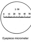
Stomata embedded in the epidermis of terrestrial plants are important for CO2 absorption and water transpiration, and are possible points of entry for pathogens. Thus, the regulation of stomatal apertures is extremely important for the survival of plants. Furthermore, stomata can respond via accurate change of stomatal apertures to a series of extracellular stimuli such as phytohormones, pathogens, ozone, drought, humidity, darkness, CO2, visible light and UV-B radiation, so stomatal bioassay is widely used to dissect signal transduction mechanisms of plant cells in responses to multiple stimuli. This protocol describes how to measure stomatal apertures in leaves of model plant Arabidopsis thaliana under multiple treatments.
Measurement of Endogenous H2O2 and NO and Cell Viability by Confocal Laser Scanning Microscopy

Measurement of Endogenous H2O2 and NO and Cell Viability by Confocal Laser Scanning Microscopy

MW Mi-Mi Wu
XM Xian-Ge Ma
Jun-Min  He Jun-Min He
13633 Views
Oct 5, 2013
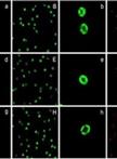
Recently, there is compelling evidence that hydrogen peroxide (H2O2) and nitric oxide (NO) function as signaling molecules in plants, mediating a range of responses including stomatal movement. Thus, the choice of sensitive methods for detection of endogenous H2O2 and NO in guard cells are very important for understanding the role of H2O2 and NO in guard cell signaling. In addition, besides stomatal closure caused by interfering guard cell signaling, it can also be caused by widespread, nonspecific damage to guard cells. To determine whether stomatal movement is caused by damage to guard cells, sensitive methods for detection of guard cell viability are often required. The oxidatively sensitive fluorophore 2′,7′-dichlorofluorecin (H2DCF) is commonly employed to measure changes in intracellular H2O2 level directly. The non-polar diacetate ester (H2DCFDA) of H2DCF enters the cell and is hydrolysed into the more polar, non-fluorescent compound H2DCF, which, therefore, is trapped. Subsequent oxidation of H2DCF by H2O2, catalysed by peroxidases, yields the highly fluorescent DCF. Similarly, the cell-permeable, NO-sensitive fluorescent probe 4,5-diaminofluorescein diacetate (DAF-2DA) is widely used for the direct detection of NO presence in both animal and plant cells. The non-polar DAF-2DA enters the cell and is hydrolyzed by cytosolic esterase into the more polar, non-fluorescent compound DAF-2, which in the presence of NO is converted to the highly fluorescent triazole derivative DAF-2T. The fluorescent indicator dyes fluorescein diacetate (FAD) and propidium iodide (PI) are widely used for detection of cell viability. FAD passes through cell membranes and is hydrolyzed by intracellular esterase to produce a polar compound that passes slowly through a living cell membrane but fast through a damaged or dead cell membrane, and thus accumulates inside the viable cells and exhibits green fluorescence when excited by blue light. In contrast, PI passes through damaged or dead cell membranes and intercalates with DNA and RNA to form a bright red fluorescent complex seen in the nuclei of dying or dead cells but not living cells. Based on the above analysis, the fluorescent indicator dyes H2DCFDA, DAF-2DA, FAD and PI load readily into guard cells, and their optical properties make them amenable to analysis by confocal laser scanning microscopy.This protocol describes how to combine confocal laser scanning microscopy with fluorescent indicator dyes H2DCFDA, DAF-2DA, FAD and PI respectively for measurement of H2O2 and NO and viability of guard cell in leaves of Arabidopsis (Arabidopsis thaliana).
MAPK Phosphorylation Assay with Leaf Disks of Arabidopsis

MAPK Phosphorylation Assay with Leaf Disks of Arabidopsis

PF Pascale Flury
DK Dominik Klauser
TB Thomas Boller
Sebastian  Bartels Sebastian Bartels
12931 Views
Oct 5, 2013
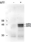
Activation of mitogen activated protein kinases (MAPKs) is involved in many abiotic and biotic stress responses including plant defense. MAPK acitvation is based on the dual phosphorylation of threonine (T) and tyrosin (Y) residues (T-x-Y motif) in the activation loop of the MAPK protein. By determination of the phosphorylation status of a specific MAPK one can detect if the MAPK has been activated or not. This protocol describes how to analyze the phosphorylation status of Arabidopsis MAPKs MPK3 and MPK6 by using leaf disks, western blotting and a specific antibody (Figure 1). It can also be used for the analysis of MAPKs in other plant systems although some alterations regarding protein extraction might be necessary.
Preparation of Arabinogalactan Glycoproteins from Plant Tissue

Preparation of Arabinogalactan Glycoproteins from Plant Tissue

Derek T. A.  Lamport Derek T. A. Lamport
11603 Views
Oct 5, 2013
This supplements an earlier protocol (Popper, 2011) for the extraction and assay of cell surface arabinogalactan proteins (AGPs). These highly glycosylated glycoproteins (~95% carbohydrate) contain numerous glycomodules with paired glucuronic acid residues that bind Ca2+ in a pH dependent manner (Lamport and Varnai, 2013). Classical AGPs comprise the bulk of cell surface glycoproteins and are thus integral components of a Ca2+ oscillator involved in a signalling pathway where calcium is a “universal signalling currency” analogous to ATP as the universal energy currency. The central role of these peripheral glycoproteins is thus reason enough for their further study. However, problems arise due to the extensive glycosylation and its apparent microheterogeneity generally assumed to preclude a simple reductionist approach.Here I describe a simple partial purification of classical AGPs based on their specific interaction with the β-D-glucosyl or galactosyl Yariv reagent, a synthetic diazo dye that precipitates AGPs as an insoluble complex in salt solutions at neutral pH. (The solubility of this complex in dilute alkali provides a rapid sensitive quantitative assay for AGPs.) Reduction of the Yariv diazo linkage releases soluble AGPs for further analysis. For example deglycosylation of AGPs in anhydrous hydrogen fluoride followed by column chromatography yields just a few major AGP polypeptides purified to homogeneity (Zhao et al., 2002). However, purification of individual AGP glycoproteins to homogeneity is rarely achieved (Darjania et al., 2002); not only do the closely related AGP glycosylation profiles vastly outweigh any contribution from the amino acid composition but the glycan polydispersity made isolation of a single molecular entity well-nigh impossible until AGPs genetically engineered with a hydrophobic green fluorescent protein tag allowed chromatographic purification (Zhao et al., 2002). New approaches to AGP fractionation into discrete classes is now also a distinct possibility based on their calcium content hitherto ignored!