- Protocols

- Articles and Issues
- For Authors
- About
- Become a Reviewer

Past Issue in 2013
Volume: 3, Issue: 17
Cancer Biology
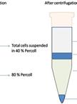
Isolation of Mouse Tumor-Infiltrating Leukocytes by Percoll Gradient Centrifugation

Three-dimensional Invasion Assay


Chemosensitivity Assay


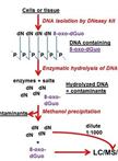
Genomic 8-oxo-7,8-dihydro-2'-deoxyguanosine Quantification
3H-Penciclovir (3H-PCV) Uptake Assay
Immunology
Mouse Macrophage Differentiation by Induction with Macrophage Colony-Stimulating Factor
C1q Binding to and Uptake of Apoptotic Lymphocytes by Human Monocyte-derived Macrophages

Microbiology
Colony Immunoblotting Assay for Detection of Bacterial Cell-surface or Extracellular Proteins


Neuroscience
Generation of Mouse Spinal Cord Injury


Preparation of Pre- and Post-synaptic Density Fraction from Mouse Cortex
Targeted Occlusion of Individual Pial Vessels of Mouse Cortex
Plant Science
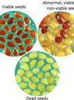
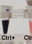
Seed Germination and Viability Test in Tetrazolium (TZ) Assay


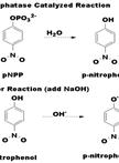
Protein Extraction, Acid Phosphatase Activity Assays, and Determination of Soluble Protein Concentration

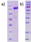
Heterologous Production and Anaerobic Purification of His- and StrepII-tagged Recombinant Proteins

Quantification of Total and Soluble Inorganic Phosphate
Pectin Methylesterase Activity Assay for Plant Material

Pyruvate:ferredoxin Oxidoreductase (PFR1) Activity Assays Using Methyl Viologen as Artificial Electron Acceptor
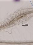
RNA Isolation From Meloidogyne Spp. Galls


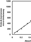
Plant Endo-β-mannanase Activity Assay

