- Protocols

- Articles and Issues
- For Authors
- About
- Become a Reviewer

Past Issue in 2016
Volume: 6, Issue: 23
Cancer Biology
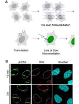
DNA Damage Induction by Laser Microirradiation


Lentiviral shRNA Screen to Identify Epithelial Integrity Regulating Genes in MCF10A 3D Culture
Cell Biology
Lymphocyte Isolation, Th17 Cell Differentiation, Activation, and Staining

Isolation of Latex Bead Phagosomes from Dictyostelium for in vitro Functional Assays


Analysis of Myosin II Minifilament Orientation at Epithelial Zonula Adherens


Developmental Biology
Vascular Smooth Muscle Cell Isolation and Culture from Mouse Aorta
Intracellular Assessment of ATP Levels in Caenorhabditis elegans


Measuring Oxygen Consumption Rate in Caenorhabditis elegans
Microbiology
Pyocyanin Extraction and Quantitative Analysis in Swarming Pseudomonas aeruginosa


Murine Leukemia Virus (MLV)-based Coronavirus Spike-pseudotyped Particle Production and Infection

Single Cell Flow Cytometry Assay for Peptide Uptake by Bacteria


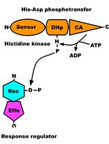
In vitro Autophosphorylation and Phosphotransfer Assay of Cyanobacterial Histidine Kinase 2
Preparation of Purified Gram-positive Bacterial Cell Wall and Detection in Placenta and Fetal Tissues


Mouse Model of Dengue Virus Infection with Serotypes 1 and 2 Clinical Isolates


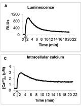
Measurement of Intracellular Calcium Concentration in Pseudomonas aeruginosa
Neuroscience
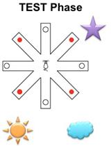
Delayed Spatial Win-shift Test on Radial Arm Maze


Various Modes of Spinal Cord Injury to Study Regeneration in Adult Zebrafish


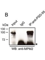
MPM-2 Mediated Immunoprecipitation of Proteins Undergoing Proline-directed Phosphorylation

Plant Science
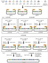
A Golden Gate-based Protocol for Assembly of Multiplexed gRNA Expression Arrays for CRISPR/Cas9


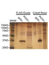
Tandem Purification of His6-3x FLAG Tagged Proteins for Mass Spectrometry from Arabidopsis

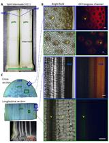
Fusarium graminearum Maize Stalk Infection Assay and Associated Microscopic Observation Protocol


Extraction and Measurement of Abscisic Acid in a Unicellular Red Alga Cyanidioschyzon merolae


Microplate Assay to Study Carboxypeptidase A Inhibition in Andean Potatoes


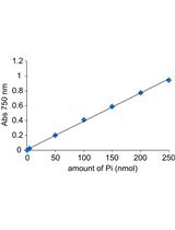
Measurement of ATP Hydrolytic Activity of Plasma Membrane H+-ATPase from Arabidopsis thaliana Leaves
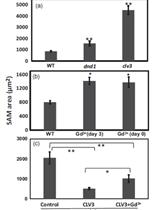
Shoot Apical Meristem Size Measurement
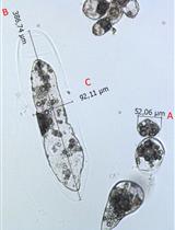
Isolating and Measuring the Growth and Morphology of Pro-embryogenic Masses in Araucaria angustifolia (Bertol.) Kuntze (Araucariaceae)


Stem Cell
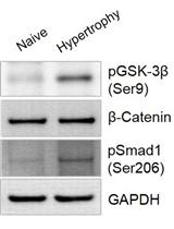
In vitro Chondrogenic Hypertrophy Induction of Mesenchymal Stem Cells

Systems Biology
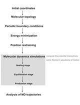
Protocol for Molecular Dynamics Simulations of Proteins