- Protocols

- Articles and Issues
- For Authors
- About
- Become a Reviewer

Past Issue in 2016
Volume: 6, Issue: 19
Biochemistry
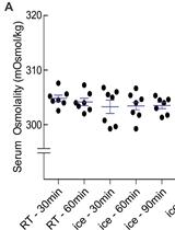
Measuring Rat Serum Osmolality by Freezing Point Osmometry

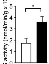
Measurement of Glucose-6-phosphate Dehydrogenase Activity in Bacterial Cell-free Extracts

Spectrophotometric Determination of Glutamine Synthetase Activity in Cultured Cells
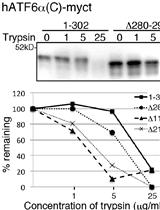
Trypsin Sensitivity Assay to Study the Folding Status of Proteins

Cell Wall-bound p-Coumaric and Ferulic Acid Analysis
PNGase Sensitivity Assay to Study the Folding Status of Proteins
Cancer Biology
Evaluation of Angiogenesis Inhibitors Using the HUVEC Fibrin Bead Sprouting Assay
Isolation of Primary Breast Cancer Cells from HER2 Transgenic Mice
Cell Biology
Detection of Reactive Oxygen Species Using MitoSOX and CellROX in Zebrafish


Mouse Corneal Stroma Fibroblast Primary Cell Culture

Developmental Biology
Isolation, Culture, and Staining of Single Myofibers


Immunology
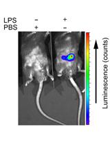
In vivo Analysis of Neutrophil Infiltration during LPS-induced Peritonitis


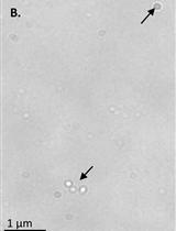
ASC-particle-induced Peritonitis
Microbiology
Measurement of Cellular Copper in Rhodobacter capsulatus by Atomic Absorption Spectroscopy


Molecular Biology
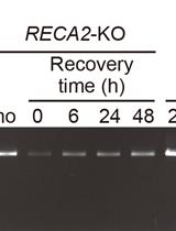
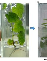
PCR-based Assay for Genome Integrity after Methyl Methanesulfonate Damage in Physcomitrella patens
Neuroscience
Sucrose Preference Test to Measure Anhedonic Behaviour in Mice

Light/Dark Transition Test to Assess Anxiety-like Behavior in Mice
Organotypic Spinal Cord Slice Cultures and a Method to Detect Cell Proliferation in These Slices


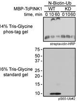
Non-radioactive in vitro PINK1 Kinase Assays Using Ubiquitin or Parkin as Substrate


Plant Science
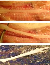
An Assay to Study Botrytis cinerea-infected Grapevine Leaves Primed with Pseudomonas fluorescens


