- Protocols

- Articles and Issues
- For Authors
- About
- Become a Reviewer

Past Issue in 2016
Volume: 6, Issue: 18
Immunology
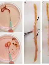
Isolation of Intestinal Mesenchymal Cells from Adult Mice


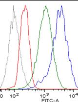
Flow Cytometry of Lung and Bronchoalveolar Lavage Fluid Cells from Mice Challenged with Fluorescent Aspergillus Reporter (FLARE) Conidia
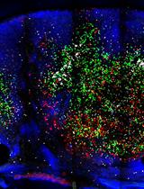
Imaging Thick Lymph Node Tissue Sections


Peptide Loading on MHC Class I Molecules of Tumor Cells

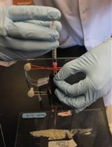
Preparation of Protein-containing Extracts from Microbiota-rich Intestinal Contents


Skin TRITC Painting to Track Dendritic Cells Migrating to the Lymph Nodes
Killer Cell Ig-like Receptors (KIR)-Binding Assay for Tumor Cells
Microbiology
Extraction and Quantification of GABA and Glutamate from Cyanobacterium Synechocystis sp. PCC 6803


Purification and Identification of Novel Host-derived Factors for Influenza Virus Replication from Human Nuclear Extracts
59Fe Uptake Assays in Paracoccidioides Species
Neuroscience
Assessment of Mechanical Allodynia in Rats Using the Electronic Von Frey Test


In vitro Assay for Dendritic Spine Retraction of Hippocampal Neurons with Sparse Labeling


Plant Science
Use of SCRI Renaissance 2200 (SR2200) as a Versatile Dye for Imaging of Developing Embryos, Whole Ovules, Pollen Tubes and Roots

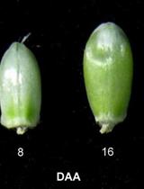
Extraction and Assays of ADP-glucose Pyrophosphorylase, Soluble Starch Synthase and Granule Bound Starch Synthase from Wheat (Triticum aestivum L.) Grains

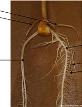
Paper Roll Culture and Assessment of Maize Root Parameters
