- Protocols

- Articles and Issues
- For Authors
- About
- Become a Reviewer

Past Issue in 2016
Volume: 6, Issue: 16
Biochemistry
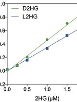
Quantification of 2-Hydroxyglutarate Enantiomers by Liquid Chromatography-mass Spectrometry


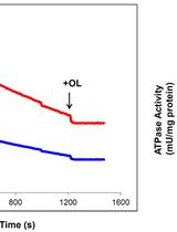
Determination of the H+-ATP Synthase and Hydrolytic Activities


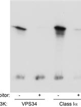
Determination of VPS34/PIK3C3 Activity in vitro Utilising 32P-γATP

Purification of Flagellin from Acidovorax avenae and Analysis of Plant Immune Responses Induced by the Purified Flagellin
Cell Biology
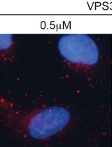
Determination of Cellular Phosphatidylinositol-3-phosphate (PI3P) Levels Using a Fluorescently Labelled Selective PI3P Binding Domain (PX)
Immunology
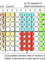
Chromium-51 (51Cr) Release Assay to Assess Human T Cells for Functional Avidity and Tumor Cell Recognition


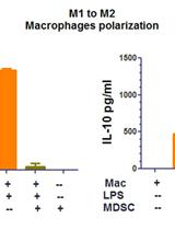
Macrophage Polarization by Tumor-induced MDSCs Assay


Adoptive Transfer of Tumor Expanded Regulatory T Cells (Tregs)
Microbiology
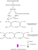
A Modified Chromogenic Assay for Determination of the Ratio of Free Intracellular NAD+/NADH in Streptococcus mutans

Neuroscience
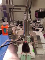
A Controlled Cortical Impact Mouse Model for Mild Traumatic Brain Injury


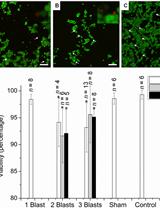
Primary Explosive Blast-induced Traumatic Brain Injury Model in PC12 Cell Culture


Plant Science
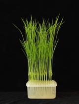
15N-nitrate Uptake Activity and Root-to-shoot Transport Assay in Rice

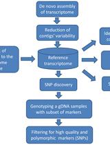
Experimental Pipeline for SNP and SSR Discovery and Genotyping Analysis of Mango (Mangifera indica L.)

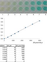
Determination of Recombinant Mannitol-1-phosphatase Activity from Ectocarpus sp.


Stem Cell
Bone Marrow-derived Endothelial Progenitor Cell Intercellular Adhesion Assay


