- Protocols

- Articles and Issues
- For Authors
- About
- Become a Reviewer

Past Issue in 2016
Volume: 6, Issue: 15
Biochemistry
Quantification of Chitinase Activity in Fusarium oxysporum


Analytical Gel Filtration for Probing Heavy Metal Transfer between Proteins

Cell Biology
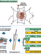
Mouse Liver Mitochondria Isolation, Size Fractionation, and Real-time MOMP Measurement


Cell Tracer Violet and CellTracker Red CMTPX Staining of Purified Mature Plasmodium-infected Red Blood Cells
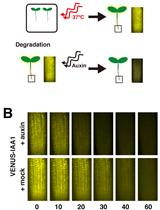
A Live-imaging, Heat Shock-inducible System to Measure Aux/IAA Degradation Rates in Planta
Immunology
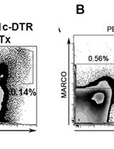
In vivo DCs Depletion with Diphtheria Toxin and MARCO+/MOMA1+ Cells Depletion with Clodronate Liposomes in B6.CD11c-DTR Mice
Microbiology
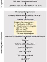
A Highly Efficient Method for Measuring Oxygen Consumption Rate in Fusarium graminearum

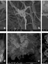
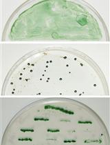
Antifungal and Zearalenone Inhibitory Activity of Ocimum sanctum L. Essential Oil on Fusarium graminearum Determined by UHPLC and RT-qPCR


Molecular Biology
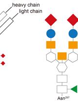
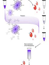
Generation of IgG-Fc Glycovariants Using Recombinant Glycosidases and Glycosyltransferases
Target Gene Inactivation in Cyanobacterium Anabaena sp. PCC 7120


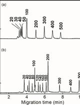
Capillary Electrophoresis in Hydroxyethylcellulose Solutions for the Analysis of dsDNA, dsRNA, and siRNA
Neuroscience
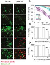
Acute Live/Dead Assay for the Analysis of Toxic Effects of Drugs on Cultured Neurons

Plant Science
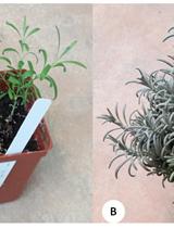
EST-SSR Analysis and Cross-species Transferability Study in Lavandula

Stem Cell
Neurosphere Co-culture Assay


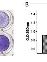
Bone Marrow Mesenchymal Stem Cells Adhesion Assay