- Protocols

- Articles and Issues
- For Authors
- About
- Become a Reviewer

Past Issue in 2016
Volume: 6, Issue: 10
Cell Biology

Isolating Liver Mitochondria by Differential Centrifugation

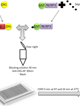
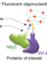
Liquid Luminescent DNA-precipitation Assay


Immunology
Proximity Ligation Assay (PLA) Protocol Using Duolink® for T Cells
Extraction and Quantification of Sphingosine 1-Phosphate (S1P)


Reconstitution of Lymphopaenic Mice with Regulatory and Conventional T cell Subsets


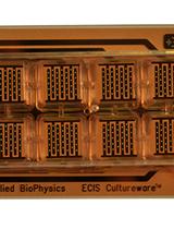
Study of Epithelium Barrier Functions by Real-time TER Measurement
Microbiology
Preparation of Respiratory Syncytial Virus with High or Low Content of Defective Viral Particles and Their Purification from Viral Stocks
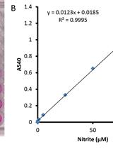
Nitrite Reduction Assay for Whole Pseudomonas Cells

Respiratory Syncytial Virus Infection in Mice and Detection of Viral Genomes in the Lung Using RT-qPCR
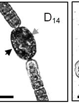
Induction, Isolation and Counting of Akinetes in Aphanizomenon ovalisporum


Molecular Biology
Preparation of Knockdown Transformants of Unicellular Charophycean Alga, Closterium peracerosum-strigosum-littorale Complex
Affymetrix Genome-wide Human SNP Assay
Neuroscience
Isolating Brain Mitochondria by Differential Centrifugation
Running Reward Conditioned Place Preference Task

Biotinylation and Purification of Plasma Membrane-associated Proteins from Rodent Cultured Neurons
