- Protocols

- Articles and Issues
- For Authors
- About
- Become a Reviewer

Past Issue in 2016
Volume: 6, Issue: 7
Biochemistry
Cloud-point PEG Glass Surfaces for Imaging of Immobilized Single Molecules by Total-internal-reflection Microscopy
Cancer Biology
Colon Cancer-associated Fibroblast Establishment and Culture Growth


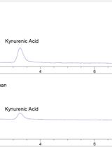
Extraction and Quantification of Tryptophan and Kynurenine from Cultured Cells and Media Using a High Performance Liquid Chromatography (HPLC) System Equipped with an Ultra-sensitive Diode Array Detector
Microbiology
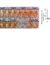
Rust Removal Experiments


Procedure for Rhamnolipids Quantification Using Methylene-blue

Methods for Detecting Microbial Methane Production and Consumption by Gas Chromatography


Molecular Biology
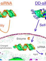
Conjugation of Duplexed siRNN Oligonucleotides with DD-HyNic Peptides for Cellular Delivery of RNAi Triggers
Neuroscience
Isolation and Primary Cell Culture of Mouse Dorsal Root Ganglion Neurons
Measurement of Inositol Triphosphate Levels from Rat Hippocampal Slices
Plant Science

Preparation of Mitotic and Meiotic Metaphase Chromosomes from Young Leaves and Flower Buds of Coccinia grandis


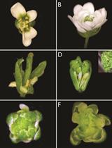
Strategies for Performing Dynamic Gene Perturbation Experiments in Flowers

Expression, Purification and Enzymatic Assay of Plant Histone Deacetylases
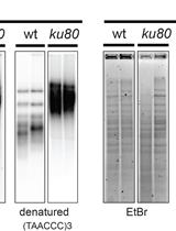
Analysis of Telomeric G-overhangs by in-Gel Hybridization
Total RNA Extraction from Grape Berry Skin for Quantitative Reverse Transcription PCR and Microarray Analysis


Stem Cell
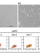
Isolation and Culture of Human CD133+ Non-adherent Endothelial Forming Cells