- Protocols

- Articles and Issues
- For Authors
- About
- Become a Reviewer

Past Issue in 2025
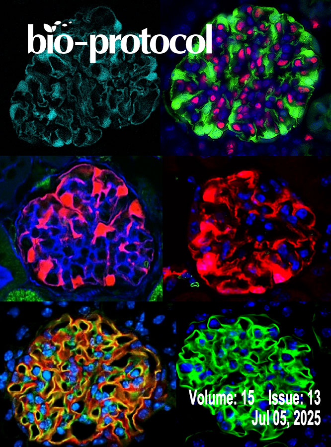
Volume: 15, Issue: 13
Biochemistry
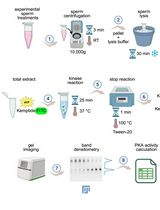
Kinase Mobility Shift Assay (KiMSA) for Assessing Protein Kinase A Activity


Bioinformatics and Computational Biology
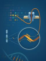
RACE-Nano-Seq: Profiling Transcriptome Diversity of a Genomic Locus

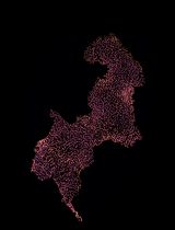
Visualization of the Evolution and Transmission of Circulating Vaccine-Derived Poliovirus (cVDPV) Outbreaks in the African Region
Cell Biology
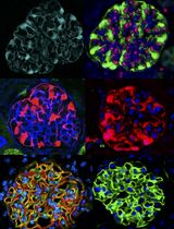
Isolation of Podocyte Cell Fractions From Mouse Kidney Using Magnetic Activated Cell Sorting (MACS)
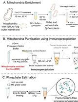
Isolation of Mitochondria From Yeast to Estimate Mitochondrial Pools of Inorganic Phosphate
Immunology
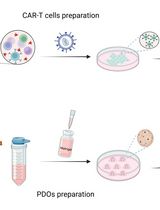
Evaluation of In Vitro Cytotoxic Activity of CAR-T Cells Using Patient-Derived Organoids
Microbiology
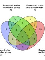
Untargeted Metabolomics of Epimastigote Forms of Trypanosoma cruzi
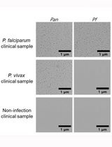
Rapid and Multiplex Diagnosis of Malaria Using Chelex-100 Extraction and LAMP-MS Assay
Molecular Biology
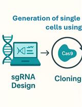
Protocol for Generation of Single-Gene Knockout in Hard-to-Transfect THP1 Cell Lines Using CRISPR/Cas9
APEX2 RNA Proximity Labeling in Mammalian Cell Lines With Low Biotin Permeability
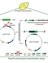
Assessing the Efficiency of Double-Strand Break Repair Mediated by Homologous Recombination and Non-homologous End-Joining Pathways in Saccharomyces cerevisiae
Neuroscience
Two-photon (2P) Microscopy to Study Ca2+ Signaling in Astrocytes From Acute Brain Slices
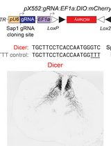
An Alternative Gene Editing Strategy Using a Single AAV Vector
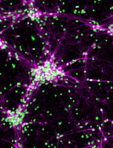
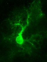
Derivation and Culture of Enriched Phrenic-Like Motor Neurons From Human iPSCs
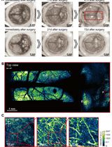
Construction of Large Cranial Windows With Nanosheet and Light-Curable Resin for Long-term Two-Photon Imaging in Mice
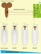
Live Cell Imaging to Monitor Axonal Pruning in Drosophila Motor Neurons
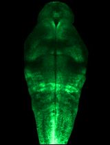
Quantification of Neural Progenitor Cells From Zika Virus-Infected Zebrafish Embryos