- Protocols

- Articles and Issues
- For Authors
- About
- Become a Reviewer

Past Issue in 2025
Volume: 15, Issue: 10
Bioinformatics and Computational Biology
From Bedside to Desktop: A Data Protocol for Normative Intracranial EEG and Abnormality Mapping
Cancer Biology
Stable 13C-glutamine Tracing Resolved Metabolomics for Cancer Metabolism Study
Cell Biology
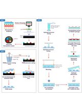
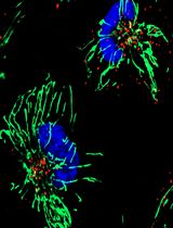
Development of Polyethylene Glycol Diacrylate-Based Micropattern Substrate to Study the Interplay Between Surface Topography and Cellular Response for Tissue Engineering Applications

Developmental Biology
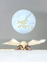
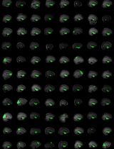
Egg Microinjection for the Silkworm Bombyx mori
Immunology
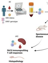
PBMC-Humanized Mouse Model for Multiple Sclerosis: Studying Immune Changes and CNS Involvement
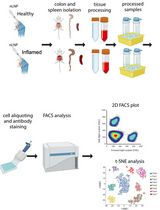
Tracking Oral Nanoparticle Uptake in Mouse Gastrointestinal Tract by Fluorescent Labeling and t-SNE Flow Cytometry
Medicine
Quantifying Thrombogenicity: A Bioanalytical Protocol for the Absorbance-Based Assessment of Vascular Implants with Plasma
Microbiology
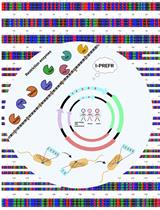
I-PREFR: Inverse PCR-Based Restriction Enzyme FRee Unidirectional Strategy for Rapid Markerless Chromosomal Gene Deletion and Reconstitution in Bacteria Using Suicide Vectors
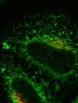
Synchronized Visualization and Analysis of Intracellular Trafficking and Maturation of Orthoflavivirus Subviral Particles
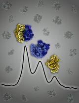
Mycobacterium smegmatis Ribosome Purification, Co-sedimentation, and Subunit Association Assay
Molecular Biology
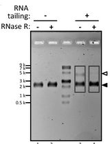
RNA PolyA Tailing Assay to Qualitatively Analyze Circular RNA Manufacturing
Neuroscience
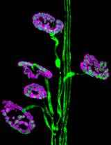
Dissection and Whole-Mount Immunofluorescent Staining of Mouse Hind Paw Muscles for Neuromuscular Junction Analysis

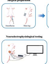
Electrophysiological Evaluation of a Sciatic Nerve Degree III Injury Model in Rats
Plant Science
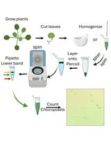
Rapid Miniprep of Intact Chloroplasts from Arabidopsis thaliana Leaves

High-Throughput Screening Identification of Chemical Compounds That Affect Cold-Regulated Gene Expression in Arabidopsis thaliana Using an Excised Single Leaf