- Protocols

- Articles and Issues
- For Authors
- About
- Become a Reviewer

Past Issue in 2025
Volume: 15, Issue: 8
Biochemistry
A Robust and Easy Protein Purification Method Using SpyDock-Modified Resin
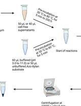
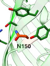
Endo-1,4-β-D-xylanase Assay Using Azo-Xylan and Variants Thereof
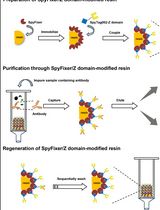
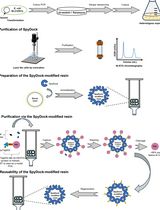
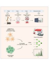
Antibody Purification Using Spy Chemistry
Bioinformatics and Computational Biology
GWAS Procedures for Gene Mapping in Diverse Populations With Complex Structures

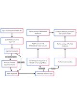
A Comprehensive Protocol for Bayesian Phylogenetic Analysis Using MrBayes: From Sequence Alignment to Model Selection and Phylogenetic Inference
Biophysics
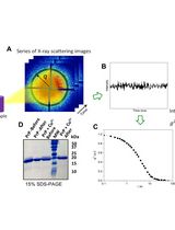
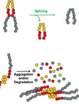
X-Ray Photon Correlation Spectroscopy, Microscopy, and Fluorescence Recovery After Photobleaching to Study Phase Separation and Liquid-to-Solid Transition of Prion Protein Condensates

Cancer Biology
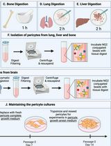
Isolation and Culture of Primary Pericytes from Mouse
Cell Biology
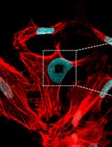
Visualization of F-Actin Through Expansion Microscopy (ExM) with Trifunctional Linker-Conjugated Phalloidin
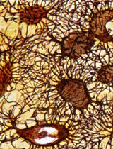
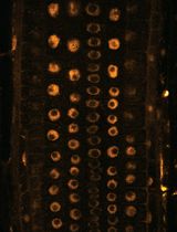
A Novel Optimized Silver Nitrate Staining Method for Visualizing and Quantifying the Osteocyte Lacuno-Canalicular System (LCS)
Immunology
In Vitro Bone Marrow–Derived Dendritic Cells (BMDC) Generation for Antigen Presentation Assay

Microbiology
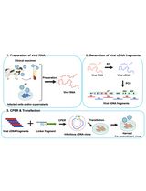
Rapid Plasmid-Free Generation of Recombinant Positive-Strand RNA Viruses That Use IRES-Mediated Translation Using an Expansion of the Circular Polymerase Extension Reaction (CPER)
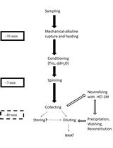
Scalable Alkaline Extraction Protocol for Microbial DNA Screening by PCR
Monitoring Protein Stability In Vivo Using an Intein-Based Biosensor
A Miniaturized Percoll Gradient Method for Isolation of Quiescent Cells of Yeast

Neuroscience
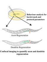
Analysis and Quantification of Functional Regeneration of Dendrite and Axon of PVD Neuron After Laser Injury in Caenorhabditis elegans
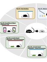
The Mouse Social Frailty Index (mSFI): A Standardized Protocol
Plant Science
Near-Infrared Autofluorescence Imaging of Nuclei in Living Plant Roots
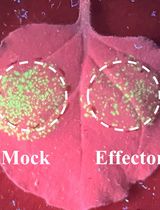
Workflow for a Functional Assay of Candidate Effectors From Phytopathogens Using a TMV-GFP-based System
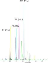
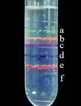
A New Approach to Detect and Semi-quantify All Molecular Species and Classes of Anionic Phospholipids Simultaneously in Plant Samples
Stem Cell
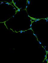
An Integrated Workflow for Three-Dimensional Visualization of Human Skeletal Muscle Stem Cell Nuclei