- Protocols

- Articles and Issues
- For Authors
- About
- Become a Reviewer

Past Issue in 2025
Volume: 15, Issue: 4
Biochemistry
Purification of Native Acetyl CoA Carboxylase From Mammalian Cells
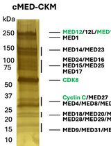
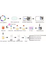
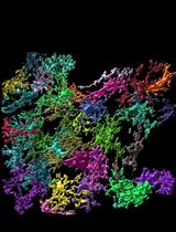
A Protocol to Purify Human Mediator Complex From Freestyle 293-F Cells

HPLC Analysis of tRNA‐Derived Nucleosides
Bioinformatics and Computational Biology
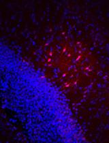
Streamlined Quantification of Microglial Morphology in Mouse Brains Using 3D Immunofluorescence Analysis

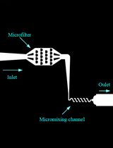
Quantifying Bacterial Chemotaxis in Controlled and Stationary Chemical Gradients With a Microfluidic Device


Biophysics
A PDMS-based Microfluidic Chip Assembly for Time-Resolved Cryo-EM (TRCEM) Sample Preparation
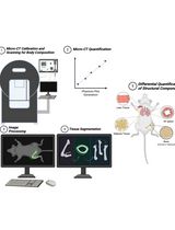
A Micro-Computed Tomography-Based Simplified Approach to Measure Body Composition, Osteoporosis, and Lung Fibrosis in Mice
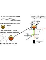
Voltage Clamp Fluorometry in Xenopus laevis Oocytes to Study the Voltage-sensing Phosphatase

Cancer Biology
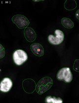
Streamlined Quantification of p-γ-H2AX Foci for DNA Damage Analysis in Melanoma and Melanocyte Co-cultures Exposed to FLASH Irradiation Using Automated Image Cytometry
Cell Biology
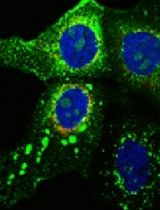
Combined FLIM, Confocal Microscopy, and STED Nanoscopy for Live-Cell Imaging
Developmental Biology
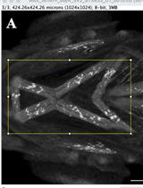
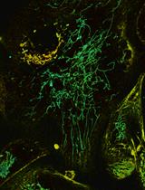
Quantification of Neuromuscular Junctions in Zebrafish Cranial Muscles
Environmental science
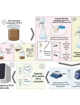
Novel Workflows for Separate Isolation of Pathogen RNA or DNA From Wastewater: Detection by Innovative and Conventional qPCR
Microbiology
Generation, Propagation, and Titering of Dicistrovirus From an Infectious Clone

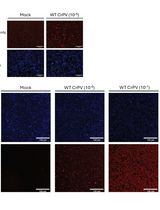
Development and Application of MLB Human Astrovirus Reverse Genetics Clones and Replicons

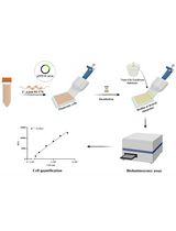
Campylobacter jejuni Biofilm Assessment by NanoLuc Luciferase Assay
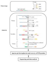
Protocol to Mine Unknown Flanking DNA Using PER-PCR for Genome Walking
Molecular Biology
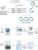
Leveraging Circular Polymerization and Extension Cloning (CPEC) Method for Construction of CRISPR Screening Libraries
Neuroscience
Visualization of Gap Junction–Mediated Astrocyte Coupling in Acute Mouse Brain Slices
Locomotor Activity Monitoring in Mice to Study the Phase Shift of Circadian Rhythms Using ClockLab (Actimetrics)
Plant Science
Closed Systems to Study Plant–Filamentous Fungi Associations: Emphasis on Microscopic Analyses

Transgene-free Genome Editing in Grapevine
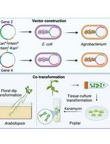
A Novel Gene Stacking Method in Plant Transformation Utilizing Split Selectable Markers
Simple Method for Efficient RNA Extraction From Arabidopsis Embryos
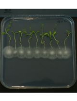
Development of a Rapid and Efficient Protocol for Seed Germination and Seedling Establishment of Oryza coarctata

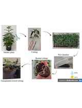
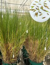
Vegetative Propagation of Cannabis sativa and Resin Obtained From its Female Inflorescences
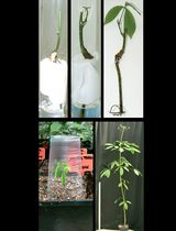
Micrografting Technique of Hevea brasiliensis In Vitro Plantlets
Stem Cell
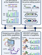
Precise Generation of Human Induced Pluripotent Stem Cell–Derived Cell Lines Harboring Disease-relevant Single Nucleotide Variants Using a Prime Editing System
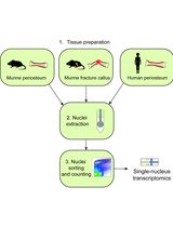
Nuclei Isolation From Murine and Human Periosteum For Transcriptomic Analyses