- Protocols

- Articles and Issues
- For Authors
- About
- Become a Reviewer

Past Issue in 2024
Volume: 14, Issue: 19
Biophysics
Fluorescence Lifetime-Assisted Probing of Protein Aggregation with sub-Organellar Resolution
Cell Biology
Construction and Application of a Static Magnetic Field Exposure Apparatus for Biological Research in Aqueous Model Systems and Cell Culture

Acutely Modifying Phosphatidylinositol Phosphates on Endolysosomes Using Chemically Inducible Dimerization Systems
Immunology
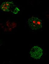
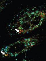
Measuring Piezo1 and Actin Polarity in Chemokine-Stimulated Jurkat Cells During Live-Cell Imaging

Microbiology
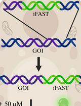
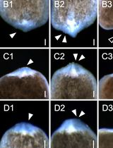
Genetic Tagging and Imaging of Proteins with iFAST in Candida albicans
Neuroscience
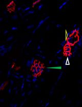
Visualization and Analysis of Neuromuscular Junctions Using Immunofluorescence
Plant Science
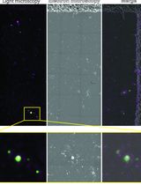
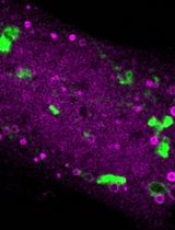
Sorghum bicolor Extracellular Vesicle Isolation, Labeling, and Correlative Light and Electron Microscopy
Stem Cell
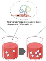
Alternative Method for Obtaining Human-Induced Pluripotent Stem Cell Lines and Three-Dimensional Growth: A Simplified, Passage-Free Approach that Minimizes Labor
