- Protocols

- Articles and Issues
- For Authors
- About
- Become a Reviewer

Past Issue in 2015
Volume: 5, Issue: 24
Cancer Biology
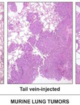
Analysis of Murine Lung Tumors by Micro PET-CT Imaging


Generation of Mouse Thyroid Calcitonin-producing Cell Tumors from Primary Mouse Tumors

Microbiology
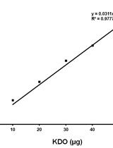
Determination of Keto-deoxy-d-manno-8-octanoic acid (KDO) from Lipopolysaccharide of Escherichia coli


Preparation and Analysis of Crude Autolytic Enzyme Extracts from Staphylococcus aureus


Transformation of the Cyanobacterium Leptolyngbya boryana by Electroporation
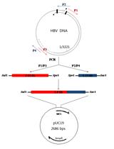
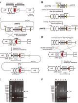
Characterization of HBV Isolates from Patient Serum Samples and Cloning
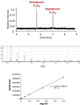
Extraction and Quantification of Alkanes in Cyanobacteria
![[14C] Linoleic Acid Uptake and Fractionation Assay in Vibrio cholerae](https://en-cdn.bio-protocol.org/imageup/arcimg/20151221041353472.jpg?t=1774097894)
[14C] Linoleic Acid Uptake and Fractionation Assay in Vibrio cholerae
Plant Science
Luminol-based Assay for Detection of Immunity Elicitor-induced Hydrogen Peroxide Production in Arabidopsis thaliana Leaves

Extraction of Apoplastic Wash Fluids and Leaf Petiole Exudates from Leaves of Arabidopsis thaliana

GC-MS-Based Analysis of Chloroform Extracted Suberin-Associated Root Waxes from Arabidopsis and Other Plant Species

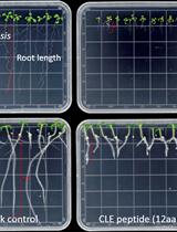
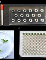
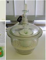
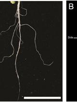
In vitro CLE Peptide Bioactivity Assay on Plant Roots


Isolation of Tonoplast Vesicles from Tomato Fruit Pericarp


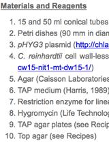
Insertional Mutagenesis of Chlamydomonas reinhardtii
Stem Cell
Porous Scaffold Seeding and Chondrogenic Differentiation of BMSC-seeded Scaffolds


