Past Issue in 2023

Volume: 13, Issue: 24

left

Dec

20

Dec

5

Nov

20

Nov

5

Oct

20

Oct

5

Sep

20

Sep

5

Aug

20

Aug

5

Jul

20

Jul

5

Jun

20

Jun

5

May

20

May

5

Apr

20

Apr

5

Mar

20

Mar

5

Feb

20

Feb

5

Jan

20

Jan

5

right

Biophysics

Correlative Light and Electron Cryo-Microscopy Workflow Combining Micropatterning, Ice Shield, and an In-Chamber Fluorescence Light Microscope

Correlative Light and Electron Cryo-Microscopy Workflow Combining Micropatterning, Ice Shield, and an In-Chamber Fluorescence Light Microscope

SB Sabrina Berkamp
DD Deniz Daviran
MS Marit Smeets
AC Alexane Caignard
Rj Riddhi A. Jani
PS Pia Sundermeyer
CJ Caspar Jonker
SG Sven Gerlach
BH Bernd Hoffmann
KL Katherine Lau
CS Carsten Sachse
3040 Views
Dec 20, 2023
In situ cryo-electron tomography (cryo-ET) is the most current, state-of-the-art technique to study cell machinery in its hydrated near-native state. The method provides ultrastructural details at sub-nanometer resolution for many components within the cellular context. Making use of recent advances in sample preparation techniques and combining this method with correlative light and electron microscopy (CLEM) approaches have enabled targeted molecular visualization. Nevertheless, the implementation has also added to the complexity of the workflow and introduced new obstacles in the way of streamlining and achieving high throughput, sample yield, and sample quality. Here, we report a detailed protocol by combining multiple newly available technologies to establish an integrated, high-throughput, optimized, and streamlined cryo-CLEM workflow for improved sample yield.Key features• PRIMO micropatterning allows precise cell positioning and maximum number of cell targets amenable to thinning with cryo focused-ion-beam–scanning electron microscopy.• CERES ice shield ensures that the lamellae remain free of ice contamination during the batch milling process.• METEOR in-chamber fluorescence microscope facilitates the targeted cryo focused-ion-beam (cryo FIB) milling of these targets.• Combining the three technologies into one cryo-CLEM workflow maximizes sample yield, throughput, and efficiency.Graphical overview

Cell Biology

Streamlined Methods for Processing and Cryopreservation of Cell Therapy Products Using Automated Systems

Streamlined Methods for Processing and Cryopreservation of Cell Therapy Products Using Automated Systems

YL Ye Li
Hazel Y. Stevens Hazel Y. Stevens
SS Srikanth Sivaraman
LP Logan N. Porter
AH Allison R. Hoffman
SG Stuart L. Gibb
SS Shivaram Selvam
AB Annie C. Bowles-Welch
2197 Views
Dec 20, 2023
Streamlined procedures for processing and cryopreservation of cell therapies using good laboratory practices are integral to biomanufacturing process development and clinical applications. The protocol herein begins with the preparation of human cell types cultured as adherent (i.e., mesenchymal stromal cells, MSCs) or suspension cells (i.e., peripheral blood mononuclear cells, PBMCs) to comprehensively demonstrate procedures that are applicable to commonly used primary cell cultures. Cell processing steps consist of preparing high yields of cells for cryopreservation using instruments routinely used in cell manufacturing, including the Finia® Fill and Finish System and a controlled-rate freezer. The final steps comprise the storage of cells at subzero temperatures in liquid nitrogen vapor phase followed by the analysis of cell phenotypes before and after processing and cryopreservation, along with cell quality metrics for validation. Additionally, the protocol includes important considerations for the implementation of quality control measures for equipment operation and cell handling, as well as Good Laboratory Practices for cell manufacturing, which are essential for the translational use of cell therapies.Key features• The protocol applies to small- or large-scale manufacturing of cell therapy products.• It includes streamlined procedures for processing and cryopreservation of cells cultured as adherent cells (MSCs) and suspension cells (PBMCs).• Provides temperature control and rapid partitioning of sample in cryopreservation solution to maintain high viability of a range of cell types throughout the procedures.• This protocol employs the Finia® Fill and Finish System and a controlled-rate freezer.Graphical overview

Developmental Biology

Quantifying Cell Proliferation Through Immunofluorescence on Whole-Mount and Cryosectioned Regenerating Caudal Fins in African Killifish

Quantifying Cell Proliferation Through Immunofluorescence on Whole-Mount and Cryosectioned Regenerating Caudal Fins in African Killifish

AO Augusto Ortega Granillo
RS Robert R. Schnittker
WW Wei Wang
AA Alejandro Sánchez Alvarado
1742 Views
Dec 20, 2023
The African killifish Nothobranchius furzeri is an attractive research organism for regeneration- and aging-related studies due to its remarkably short generation time and rapid aging. Dynamic changes in cell proliferation are an essential biological process involved in development, regeneration, and aging. Quantifying the dynamics of cell proliferation in these contexts facilitates the elucidation of the attendant underlying mechanisms. Whole-mount and cryosectioning sample preparation are the preferred approaches to investigate the distribution of cellular structures, cell–cell communication, and spatial gene expression within tissues. Using African killifish caudal fin regeneration as an example, we describe an efficient and detailed protocol to investigate cell proliferation dynamics in both space and time during caudal fin regeneration. The quantification of cell proliferation was achieved through high-resolution immunofluorescence of the proliferation marker Phospho-Histone H3 (H3P). We focused on the characterization of epithelial and mesenchymal proliferation in three-dimensional space at two regeneration time points. Our protocol provides a reliable tool for comparing cell proliferation under different biological contexts.Key features• Elaborates in detail the method used by Wang et al. (2020) to quantify whole-organ mitotic events during tail fin regeneration in vertebrates.• Enables proliferation analysis of millimeter-sized homeostatic and regenerating tissues.• Three-day alternative method to whole mount using cryosections.• Allows automatic quantification using ImageJ macros and R scripts.Graphical overview

Immunology

In vitro Assessment of Efferocytic Capacity of Human Macrophages Using Flow Cytometry

In vitro Assessment of Efferocytic Capacity of Human Macrophages Using Flow Cytometry

AS Ana C.G. Salina
MF Marlon Fortes-Rocha
LC Larissa D. Cunha
3651 Views
Dec 20, 2023
Clearance of dying cells, named efferocytosis, is a pivotal function of professional phagocytes that impedes the accumulation of cell debris. Efferocytosis can be experimentally assessed by differentially tagging the target cells and professional phagocytes and analyzing by cell imaging or flow cytometry. Here, we describe an assay to evaluate the uptake of apoptotic cells (ACs) by human macrophages in vitro by labeling the different cells with commercially available dyes and analysis by flow cytometry. We detail the methods to prepare and label human macrophages and apoptotic lymphocytes and the in vitro approach to determine AC uptake. This protocol is based on previously published literature and allows for in vitro modeling of the efficiency of AC engulfment during continual efferocytosis process. Also, it can be modified to evaluate the clearance of different cell types by diverse professional phagocytes.Graphical overview
An Optimised Indirect ELISA Protocol for Detection and Quantification of Anti-viral Antibodies in Human Plasma or Serum: A Case Study Using SARS-CoV-2

An Optimised Indirect ELISA Protocol for Detection and Quantification of Anti-viral Antibodies in Human Plasma or Serum: A Case Study Using SARS-CoV-2

CB Claire Baine
SJ Jackson Sembera
GO Gerald Kevin Oluka
JK Joseph Ssebwana Katende
VA Violet Ankunda
JS Jennifer Serwanga
2331 Views
Dec 20, 2023
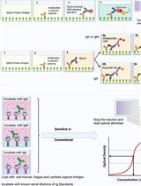
Advanced immunoassays are crucial in assessing antibody responses, serving immune surveillance goals, characterising immunological responses to evolving viral variants, and guiding subsequent vaccination initiatives. This protocol outlines an indirect ELISA protocol to detect and quantify virus-specific antibodies in plasma or serum after exposure to viral antigens. The assay enables the measurement of IgG, IgA, and IgM antibodies specific to the virus of interest, providing qualitative and quantitative optical densities and concentration data. Although this protocol refers to SARS-CoV-2, its methodology is versatile and can be modified to assess antibody responses for various viral infections and to evaluate vaccine trial outcomes.Key features• This protocol builds upon previously described methodology [1] explicitly tailored for SARS-CoV-2 and broadens its applicability to other viral infections.• The protocol outlines establishing antibody responses to SARS-CoV-2 infections by determining optical densities and concentrations from blood plasma or serum.Graphical overview Summary of the conventional ELISA (A) vs. sensitive ELISA (B) procedures. In both A and B, wells are coated with a capture antigen, such as the spike protein, while in (C) they are coated with human Kappa and Lambda capture antibodies. For the conventional ELISA (A), wells with immobilised capture antigens receive serum/plasma containing the target antibody (A1 and B1). This is followed by an HRP-conjugated detection antibody specific to the captured antibody (A2 and B2) and then a substrate solution that reacts with the HRP, producing a colour proportional to the concentration of the antibody in the serum/plasma (A3 and B3). The reaction is halted, and absorbance is measured. In the sensitive ELISA (B), after the serum/plasma addition (A1 and B1), a Biotin-conjugated primary detection antibody is introduced (A2 and B2). Depending on the target antibody, a secondary streptavidin-HRP conjugated detection antibody is added for IgG or IgM (3a) or a poly-HRP 40 detection antibody for IgA (3b). A substrate is introduced, producing a colour change proportional to the antibody concentration (A4 and B4). The reaction is then stopped, and absorbance is measured. In Panel C, wells are coated with human Kappa and Lambda capture antibodies. Serial dilutions of a known antibody standard are introduced. After undergoing the standard ELISA steps, a detection antibody is added, specifically binding to the Ig standard antibody. Subsequently, a substrate solution causes a colour change proportional to the antibody concentration in the serum/plasma. The reaction is halted, and the absorbance of each well is measured. The resulting optical densities from the coated wells form the standard curve, plotting the absorbance against concentrations.

Microbiology

Identifying Antigenic Switching by Clonal Cell Barcoding and Nanopore Sequencing in Trypanosoma brucei

Identifying Antigenic Switching by Clonal Cell Barcoding and Nanopore Sequencing in Trypanosoma brucei

AT Abdoulie O. Touray
TS Tamara Sternlieb
TI Tony Isebe
IC Igor Cestari
1567 Views
Dec 20, 2023
Many organisms alternate the expression of genes from large gene sets or gene families to adapt to environmental cues or immune pressure. The single-celled protozoan pathogen Trypanosoma brucei spp. periodically changes its homogeneous surface coat of variant surface glycoproteins (VSGs) to evade host antibodies during infection. This pathogen expresses one out of ~2,500 VSG genes at a time from telomeric expression sites (ESs) and periodically changes their expression by transcriptional switching or recombination. Attempts to track VSG switching have previously relied on genetic modifications of ES sequences with drug-selectable markers or genes encoding fluorescent proteins. However, genetic modifications of the ESs can interfere with the binding of proteins that control VSG transcription and/or recombination, thus affecting VSG expression and switching. Other approaches include Illumina sequencing of the VSG repertoire, which shows VSGs expressed in the population rather than cell switching; the Illumina short reads often limit the distinction of the large set of VSG genes. Here, we describe a methodology to study antigenic switching without modifications of the ES sequences. Our protocol enables the detection of VSG switching at nucleotide resolution using multiplexed clonal cell barcoding to track cells and nanopore sequencing to identify cell-specific VSG expression. We also developed a computational pipeline that takes DNA sequences and outputs VSGs expressed by cell clones. This protocol can be adapted to study clonal cell expression of large gene families in prokaryotes or eukaryotes.Key features• This protocol enables the analysis of variant surface glycoproteins (VSG) switching in T. brucei without modifying the expression site sequences.• It uses a streamlined computational pipeline that takes fastq DNA sequences and outputs expressed VSG genes by each parasite clone.• The protocol leverages the long reads sequencing capacity of the Oxford nanopore sequencing technology, which enables accurate identification of the expressed VSGs.• The protocol requires approximately eight to nine days to complete.Graphical overview

Neuroscience

Headplate Installation and Craniotomy for Awake In Vivo Electrophysiological Recordings or Two-Photon Imaging of the Mouse Inferior Colliculus

Headplate Installation and Craniotomy for Awake In Vivo Electrophysiological Recordings or Two-Photon Imaging of the Mouse Inferior Colliculus

BK Blom Kraakman
SS Sofja Solovjova
JB J. Gerard G. Borst
AW Aaron Benson Wong
1716 Views
Dec 20, 2023
The inferior colliculus (IC) is an important processing center in the auditory system, which also receives non-auditory sensory input. The IC consists of several subnuclei whose functional role in (non-) auditory processing and plastic response properties are best approached by studying awake animals, preferably in a longitudinal fashion. The increasing use of mice in auditory research, the availability of genetic models, and the superficial location of the IC in the mouse have made it an attractive species for studying IC function. Here, we describe a protocol for exposing the mouse IC for up to a few weeks for in vivo imaging or electrophysiology in a stable manner. This method allows for a broader sampling of the IC while maintaining the brain surface in good quality and without reopening the craniotomy. Moreover, as it is adaptable for both electrophysiological recordings of the entire IC and imaging of the dorsal IC surface, it can be applied to answer a multitude of questions.Key features• A surgical protocol for long-term physiological recordings from the same or separate neuronal populations in the inferior colliculus.• Optimized for awake in vivo experiments in the house mouse (Mus musculus).
Dissociation and Culture of Adult Mouse Satellite Glial Cells

Dissociation and Culture of Adult Mouse Satellite Glial Cells

RT Raquel Tonello
SD Steve Davidson
TB Temugin Berta
1406 Views
Dec 20, 2023
Satellite glial cells (SGCs) are a type of glial cell population that originates from neural crest cells. They ultimately migrate to surround the cell bodies of neurons in the ganglia of the peripheral nervous system. Under physiological conditions, SGCs perform homeostatic functions by modifying the microenvironment around nearby neurons and provide nutrients, structure, and protection. In recent years, they have gained considerable attention due to their involvement in peripheral nerve regeneration and pain. Although methods for culturing neonatal or rat SGCs have long existed, a well-characterized method for dissociating and culturing adult SGCs from mouse tissues has been lacking until recently. This has impeded further studies of their function and the testing of new therapeutics. This protocol provides a detailed description of how to obtain primary cultures of adult SGCs from mouse dorsal root ganglia in approximately two weeks with over 90% cell purity. We also demonstrate cell purity of these cultures using quantitative real-time RT-PCR and their functional integrity using calcium imaging.Key features• Detailed and simplified protocol to dissociate and culture primary satellite glial cells (SGCs) from adult mice.• Cells are dissociated in approximately 2–3 h and cultured for approximately two weeks.• These SGC cultures allow both molecular and functional studies.Graphical overviewDissociation and culture of mouse satellite glial cells