Past Issue in 2023

Volume: 13, Issue: 11

left

Dec

20

Dec

5

Nov

20

Nov

5

Oct

20

Oct

5

Sep

20

Sep

5

Aug

20

Aug

5

Jul

20

Jul

5

Jun

20

Jun

5

May

20

May

5

Apr

20

Apr

5

Mar

20

Mar

5

Feb

20

Feb

5

Jan

20

Jan

5

right

Biophysics

Synthesis and Purification of Lipid-conjugated Fluorescent pH Sensors

Synthesis and Purification of Lipid-conjugated Fluorescent pH Sensors

WW Wiebke Wiesner
RK Ronja Marie Kühnel
TP Thomas Guenther Pomorski
1209 Views
Jun 5, 2023
Lipid-conjugated pH sensors based on fluorophores coupled to lipids are a powerful tool for monitoring pH gradients in biological microcompartments and reconstituted membrane systems. This protocol describes the synthesis of pH sensors based on amine-reactive pHrodo esters and the amino phospholipid phosphatidylethanolamine. The major features of this sensor include efficient partitioning into membranes and strong fluorescence under acidic conditions. The protocol described here can be used as a template to couple other amine-reactive fluorophores to phosphatidylethanolamines.Graphical overviewSynthesis of lipid-conjugated pH sensors based on amine-reactive fluorophore esters and the aminophospholipid phosphoethanolamine (PE)

Cell Biology

Cycloheximide (CHX) Chase Assay to Examine Protein Half-life

Cycloheximide (CHX) Chase Assay to Examine Protein Half-life

YM Ying Miao
QD Qian Du
HZ Hong-Guang Zhang
YY Yukang Yuan
YZ Yibo Zuo
HZ Hui Zheng
12838 Views
Jun 5, 2023
Cycloheximide (CHX) is a small molecule derived from Streptomyces griseus that acts as fungicide. As a ribosome inhibitor, CHX can restrict the translation elongation of eukaryotic protein synthesis. Once protein synthesis is inhibited by CHX, the level of intracellular proteins decreases by degradation through the proteasome or lysosome system. Thus, the CHX chase assay is widely recognized and used to observe intracellular protein degradation and to determine the half-life of a given protein in eukaryotes. Here, we present a complete experimental procedure of the CHX chase assay.Graphical overview

Developmental Biology

Polysome Profiling in Adult Mouse Testes

Polysome Profiling in Adult Mouse Testes

JK Jun-Yan Kang
AZ Ai Zhong
ZW Ze Wen
XY Xinghai Yu
YZ Yu Zhou
ML Mo-Fang Liu
1567 Views
Jun 5, 2023
Polysome profiling is widely used to isolate and analyze polysome fractions, which consist of actively translating mRNAs and ribosomes. Compared to ribosome profiling and translating ribosome affinity purification, polysome profiling is simpler and less time consuming in sample preparation and library constructions. Spermiogenesis, i.e., the post-meiotic phase of male germ cell development, is a highly coordinated developmental process in which transcription and translation are decoupled because of nuclear condensation, resulting in translation regulation as the major mode for the regulation of gene expression in post-meiotic spermatids. To understand the translation regulation during spermiogenesis, an overview of translational state of spermiogenic mRNAs is required. Here, we describe a protocol to identify translating mRNAs using polysome profiling. Briefly, mouse testes are gently homogenized to release polysomes containing translating mRNAs, following polysome-bound mRNAs isolated by sucrose density gradient purification and characterized by RNA-seq. This protocol allows to quickly isolate translating mRNAs from testes and analyze the discrepancy of translational efficiency in mouse testes from different mouse lines.Key features• Quickly obtain polysome RNAs from testes.• Omit RNase digestion and RNA recovery from gel.• High efficiency and robustness compared to ribo-seq.Graphical overviewSchematic illustrating the experimental design for polysome profiling in mouse testes. Mouse testes are homogenized and lysed in Sample preparation, and polysome RNAs are enriched by sucrose gradient centrifugation and used to calculate translation efficiency in Sample analysis.

Immunology

Large-scale Isolation of Exosomes Derived from NK Cells for Anti-tumor Therapy

Large-scale Isolation of Exosomes Derived from NK Cells for Anti-tumor Therapy

HL Heyong Luo
JZ Jing Zhang
AY Anqing Yang
WO Weiwei Ouyang
SL Shiqi Long
XL Xiaojin Lin
NY Na Yang
ZY Zhiru Yang
YZ Yingchun Zhang
YW Wei Yang
QC Qiyuan Che
YY Yuxin Yang
TG Ting Guo
XZ Xing Zhao
3306 Views
Jun 5, 2023
Exosomes are lipid bilayer–enclosed vesicles, actively secreted by cells, containing proteins, lipids, nucleic acids, and other substances with multiple biological functions after entering target cells. Exosomes derived from NK cells have been shown to have certain anti-tumor effects and potential applications as chemotherapy drug carriers. These developments have resulted in high demand for exosomes. Although there has been large-scale industrial preparation of exosomes, they are only for generally engineered cells such as HEK 293T. The large-scale preparation of specific cellular exosomes is still a major problem in laboratory studies. Therefore, in this study, we used tangential flow filtration (TFF) to concentrate the culture supernatants isolated from NK cells and isolated NK cell–derived exosomes (NK-Exo) by ultracentrifugation. Through a series of characterization and functional verification of NK-Exo, the characterization, phenotype, and anti-tumor activity of NK-Exo were verified. Our study provides a considerably time- and labor-saving protocol for the isolation of NK-Exo.

Microbiology

Detecting Photoactivatable Cre-mediated Gene Deletion Efficiency in Escherichia coli

Detecting Photoactivatable Cre-mediated Gene Deletion Efficiency in Escherichia coli

YK Yuta Koganezawa
YW Yuichi Wakamoto
MS Moritoshi Sato
MU Miki Umetani
1312 Views
Jun 5, 2023
Gene deletion is one of the standard approaches in genetics to investigate the roles and functions of target genes. However, the influence of gene deletion on cellular phenotypes is usually analyzed sometime after the gene deletion was introduced. Such lags from gene deletion to phenotype evaluation could select only the fittest fraction of gene-deleted cells and hinder the detection of potentially diverse phenotypic consequences. Therefore, dynamic aspects of gene deletion, such as real-time propagation and compensation of deletion effects on cellular phenotypes, still need to be explored. To resolve this issue, we have recently introduced a new method that combines a photoactivatable Cre recombination system and microfluidic single-cell observation. This method enables us to induce gene deletion at desired timings in single bacterial cells and to monitor their dynamics for prolonged periods. Here, we detail the protocol for estimating the fractions of gene-deleted cells based on a batch-culture assay. The duration of blue light exposure significantly affects the fractions of gene-deleted cells. Therefore, gene-deleted and non-deleted cells can coexist in a cellular population by adjusting the duration of blue light exposure. Single-cell observations under such illumination conditions allow the comparison of temporal dynamics between gene-deleted and non-deleted cells and unravel phenotypic dynamics provoked by gene deletion.

Molecular Biology

Revised iCLIP-seq Protocol for Profiling RNA–protein Interaction Sites at Individual Nucleotide Resolution in Living Cells

Revised iCLIP-seq Protocol for Profiling RNA–protein Interaction Sites at Individual Nucleotide Resolution in Living Cells

SN Syed Nabeel-Shah
JG Jack F. Greenblatt
3638 Views
Jun 5, 2023
Individual nucleotide resolution UV cross-linking and immunoprecipitation followed by high-throughput sequencing (iCLIP-seq) is a powerful technique that is used to identify RNA-binding proteins’ (RBP) binding sites on target RNAs and to characterize the molecular basis of posttranscriptional regulatory pathways. Several variants of CLIP have been developed to improve its efficiency and simplify the protocol [e.g., iCLIP2 and enhanced CLIP (eCLIP)]. We have recently reported that transcription factor SP1 functions in the regulation of alternative cleavage and polyadenylation through direct RNA binding. We utilized a modified iCLIP method to identify RNA-binding sites for SP1 and several of the cleavage and polyadenylation complex subunits, including CFIm25, CPSF7, CPSF100, CPSF2, and Fip1. Our revised protocol takes advantage of several features of the eCLIP procedure and also improves on certain steps of the original iCLIP method, including optimization of circularization of cDNA. Herein, we describe a step-by-step procedure for our revised iCLIP-seq protocol, that we designate as iCLIP-1.5, and provide alternative approaches for certain difficult-to-CLIP proteins.Key features• Identification of RNA-binding sites of RNA-binding proteins (RBPs) at nucleotide resolution.• iCLIP-seq provides precise positional and quantitative information on the RNA-binding sites of RBPs in living cells.• iCLIP facilitates the identification of sequence motifs recognized by RBPs.• Allows quantitative analysis of genome-wide changes in protein-RNA interactions.• Revised iCLIP-1.5 protocol is more efficient and highly robust; it provides higher coverage even for low-input samples.Graphical overview

Neuroscience

Effective Hand Rearing of Neonatal Mice for Developmental Studies

Effective Hand Rearing of Neonatal Mice for Developmental Studies

LM Marcus N. Leiwe
IT Satoshi Fujimoto
TI Takeshi Imai
1499 Views
Jun 5, 2023
Chronic manipulation in neonatal mice is a technical challenge, but it can achieve greater insights into how mice develop immediately after birth. However, these manipulations can often result in maternal rejection and consequently serious malnourishment and occasional death. Here, we describe a method to effectively hand rear mice to develop normally during the first post-natal week. In our experiments, we were able to negate the feeding deficiencies of anosmic mutant mice when compared to littermate controls. As a result, the delayed neuronal remodeling seen in maternally reared mutant mice was not seen in the hand-reared mutant mice. This methodology is user intensive but can be useful for a broad range of studies either requiring many interventions or one intervention that can result in maternal rejection or being outcompeted by healthy littermates.

Plant Science

Transformation and Detection of Soybean Hairy Roots

Transformation and Detection of Soybean Hairy Roots

XX Xing Xu
TY Tai-Fei Yu
JM Jian Ma
JC Jun Chen
YZ Yong-Bin Zhou
MC Ming Chen
ZC Zhan-Yu Chen
YM You-Zhi Ma
ZX Zhao-Shi Xu
ZZ Zhi-An Zhang
1657 Views
Jun 5, 2023
Agrobacterium rhizogenes is a soil bacteria with extensive infectivity, which can infect almost all dicotyledonous plants and a few monocotyledonous plants to induce root nodules. This is caused by the root-inducing plasmid, which contains genes responsible for the autonomous growth of root nodules and crown gall base synthesis. Structurally, it is similar to the tumor-inducing plasmid in that it mainly contains the Vir region, the T-DNA region, and the functional region of crown gall base synthesis. Its T-DNA is integrated into the nuclear genome of the plant with the assistance of Vir genes, causing hairy root disease in the host plant and the formation of hairy roots. The roots produced by Agrobacterium rhizogenes–infested plants are characterized by a fast growth rate, high degree of differentiation, physiological, biochemical, and genetic stability, and ease of manipulation and control. In particular, the hairy root system is an efficient and rapid research tool for plants that have no affinity for transformation by Agrobacterium rhizogenes and low transformation efficiency. The establishment of germinating root culture system for the production of secondary metabolites in the original plants through the genetic transformation of natural plants mediated by root-inducing plasmid in Agrobacterium rhizogenes has become a new technology combining plant genetic engineering and cell engineering. It has been widely used in a variety of plants for different molecular purposes, such as pathological analysis, gene function verification, and secondary metabolite research. Chimeric plants obtained by induction of Agrobacterium rhizogenes that can be expressed instantaneously and contemporarily are more rapidly obtained, compared to tissue culture and stably inheritable transgenic strains. In general, transgenic plants can be obtained in approximately one month.
Measurement of Total Phosphorus and Polyphosphate in Chlamydomonas reinhardtii

Measurement of Total Phosphorus and Polyphosphate in Chlamydomonas reinhardtii

YY Yujie Yang
SR Suna Ren
XJ Xianqing Jia
HZ Houqing Zeng
LW Long Wang
YZ Yiyong Zhu
KY Keke Yi
879 Views
Jun 5, 2023
Phosphorus is an essential nutrient for plants. Green algae usually store excess P as polyphosphate (polyP) in the vacuoles. PolyP, a linear chain of three to hundreds of phosphate residues linked by phosphoanhydride bonds, is important for cell growth. Based on the previous method of polyP purification with silica gel columns (Werner et al., 2005; Canadell et al., 2016) in yeast cells, we developed a protocol to purify and determine the total P and polyP in Chlamydomonas reinhardtii by a quick, simplified, and quantitative method. We use hydrochloric acid or nitric acid to digest polyP or total P in dried cells and analyze P content using the malachite green colorimetric method. This method may be applied to other microalgae.
An Improved System to Measure Leaf Gas Exchange on Adaxial and Abaxial Surfaces

An Improved System to Measure Leaf Gas Exchange on Adaxial and Abaxial Surfaces

DM D. A. Márquez
HS H. Stuart-Williams
SW S. C. Wong
GF G. D. Farquhar
796 Views
Jun 5, 2023
Measurement of leaf carbon gain and water loss (gas exchange) in planta is a standard procedure in plant science research for attempting to understand physiological traits related to water use and photosynthesis. Leaves carry out gas exchange through the upper (adaxial) and lower (abaxial) surfaces at different magnitudes, depending on the stomatal density, stomatal aperture, cuticular permeability, etc., of each surface, which we account for in gas exchange parameters such as stomatal conductance. Most commercial devices measure leaf gas exchange by combining the adaxial and abaxial fluxes and calculating bulk gas exchange parameters, missing details of the plant's physiological response on each side. Additionally, the widely used equations to estimate gas exchange parameters neglect the contribution of small fluxes such as cuticular conductance, adding extra uncertainties to measurements performed in water-stress or low-light conditions. Accounting for the gas exchange fluxes from each side of the leaf allows us to better describe plants' physiological traits under different environmental conditions and account for genetic variability. Here, apparatus and materials are presented for adapting two LI-6800 Portable Photosynthesis Systems to work as one gas exchange system to measure adaxial and abaxial gas exchange simultaneously. The modification includes a template script with the equations to account for small fluxes. Instructions are provided for incorporating the add-on script into the device's computational sequence, display, variables, and spreadsheet results. We explain the method to obtain an equation to estimate boundary layer conductance to water for the new setup and how to embed this equation in the devices' calculations using the provided add-on script. The apparatus, methods, and protocols presented here provide a simple adaptation combining two LI-6800s to obtain an improved system to measure leaf gas exchange on adaxial and abaxial surfaces.Graphical overviewFigure 1. Diagram of the connection of two LI-6800s. Figure adapted from Márquez et al. (2021).

Stem Cell

Automated 384-well SYBR Green Expression Array for Optimization of Human Induced Pluripotent Stem Cell Differentiation

Automated 384-well SYBR Green Expression Array for Optimization of Human Induced Pluripotent Stem Cell Differentiation

MC Max Y. Chen
LH Laurin Heinrich
FZ Faria Zafar
KS Kamilla Sedov
BS Birgitt Schuele
1381 Views
Jun 5, 2023
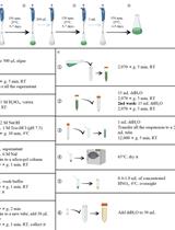
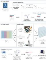
Cell populations and tissues exhibit unique gene expression profiles, which allow for characterizing and distinguishing cellular subtypes. Monitoring gene expression of cell type–specific markers can indicate cell status such as proliferation, stress, quiescence, or maturation. Quantitative reverse transcriptase PCR (qRT-PCR) allows quantifying RNA expression of cell type–specific markers and distinguishing one cell type from another. However, qRT-PCR methods such as TaqMan technology require fluorescent reporters to characterize target genes and are challenging to scale up as they need different probes for each reaction. Bulk or single-cell RNA transcriptomics is time-consuming and expensive. Processing RNA sequencing data can take several weeks, which is not optimal for quality control and monitoring gene expression, e.g., during a differentiation paradigm of induced pluripotent stem cells (iPSCs) into a specialized cell type.A more cost-effective assay is based on SYBR Green technology. SYBR Green is a nucleic acid dye that binds to double-stranded DNA, absorbs blue light at 497 nm, and emits green light at 520 nm up to 1,000-fold upon intercalation with double-stranded DNA. Amplification of a region of interest can be quantified based on the level of fluorescence intensity when normalized to a housekeeping gene and compared to control conditions. Previously, we established a SYBR Green qRT-PCR protocol to characterize samples using a limited set of markers plated on a 96-well plate.Here, we optimize the process and increase throughput to a 384-well format and compare mRNA expression to distinguish iPSC-derived neuronal subtypes from each other by increasing the number of genes, cell types, and differentiation time points. In this protocol, we develop the following: i) using the command-line version of Primer3 software, we design primers more easily and quickly for the gene of interest; ii) using a 384-well plate format, electronic multichannel pipettes, and pipetting robots, we analyze four times more genes on a single plate while using the same volume of reagents as in a 96-well plate. The advantages of this protocol are the increased throughput of this SYBR Green assay while limiting pipetting errors/inconsistencies, reagent use, cost, and time.Graphical overviewFigure 1. Overall optimized SYBR Green qRT-PCR workflow. (A) Primers are designed through the command-line version of Primer3. The program takes a couple of files as arguments: 1) an input file containing a sequence of the region of interest and a target, and 2) settings file with custom settings and primer picking conditions. The results are saved to a text file, checked for secondary and tertiary structures, then synthesized. (B) Primers are then plated using either multichannel pipettes with a pipetting aid or an automated pipetting robot. Plates are left to dry at room temperature and can be stored for an indefinite time. (C) Meanwhile, RNA is extracted from cell samples, reverse-transcribed into cDNA, then plated onto pre-coated 384-well plates. SYBR Green qRT-PCR is run and analyzed with QuantStudio software and Microsoft Excel.

Correction

Correction Notice: Assessment of Chemosensory Response to Volatile Compounds in Healthy, Aged, and Neurodegenerative Caenorhabditis elegans Models

Correction Notice: Assessment of Chemosensory Response to Volatile Compounds in Healthy, Aged, and Neurodegenerative Caenorhabditis elegans Models

CC Cira Crespo
RG Roberto Grau
313 Views
Jun 5, 2023