- Protocols

- Articles and Issues
- For Authors
- About
- Become a Reviewer

Past Issue in 2015
Volume: 5, Issue: 21
Cancer Biology
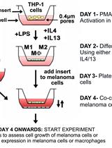
Differentiation of THP1 Cells into Macrophages for Transwell Co-culture Assay with Melanoma Cells
Isolation and Flow-cytometric Analysis of Mouse Intestinal Crypt Cells
Capturing the Driving Role of Tumor-host Crosstalk in a Dynamical Model of Tumor Growth

Cell Biology
Culture of Megakaryocytes from Human Peripheral Blood Mononuclear Cells


Assessment of Brown Adipocyte Thermogenic Function by High-throughput Respirometry
Microbiology
Chromatin Immunoprecipitation (ChIP) Assay for Detecting Direct and Indirect Protein – DNA Interactions in Magnaporthe oryzae


Determination of Fructokinase Activity from Zobellia galactanivorans


Determination of Mannitol-2-dehydrogenase Activity
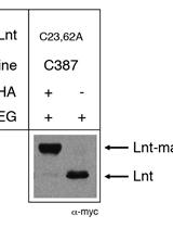
Substituted Cysteine Accessibility Method for Topology and Activity Studies of Membrane Enzymes Forming Thioester Acyl Intermediates in Bacteria
Neuroscience

Electroretinogram (ERG) Recordings from Drosophila


Plant Science
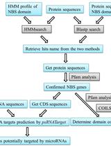
Computational Identification of MicroRNA-targeted Nucleotide-binding Site-leucine-rich Repeat Genes in Plants


Observation of Chloroplast Movement in Vallisneria
In vitro Phosphorylation Assay of Putative Blue-light Receptor Phototropins Using Microsomal and Plasma-membrane Fractions Prepared from Vallisneria Leaves
Stem Cell

Isolation of Murine Adipose Tissue-derived Mesenchymal Stromal Cells (mASCs) and the Analysis of Their Proliferation in vitro

Skeletal Myogenesis in vitro