- Protocols

- Articles and Issues
- For Authors
- About
- Become a Reviewer

Past Issue in 2023
Volume: 13, Issue: 4

Biochemistry
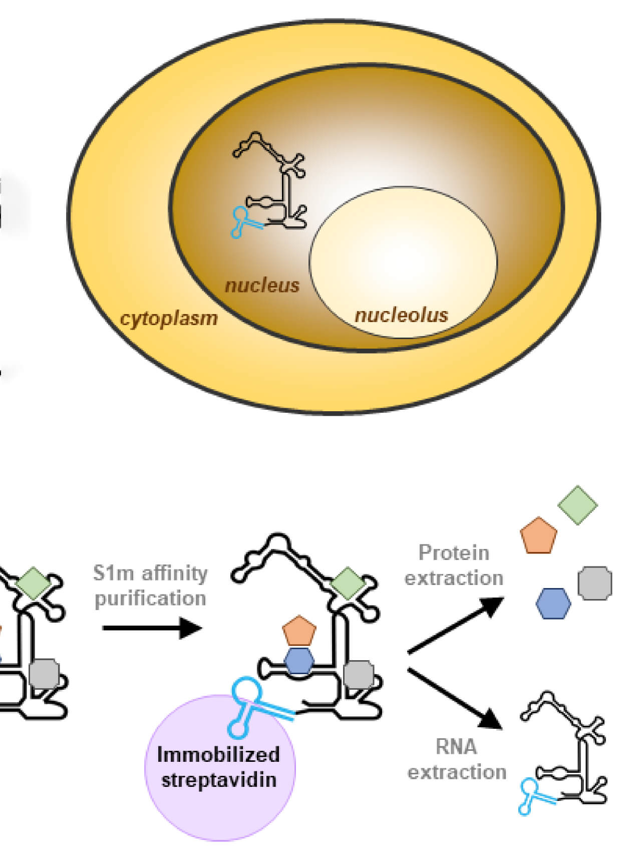
A Novel Method to Isolate RNase MRP Using RNA Streptavidin Aptamer Tags
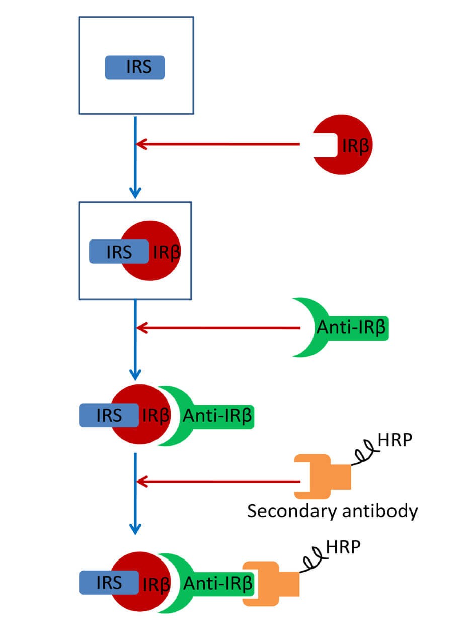
Far-western Blotting Detection of the Binding of Insulin Receptor Substrate to the Insulin Receptor
Biological Sciences
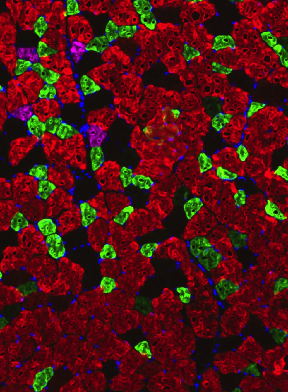
Comprehensive Analyses of Muscle Function, Lean and Muscle Mass, and Myofiber Typing in Mice
Biophysics
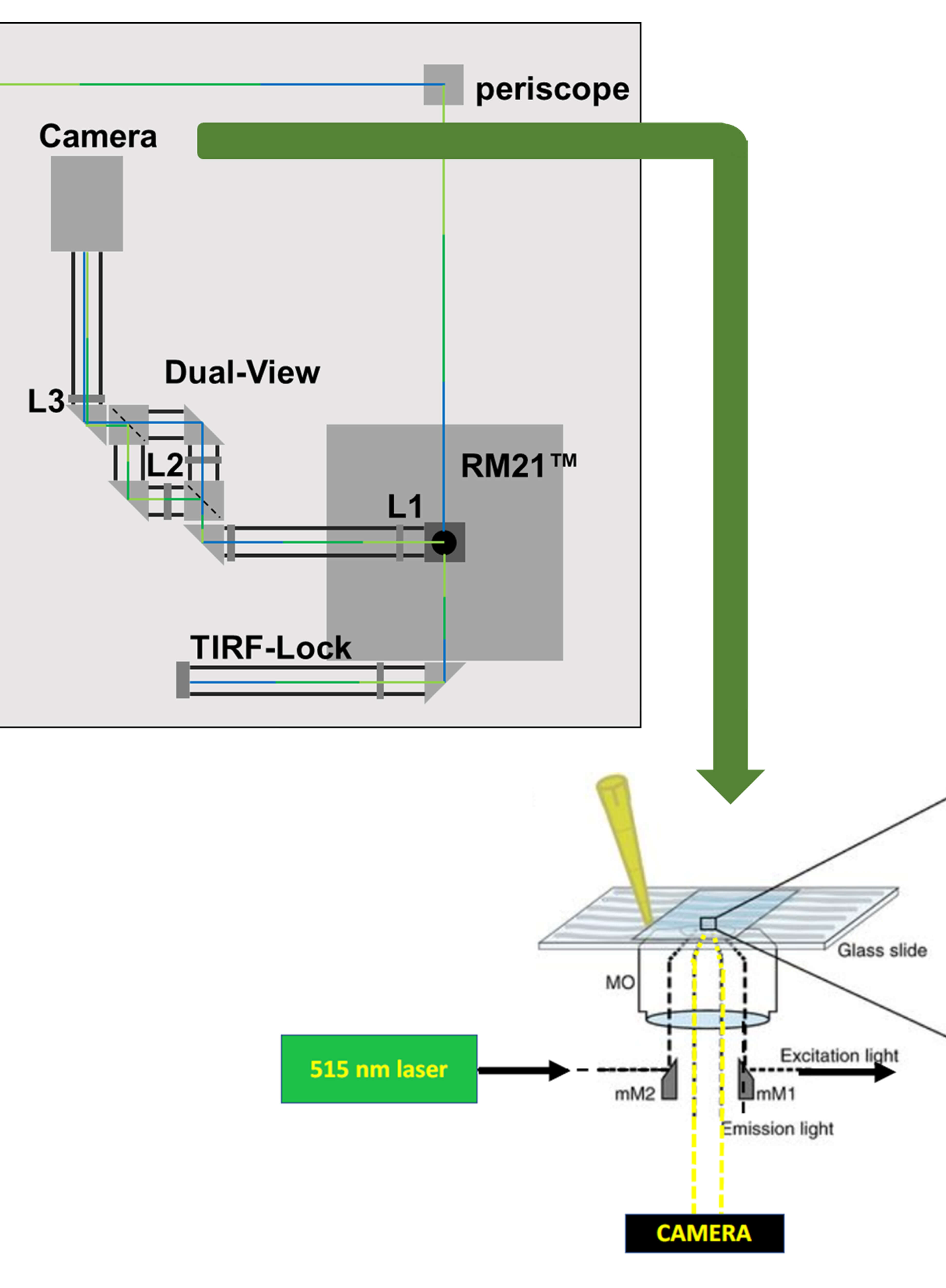
Imaging Membrane Proteins Using Total Internal Reflection Fluorescence Microscopy (TIRFM) in Mammalian Cells
Cell Biology
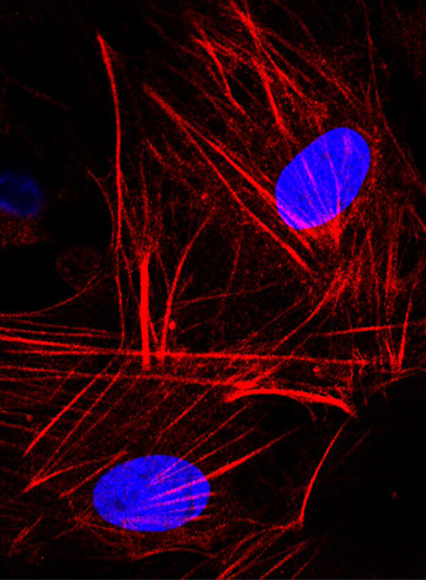
Isolation and Culture of Primary Fibroblasts from Neonatal Murine Hearts to Study Cardiac Fibrosis
Immunology
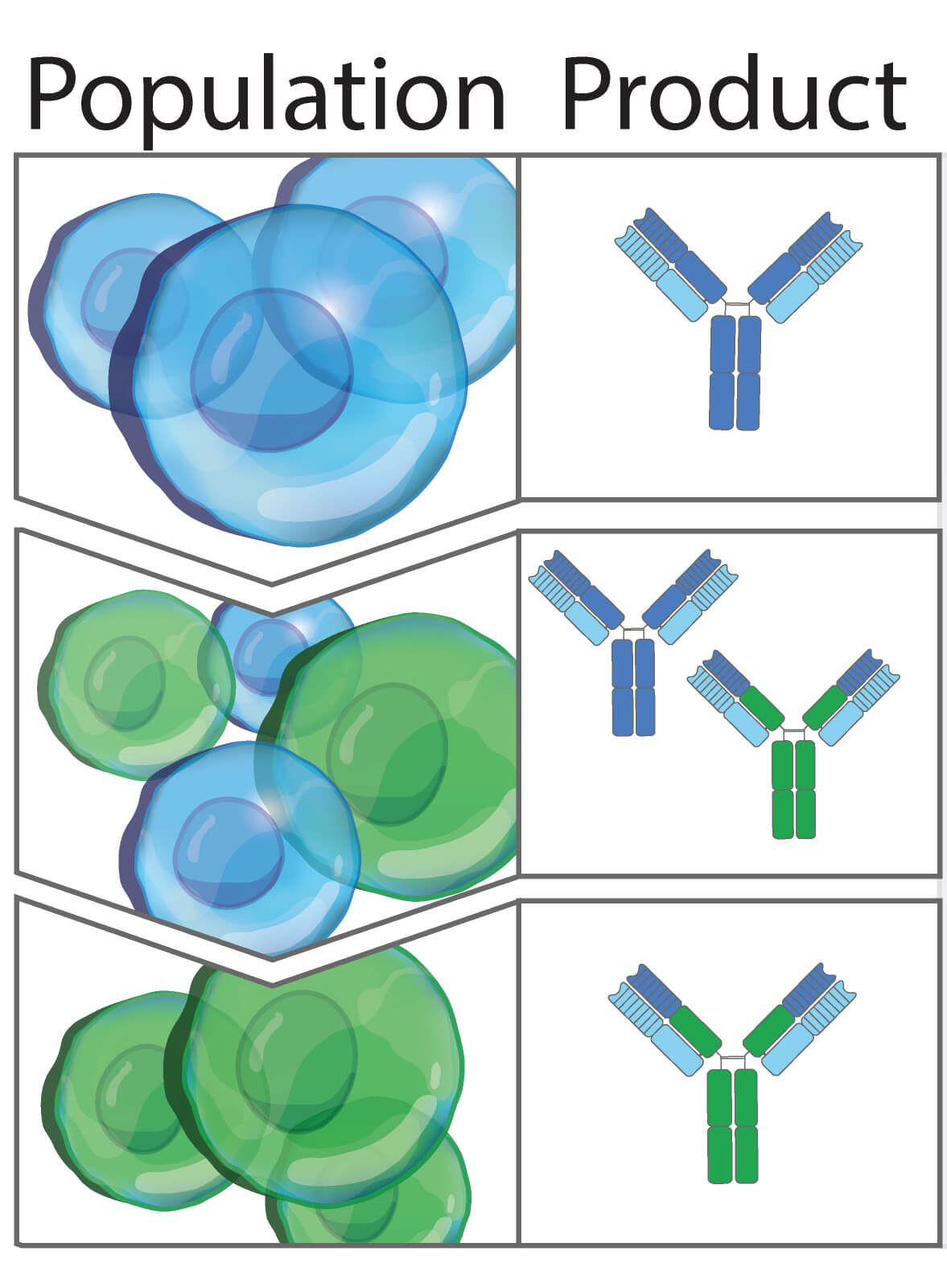
CRISPR/Cas9-based Engineering of Immunoglobulin Loci in Hybridoma Cells
Neuroscience
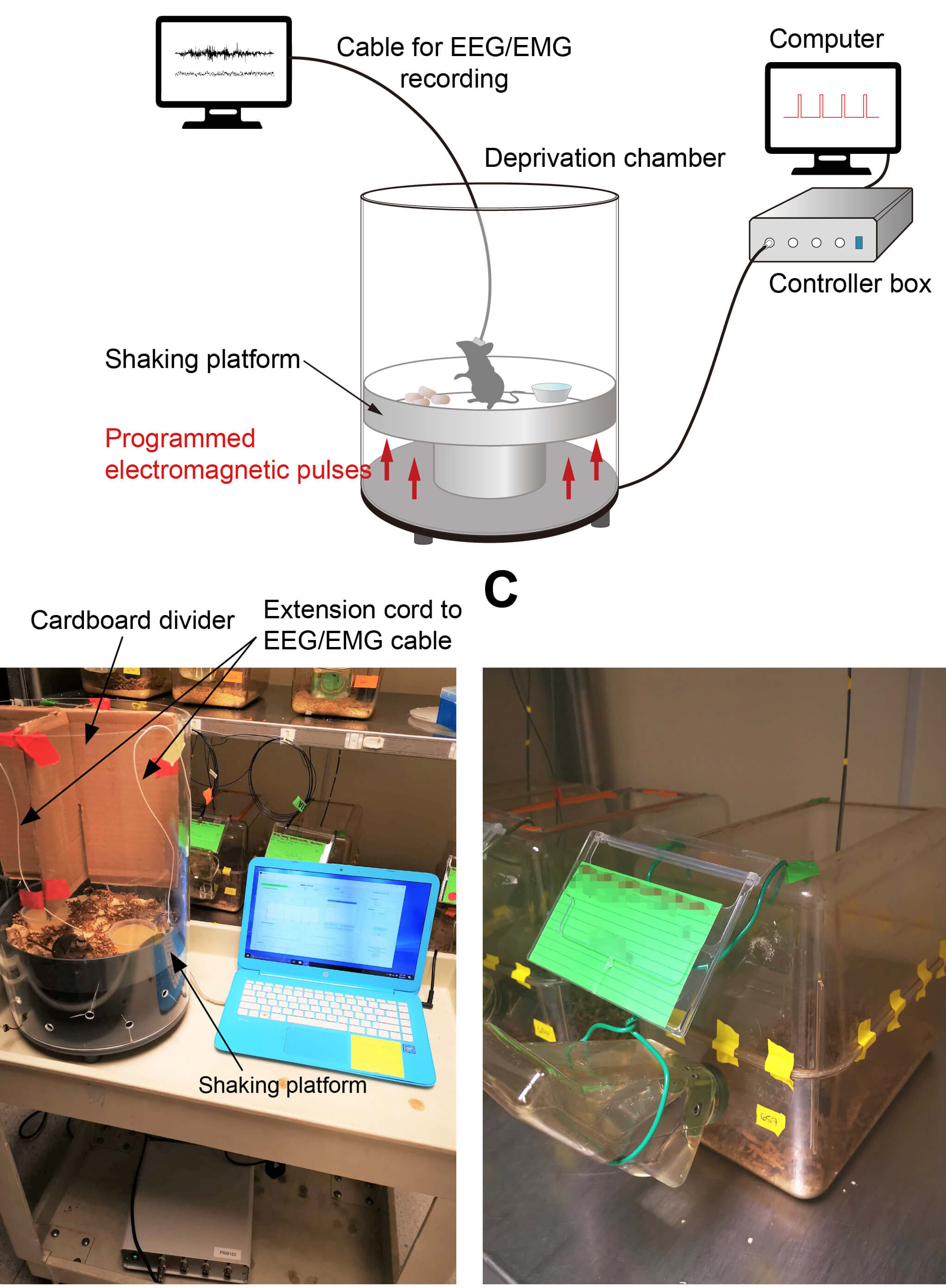
Automated Sleep Deprivation Setup Using a Shaking Platform in Mice

Detection of Zebrafish Retinal Proteins by Infrared Western Blotting
Plant Science
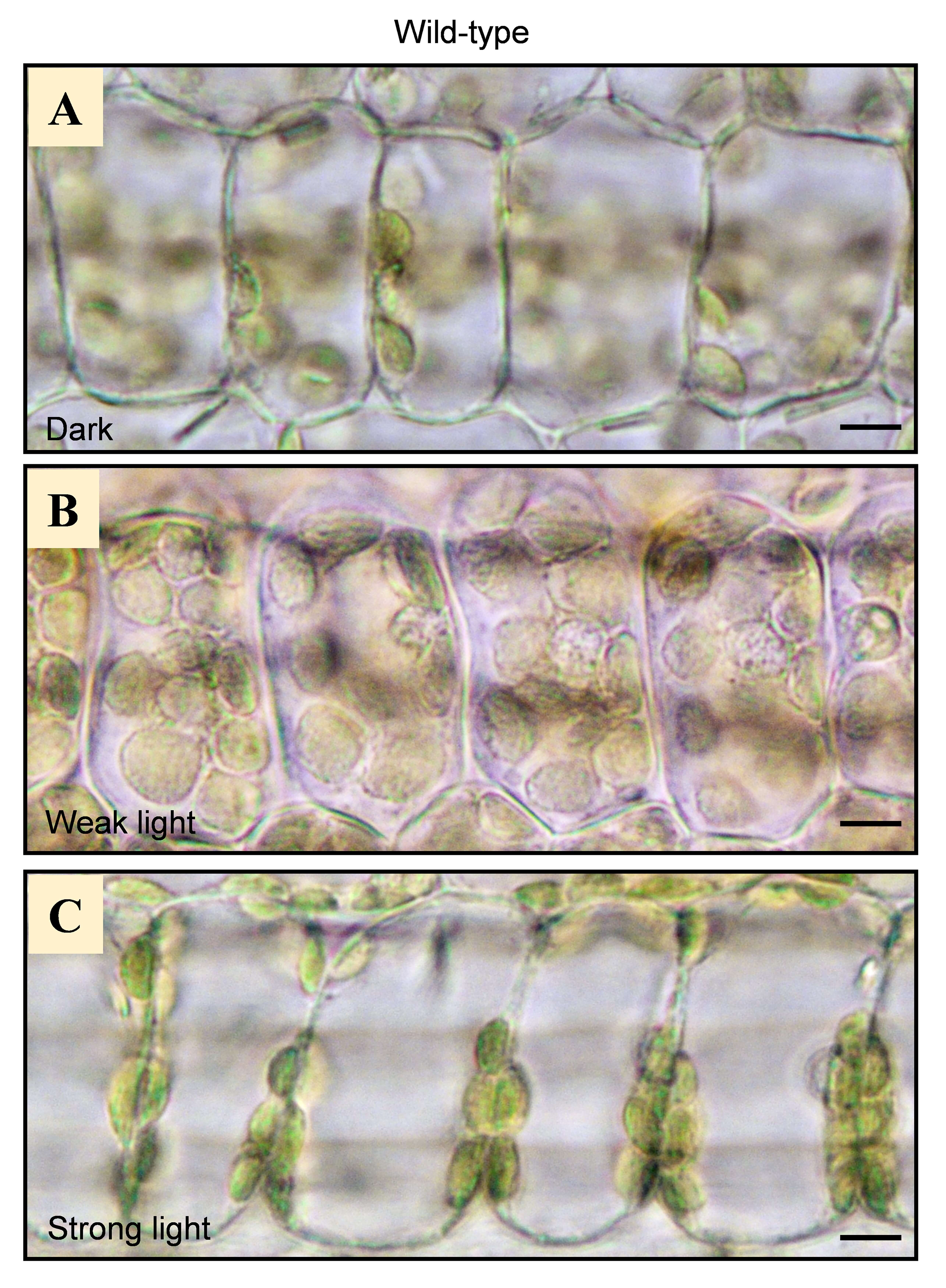
Imaging of Chloroplast Movement Responses to Light Stimulation in Different Intensities in Rice