- Protocols

- Articles and Issues
- For Authors
- About
- Become a Reviewer

Past Issue in 2022
Volume: 12, Issue: 11
Biochemistry
H2O2 Release Assay
Biological Engineering
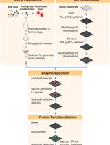
Patterned Substrate of Mobile and Immobile Ligands to Probe EphA2 Receptor Clustering
Cancer Biology
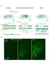
Plasma Membrane Wounding and Repair Assays for Eukaryotic Cells
Cell Biology
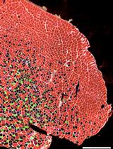
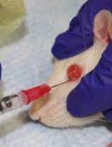
Electroporation of Small Interfering RNAs into Tibialis Anterior Muscles of Mice
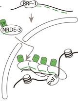
Labelling of Active Transcription Sites with Argonaute NRDE-3—Image Active Transcription Sites in vivo in Caenorhabditis elegans
Developmental Biology

Simple Methods for Permanent or Transient Denervation in Mouse Sciatic Nerve Injury Models

Efficient Superovulation and Egg Collection from Mice
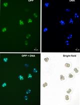
Click-iT® Plus OPP Alexa Fluor® Protein Synthesis Assay in Embryonic Cells
Immunology
Assessing the Presence of Hematopoietic Stem and Progenitor Cells in Mouse Spleen
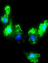
Flow Cytometric Characterization of Macrophages Infected in vitro with Salmonella enterica Serovar Typhimurium Expressing Red Fluorescent Protein
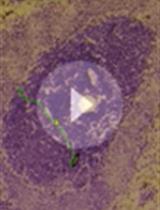
Protocol to Isolate Germinal Centers by Laser Microdissection
Microbiology
A Modified Fluctuation Assay with a CAN1 Reporter in Yeast
Plant Science
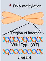
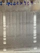
Investigation of Transposon DNA Methylation and Copy Number Variation in Plants Using Southern Hybridisation