- Protocols

- Articles and Issues
- For Authors
- About
- Become a Reviewer

Past Issue in 2022
Volume: 12, Issue: 1
Biochemistry
A Simple Technique for Direct Immobilization of Target Enzymes from Cell Lysates Based on the SpyTag/SpyCatcher Spontaneous Reaction
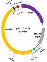
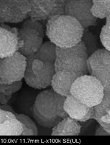
Heterologous Expression and High Degree Purification of the Restriction Endonuclease SauUSI
Developmental Biology
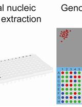
Total Nucleic Acid Extraction from Single Zebrafish Embryos for Genotyping and RNA-seq
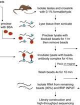
Identification of Protein-RNA Interactions in Mouse Testis Tissue Using fRIP
Immunology
Evaluating Human Natural Killer Cells Antibody-dependent Cellular Cytotoxicity (ADCC) Using Plate-bound Anti-CD16 Antibodies
Induction of Acute or Disseminating Bacterial Pneumonia in Mice and Sampling of Infected Organs for Studying the Host Response to Bacterial Pneumonia
Static Adhesion Assay for Human Peripheral Blood Mononuclear Cells
Medicine
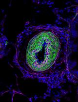
Chronic Daily House Dust Mite Exposure in Mice is an Effective Model to Quantify the Effect of Pharmacologic Agents on Discrete Stages of Artery Remodeling in Pulmonary Hypertension

Microbiology
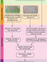
Real-Time Analysis of Mitochondrial Electron Transport Chain Function in Toxoplasma gondii Parasites Using a Seahorse XFe96 Extracellular Flux Analyzer
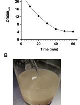
Isolation of Mitochondria from Ustilago maydis Protoplasts
Molecular Biology
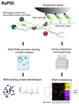
An Aptamer-based mRNA Affinity Purification Procedure (RaPID) for the Identification of Associated RNAs (RaPID-seq) and Proteins (RaPID-MS) in Yeast
Neuroscience
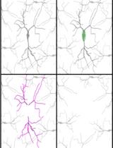
Subcellular RNA-seq for the Analysis of the Dendritic and Somatic Transcriptomes of Single Neurons
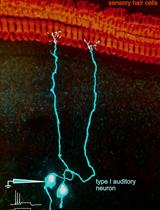
Patch-clamp Recordings and Single Fiber Labeling from Spiral Ganglion Somata in Acutely Prepared Semi-intact Cochleae from Neonatal Rats
Stem Cell
Live-cell Imaging and Analysis of Germline Stem Cell Mitosis in Caenorhabditis elegans
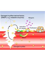
An in vitro Mechanical Damage Model of Isolated Myofibers in a Floating Culture Condition
Update