- Protocols

- Articles and Issues
- For Authors
- About
- Become a Reviewer

Past Issue in 2021
Volume: 11, Issue: 7
Biochemistry
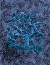
Expression and Purification of the Human Cation-chloride Cotransporter KCC1 from HEK293F Cells for Structural Studies
Biophysics
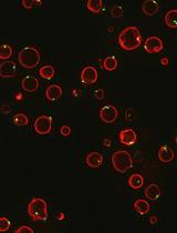
FRET-based Microscopy Assay to Measure Activity of Membrane Amino Acid Transporters with Single-transporter Resolution
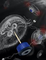
Developing Biohybrid Robotic Jellyfish (Aurelia aurita) for Free-swimming Tests in the Laboratory and in the Field
Cancer Biology
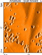
Atomic Force Microscopy to Characterize Ginger Lipid-Derived Nanoparticles (GLDNP)
Developmental Biology
A Workflow for High-pressure Freezing and Freeze Substitution of the Caenorhabditis elegans Embryo for Ultrastructural Analysis by Conventional and Volume Electron Microscopy
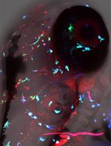
Analysis of TORC1-body Formation in Budding Yeast
Immunology
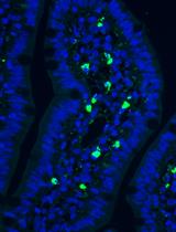
Brain-localized and Intravenous Microinjections in the Larval Zebrafish to Assess Innate Immune Response
A Potent Vaccine Delivery System
Using the Cecal Ligation and Puncture Model of Sepsis to Induce Rats to Multiple Organ Dysfunction
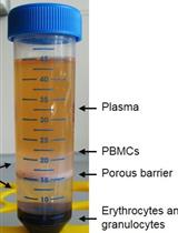
Ex vivo Assessment of Mitochondrial Function in Human Peripheral Blood Mononuclear Cells Using XF Analyzer
Molecular Biology
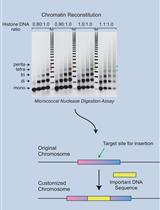
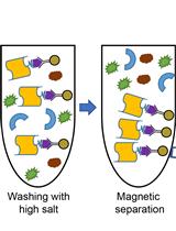
Reconstitution of Chromatin by Stepwise Salt Dialysis
Neuroscience
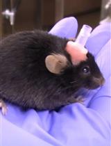
Cranioplastic Surgery and Acclimation Training for Awake Mouse fMRI
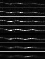
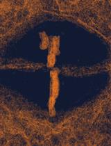
Imaging Microtubules in vitro at High Resolution while Preserving their Structure
Plant Science
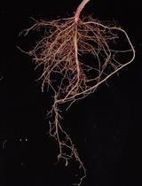
Measurements of Root Colonized Bacteria Species
In vitro Reconstitution Assays of Arabidopsis 20S Proteasome
A Novel Method to Construct Binary CRISPR Vectors for Plant Transformation by Single Round of PCR Amplification