- Protocols

- Articles and Issues
- For Authors
- About
- Become a Reviewer

Past Issue in 2021
Volume: 11, Issue: 6
Biochemistry

Monitoring Real-time Temperature Dynamics of a Short RNA Hairpin Using Förster Resonance Energy Transfer and Circular Dichroism
Biological Engineering
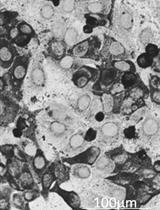
Multilayered Fabrication Assembly Technique to Engineer a Corneal Stromal Equivalent
Cancer Biology
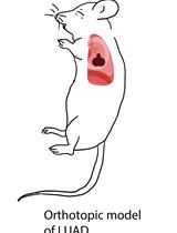
Preparation of an Orthotopic, Syngeneic Model of Lung Adenocarcinoma and the Testing of the Antitumor Efficacy of Poly(2-oxazoline) Formulation of Chemo-and Immunotherapeutic Agents
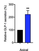
Intestinal Co-culture System to Study TGR5 Agonism and Gut Restriction
Preparation and Characterization of Poly(2-oxazoline) Micelles for the Solubilization and Delivery of Water Insoluble Drugs
Cell Biology

Retention Using Selective Hooks (RUSH) Cargo Sorting Assay for Live-cell Vesicle Tracking in the Secretory Pathway Using HeLa Cells
Immunology
Murine Monocyte and Macrophage Culture
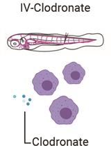
Liposomal Clodronate-mediated Macrophage Depletion in the Zebrafish Model
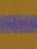
An Image-based Dynamic High-throughput Analysis of Adherent Cell Migration

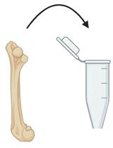
Quantitative Measurement of Mucolytic Enzymes in Fecal Samples
Microbiology
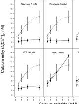
A Spectrofluorophotometrical Method Based on Fura-2-AM Probe to Determine Cytosolic Ca2+ Level in Pseudomonas syringae Complex Bacterial Cells
Detection and Quantification of African Swine Fever Virus in MA-104 Cells

Molecular Biology
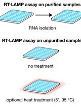
Colorimetric RT-LAMP and LAMP-sequencing for Detecting SARS-CoV-2 RNA in Clinical Samples
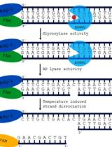
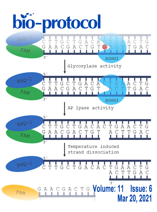
Real-time Base Excision Repair Assay to Measure the Activity of the 8-oxoguanine DNA Glycosylase 1 in Isolated Mitochondria of Human Skin Fibroblasts
Neuroscience
Investigate Synaptic Vesicles Mobility in Neuronal Culture Axons by FRAP Imaging
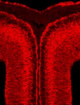
Ligand and Carbohydrate Engagement (LACE) Assay and Fluorescence Quantification on Murine Neural Tissue
A Time Duration Discrimination Task for the Study of Elapsed Time Processing in Rats
