- Protocols

- Articles and Issues
- For Authors
- About
- Become a Reviewer

Past Issue in 2020
Volume: 10, Issue: 9
Biochemistry
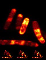
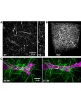
Confocal and Super-resolution Imaging of RNA in Live Bacteria Using a Fluorogenic Silicon Rhodamine-binding Aptamer


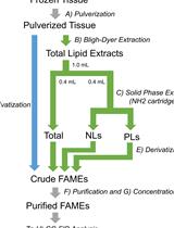
Quantification of Fatty Acids in Mammalian Tissues by Gas Chromatography–Hydrogen Flame Ionization Detection
Cancer Biology
Colorimetric RhoB GTPase Activity Assay
Developmental Biology
Live Cell Imaging of Male Meiosis in Arabidopsis by a Landmark-based System
Application of Mechanical Forces on Drosophila Embryos by Manipulation of Microinjected Magnetic Particles
A Modified Semisolid Clonal Culture for Identification of B-1 and B-2 Progenitor Colony Forming Ability of Mouse Embryonic Hemogenic Endothelial Cells

Immunology
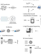
HIV-CRISPR: A CRISPR/Cas9 Screening Method to Identify Genes Affecting HIV Replication
Evaluation of B Cell Proliferation in vivo by EdU Incorporation Assay
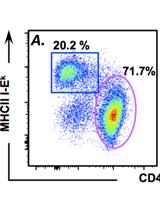
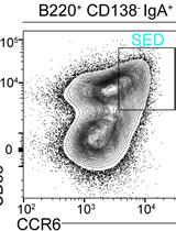
Assessing in vitro and in vivo Trogocytosis By Murine CD4+ T cells

Microbiology
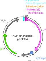
A Sensitive Coupled Enzyme Assay for Measuring Kinase and ATPase Kinetics Using ADP-Specific Hexokinase

Viral Double-Stranded RNA Detection by DNase I and Nuclease S1 digestions in Leishmania parasites
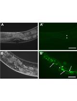
Quantification of Bacteria Residing in Caenorhabditis elegans Intestine

Molecular Biology
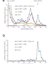
Split Nano Luciferase-based Assay to Measure Assembly of Japanese Encephalitis Virus
Neuroscience
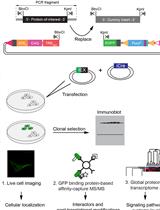
Rapid Generation of Human Neuronal Cell Models Enabling Inducible Expression of Proteins-of-interest for Functional Studies
Systems Biology
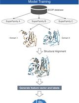
Sequence Alignment Using Machine Learning for Accurate Template-based Protein Structure Prediction


Correction