- Protocols

- Articles and Issues
- For Authors
- About
- Become a Reviewer

Past Issue in 2020
Volume: 10, Issue: 5
Biochemistry
Assembly of Genetic Circuits with the Mammalian ToolKit

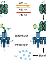
Production, Purification and Characterization of Recombinant Biotinylated Phytochrome B for Extracellular Optogenetics
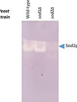
Manganese Superoxide Dismutase Activity Assay in the Yeast Saccharomyces cerevisiae
Developmental Biology
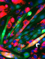
Lipid Mixing Assay for Murine Myoblast Fusion and Other Slow Cell-cell Fusion Processes
Immunology
Optogenetic Tuning of Ligand Binding to The Human T cell Receptor Using The opto-ligand-TCR System
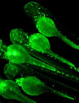
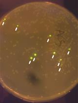
Zebrafish Bacterial Infection Assay to Study Host-Pathogen Interactions
Microbiology
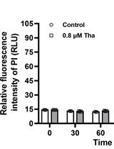
Contemporaneous Measurement of Outer and Inner Membrane Permeability in Gram-negative Bacteria

Growth Recovery Assay and FACS-based Population Sorting Following Territorial Exclusion in Proteus mirabilis
Molecular Biology
Cell-free Reconstitution of the Packaging of Cargo Proteins into Vesicles at the trans Golgi Network

Neuroscience

Delayed Alternation Task for the Study of Spatial Working and Long Term Memory in Rats
Plant Science

Tandem Tag Assay Optimized for Semi-automated in vivo Autophagic Activity Measurement in Arabidopsis thaliana roots


Quantification of Protein Enrichment at Plasmodesmata

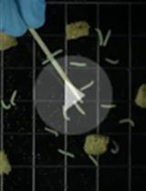
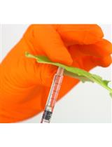
Insect Feeding Assays with Spodoptera exigua on Arabidopsis thaliana

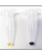
Single-run HPLC Quantification of Plant Cell Wall Monosaccharides
Systems Biology
mRNA Extraction from Gill Tissue for RNA-sequencing