- Protocols

- Articles and Issues
- For Authors
- About
- Become a Reviewer

Past Issue in 2020
Volume: 10, Issue: 4
Biochemistry
Photoactivable Cholesterol as a Tool to Study Interaction of Influenza Virus Hemagglutinin with Cholesterol
Cell Biology
CRISPR-Cas9 Genome Editing of Plasmodium knowlesi
Estimating Cellular Abundances of Halo-tagged Proteins in Live Mammalian Cells by Flow Cytometry
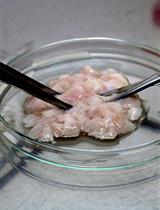
Preparation of Single Epithelial Cells Suspension from Mouse Mammary Glands
ChIP-Seq from Limited Starting Material of K562 Cells and Drosophila Neuroblasts Using Tagmentation Assisted Fragmentation Approach
Developmental Biology
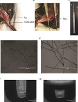
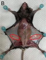
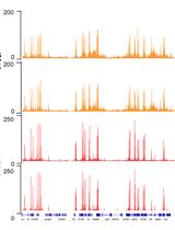
RNA Sequencing of Single Myofibers from Mus musculus
Immunology
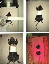
Skin Transplantation and Lymphoid Organ Analysis in Mice
Molecular Biology
Assessing Self-interaction of Mammalian Nuclear Proteins by Co-immunoprecipitation

Surface Plasmon Resonance Analysis of the Protein-protein Binding Specificity Using Autolab ESPIRIT


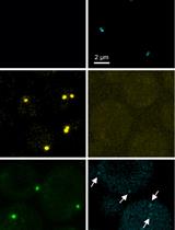
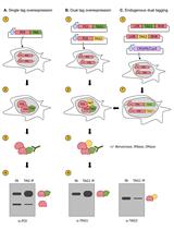
Super-resolution Microscopy-based Bimolecular Fluorescence Complementation to Study Protein Complex Assembly and Co-localization

Neuroscience
Induction of Temporal Lobe Epilepsy in Mice with Pilocarpine


Method for Primary Epithelial Cell Culture from the Rat Choroid Plexus


Hippocampal Unicellular Recordings and Hippocampal-dependent Innate Behaviors in an Adolescent Mouse Model of Alzheimer’s disease
Plant Science
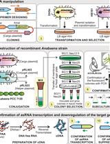
Construction of Antisense RNA-mediated Gene Knock-down Strains in the Cyanobacterium Anabaena sp. PCC 7120
Stem Cell
Mesenchymal Stromal Cells Derived from Bone Marrow and Adipose Tissue: Isolation, Culture, Characterization and Differentiation


