- Protocols

- Articles and Issues
- For Authors
- About
- Become a Reviewer

Past Issue in 2020
Volume: 10, Issue: 2
Biochemistry
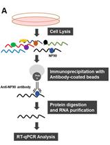
Ribonucleoprotein Immunoprecipitation (RIP) Analysis
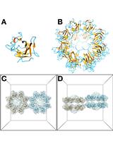
Protocols for Processing and Interpreting cryoEM Data Using Bsoft: A Case Study of the Retinal Adhesion Protein, Retinoschisin
Biophysics
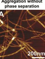
Studying Protein Aggregation in the Context of Liquid-liquid Phase Separation Using Fluorescence and Atomic Force Microscopy, Fluorescence and Turbidity Assays, and FRAP
Characterizing the Two-photon Absorption Properties of Fluorescent Molecules in the 680-1300 nm Spectral Range
Cancer Biology
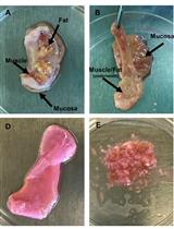
Bone-in-culture Array to Model Bone Metastasis in ex vivo Condition
Isolation of Stem Cells, Endothelial Cells and Pericytes from Human Infantile Hemangioma
Immunology
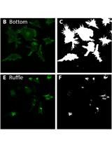
Automated Analysis of Cell Surface Ruffling: Ruffle Quantification Macro
.jpeg)


Quantifying HIV-1-Mediated Gut CD4+ T Cell Death in the Lamina Propria Aggregate Culture (LPAC) Model
Microbiology
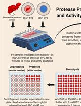
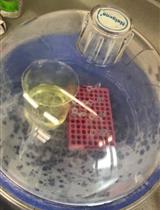
Study of Microbial Extracellular Vesicles: Separation by Density Gradients, Protection Assays and Labelling for Live Tracking
Sulfatase Assay to Determine Influence of Plants on Microbial Activity in Soil
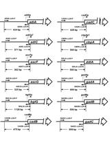
Measurement of the Promoter Activity in Escherichia coli by Using a Luciferase Reporter
A Radioactive-free Kinase Inhibitor Discovery Assay Against the Trypanosoma brucei Glycogen Synthase Kinase-3 short (TbGSK-3s)

Neuroscience
A Single Test to Study Social Behavior and Repetitive Self-grooming in Mice
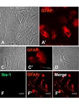
A Simple and Efficient Method for Concomitant Isolation and Culture of Enriched Astroglial and Microglial Cells from the Rat Spinal Cord
Protocol for Measuring Free (Low-stress) Exploration in Rats
