- Protocols

- Articles and Issues
- For Authors
- About
- Become a Reviewer

Past Issue in 2019
Volume: 9, Issue: 18
Biochemistry
Subcellular Fractionation of Hela Cells for Lysosome Enrichment Using a Continuous Percoll-density Gradient
Cancer Biology
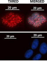
SIRF: A Single-cell Assay for in situ Protein Interaction with Nascent DNA Replication Forks
A Cell Culture Model that Mimics Physiological Tissue Oxygenation Using Oxygen-permeable Membranes

Cell Biology
Conjugation of Fab’ Fragments with Fluorescent Dyes for Single-molecule Tracking on Live Cells
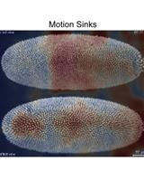
Characterization of Biological Motion Using Motion Sensing Superpixels
Supported Cell Membrane Sheets to Monitor Protein Assembly
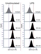
Quantitation of TLR4 Internalization in Response to LPS in Thioglycollate Elicited Peritoneal Mouse Macrophages by Flow Cytometry
Immunology
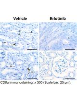
Immunohistochemical Staining of CD8α in Diabetic Mouse Kidney
Microbiology
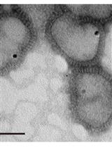
Cryo-transmission Electron Microscopy of Outer-inner Membrane Vesicles Naturally Secreted by Gram-negative Pathogenic Bacteria

Production of Quantum Dots-containing Influenza Virus Particles for Studying Viral Uncoating Processes
Molecular Biology
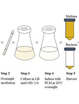
A PhoA-STII Based Method for Efficient Extracellular Secretion and Purification of Fab from Escherichia coli
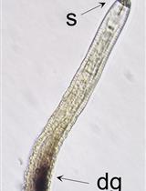
In situ Hybridization of Plant-parasitic Nematode Globodera pallida Juveniles to Detect Gene Expression


Using indCAPS to Detect CRISPR/Cas9 Induced Mutations
Neuroscience
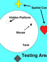
Simple Protocol for Distinguishing Drug-induced Effects on Spatial Memory Acquisition, Consolidation and Retrieval in Mice Using the Morris Water Maze


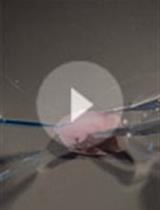
Explant Culture of the Embryonic Mouse Spinal Cord and Gene Transfer by ex vivo Electroporation


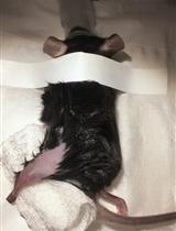
Sciatic Nerve Cut and Repair Using Fibrin Glue in Adult Mice
