- Protocols

- Articles and Issues
- For Authors
- About
- Become a Reviewer

Past Issue in 2018
Volume: 8, Issue: 9
Biochemistry
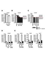
The Long-lived Protein Degradation Assay: an Efficient Method for Quantitative Determination of the Autophagic Flux of Endogenous Proteins in Adherent Cell Lines


Cancer Biology
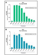
An Image-based Assay for High-throughput Analysis of Cell Proliferation and Cell Death of Adherent Cells

Cell Biology
Quantifying Podocytes and Parietal Epithelial Cells in Human Urine Using Liquid-based Cytology and WT1 Immunoenzyme Staining
Immunology
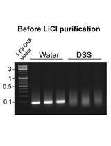
Purification of Total RNA from DSS-treated Murine Tissue via Lithium Chloride Precipitation

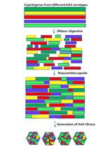
Nab Escaping AAV Mutants Isolated from Mouse Muscles
Microbiology
Heterologous Expression and Purification of the CRISPR-Cas12a/Cpf1 Protein


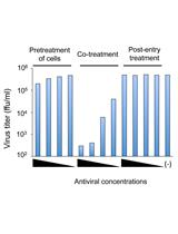
Time-of-addition and Temperature-shift Assays to Determine Particular Step(s) in the Viral Life Cycle that is Blocked by Antiviral Substance(s)

Infection Process Observation of Magnaporthe oryzae on Barley Leaves

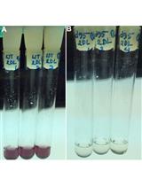
Glycogen and Extracellular Glucose Estimation from Cyanobacteria Synechocystis sp. PCC 6803
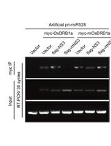
Detecting the Interaction of Double-stranded RNA Binding Protein, Viral Protein and Primary miRNA Transcript by Co-immunoprecipitation in planta

Assessing the Efficacy of Small Molecule Inhibitors in a Mouse Model of Persistent Norovirus Infection


A Modified Low-quantity RNA-Seq Method for Microbial Community and Diversity Analysis Using Small Subunit Ribosomal RNA

Molecular Biology
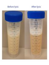
Mammalian Cell-derived Vesicles for the Isolation of Organelle Specific Transmembrane Proteins to Conduct Single Molecule Studies
Plant Science
Functional Evaluation of the Signal Peptides of Secreted Proteins

Evaluation of the Condition of Respiration and Photosynthesis by Measuring Chlorophyll Fluorescence in Cyanobacteria
Stem Cell
Cobblestone Area-forming Cell Assay of Mouse Bone Marrow Hematopoietic Stem Cells